113
Limbic system emotions + memory Veronika Němcová
Limbic systememotions + memory
Veronika Němcová
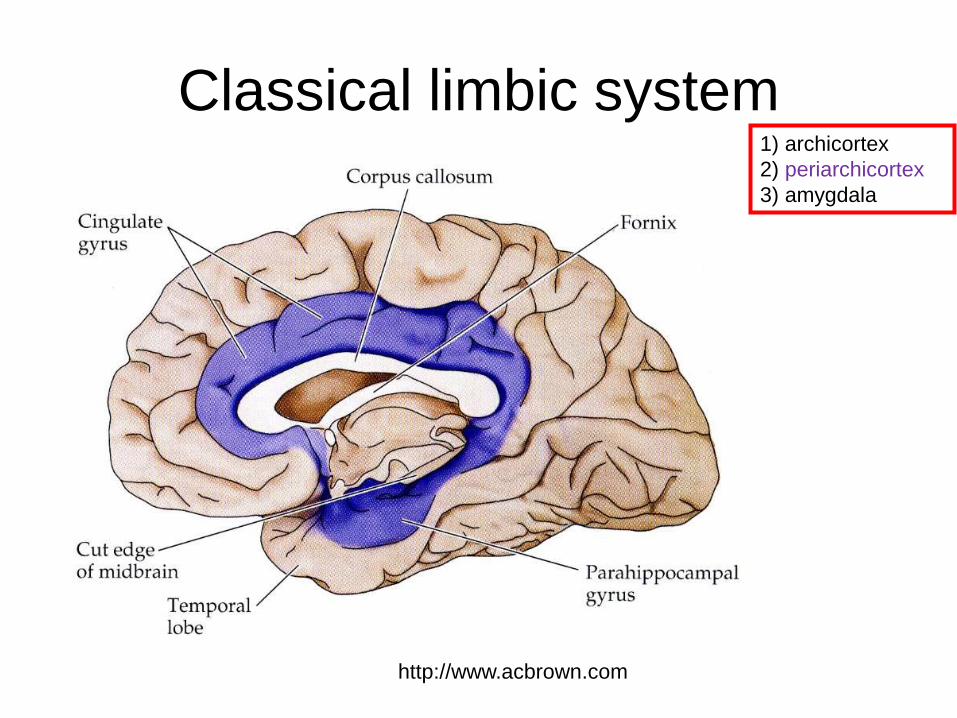
Classical limbic system
http://www.acbrown.com
1) archicortex
2) periarchicortex
3) amygdala
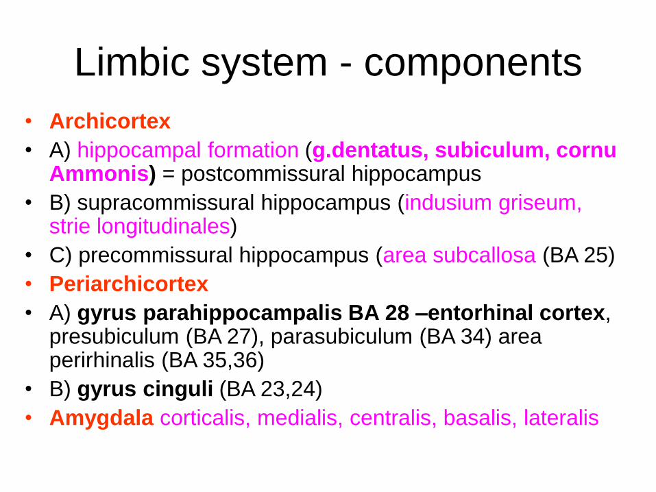
Limbic system - components
• Archicortex
• A) hippocampal formation (g.dentatus, subiculum, cornu Ammonis) = postcommissural hippocampus
• B) supracommissural hippocampus (indusium griseum, strie longitudinales)
• C) precommissural hippocampus (area subcallosa (BA 25)
• Periarchicortex
• A) gyrus parahippocampalis BA 28 –entorhinal cortex, presubiculum (BA 27), parasubiculum (BA 34) area perirhinalis (BA 35,36)
• B) gyrus cinguli (BA 23,24)
• Amygdala corticalis, medialis, centralis, basalis, lateralis
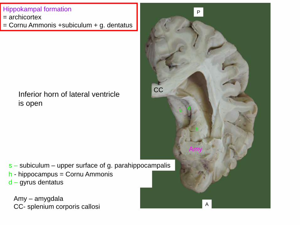
s d
h
Hippokampal formation
= archicortex
= Cornu Ammonis +subiculum + g. dentatus
P
A
Inferior horn of lateral ventricle
is open
h - hippocampus = Cornu Ammonis
d – gyrus dentatus
s – subiculum – upper surface of g. parahippocampalis
Amy
Amy – amygdala
CC- splenium corporis callosi
CC
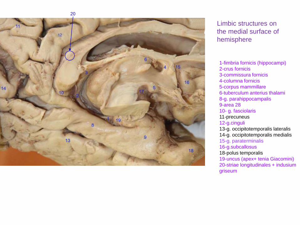
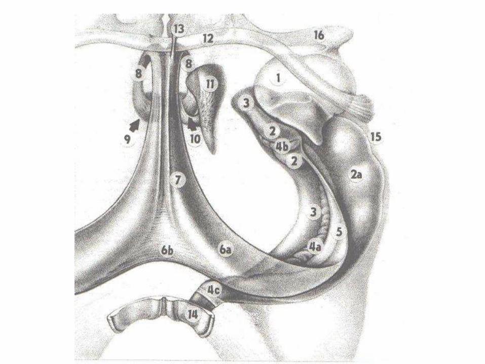
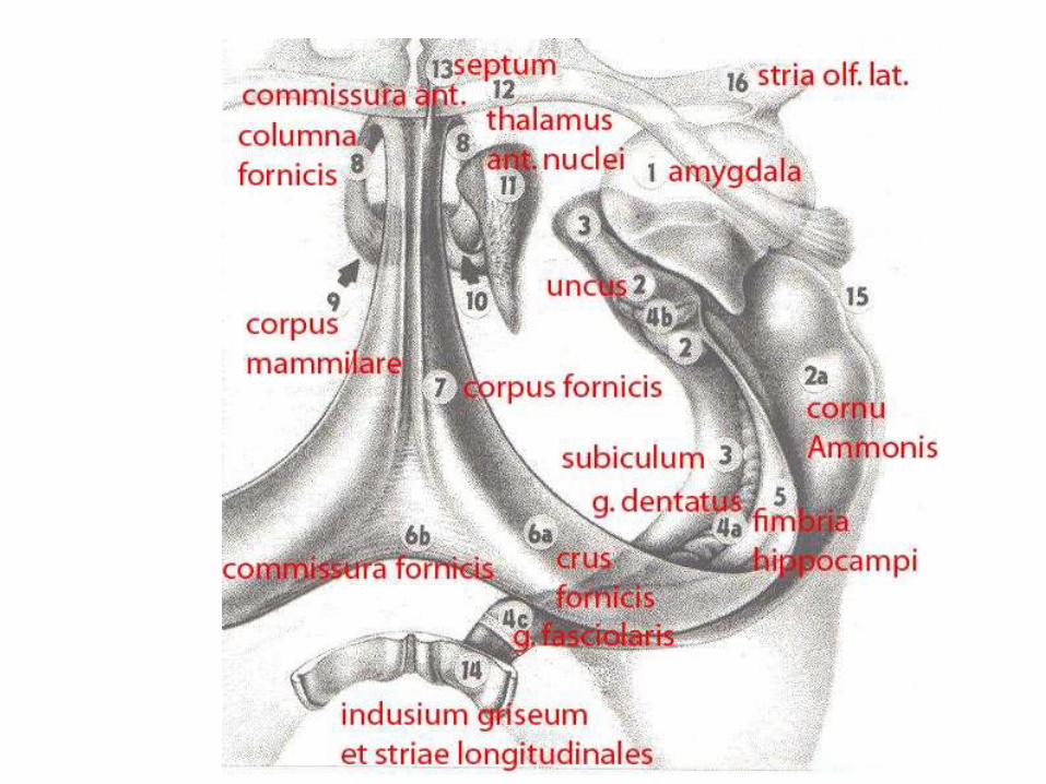
1-fimbria fornicis (hippocampi)
2-crus fornicis
3-commissura fornicis
4-columna fornicis
5-corpus mammillare
6-tuberculum anterius thalami
8-g. parahippocampalis
9-area 28
10- g. fasciolaris
11-precuneus
12-g.cinguli
13-g. occipitotemporalis lateralis
14-g. occipitotemporalis medialis
15-g. paraterminalis
16-g.subcallosus
18-polus temporalis
19-uncus (apex+ tenia Giacomini)
20-striae longitudinales + indusium
griseum
Limbic structures on
the medial surface of
hemisphere
20
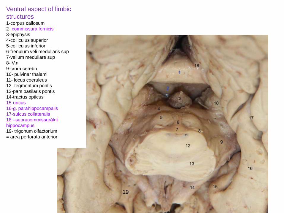
Ventral aspect of limbic
structures1-corpus callosum
2- commissura fornicis
3-epiphysis
4-colliculus superior
5-colliculus inferior
6-frenulum veli medullaris sup
7-vellum medullare sup
8-IV.n
9-crura cerebri
10- pulvinar thalami
11- locus coeruleus
12- tegmentum pontis
13-pars basilaris pontis
14-tractus opticus
15-uncus
16-g. parahippocampalis
17-sulcus collateralis
18 –supracommissurální
hippocampus
19- trigonum olfactorium
= area perforata anterior
19
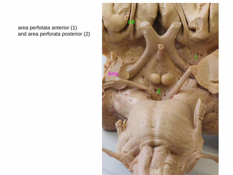
area perfotata anterior (1)
and area perforata posterior (2)
1
2
Amy
Olf
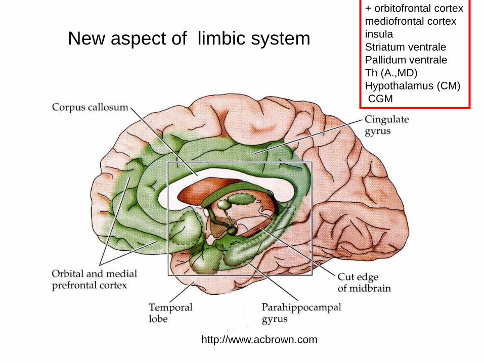
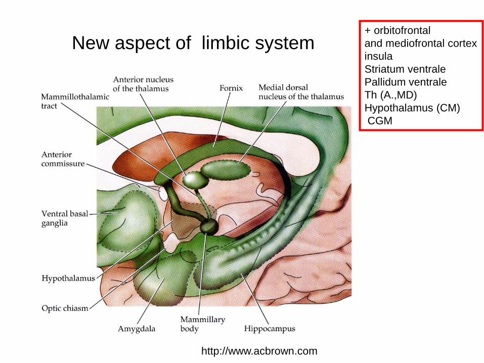
New aspect of limbic system
http://www.acbrown.com
+ orbitofrontal cortex
mediofrontal cortex
insula
Striatum ventrale
Pallidum ventrale
Th (A.,MD)
Hypothalamus (CM)
CGM
New aspect of limbic system
http://www.acbrown.com
+ orbitofrontal
and mediofrontal cortex
insula
Striatum ventrale
Pallidum ventrale
Th (A.,MD)
Hypothalamus (CM)
CGM
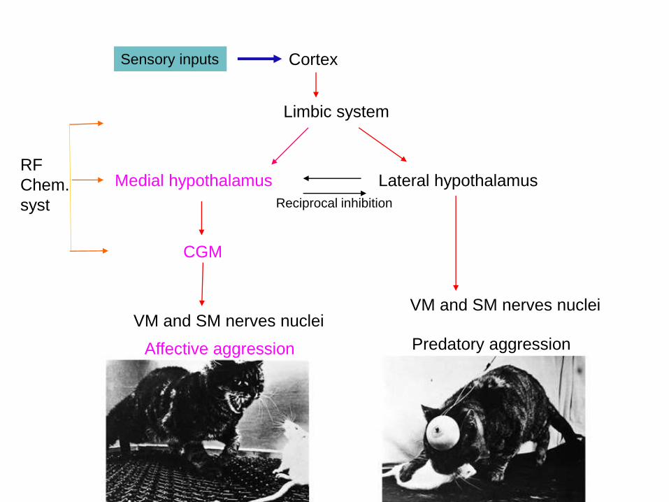
Medial hypothalamus Lateral hypothalamus
CGM
Cortex
Limbic system
VM and SM nerves nucleiVM and SM nerves nuclei
Reciprocal inhibition
Sensory inputs
Predatory aggressionAffective aggression
RF
Chem.
syst
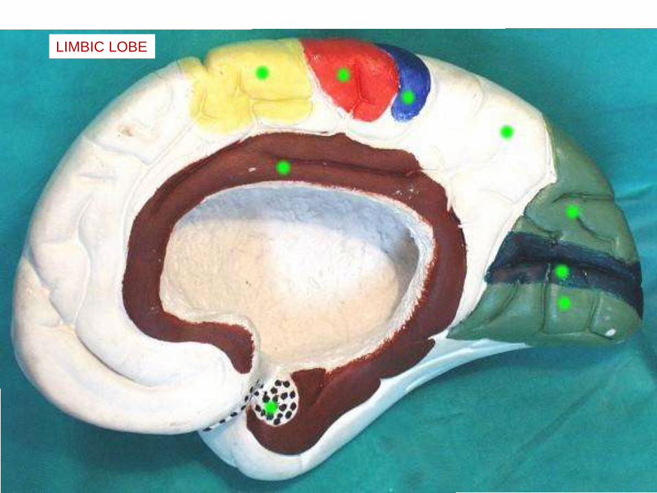
LIMBIC LOBE
A
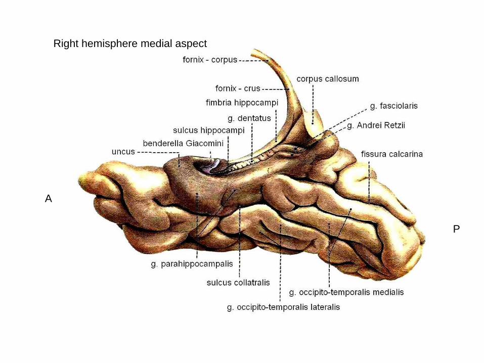
P
Right hemisphere medial aspect
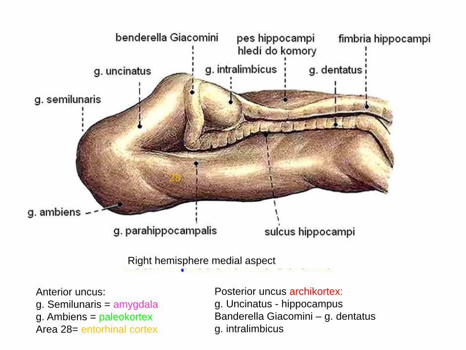
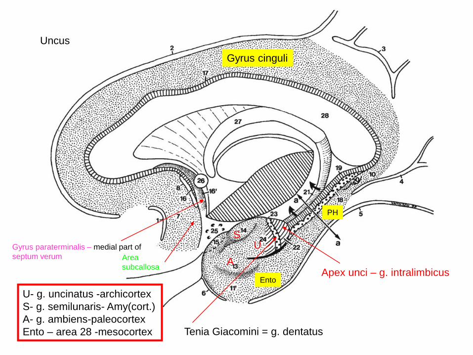
Anterior uncus:
g. Semilunaris = amygdala
g. Ambiens = paleokortex
Area 28= entorhinal cortex
Posterior uncus archikortex:
g. Uncinatus - hippocampus
Banderella Giacomini – g. dentatus
g. intralimbicus
Right hemisphere medial aspect
28
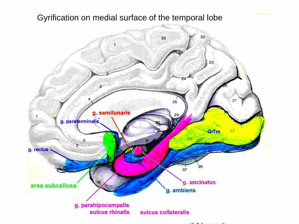
Gyrification on medial surface of the temporal lobe
Gyrification on medial surface of the temporal lobe
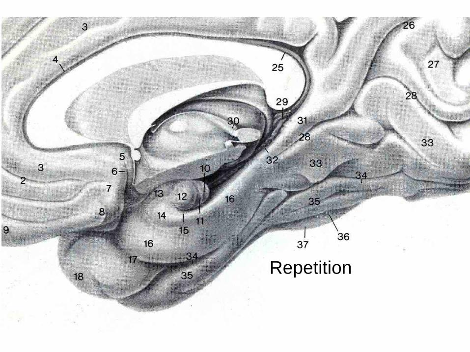
Repetition
Gyrus cinguli
US
A
Ento
PH
Area
subcallosa
Gyrus paraterminalis – medial part of
septum verum
Apex unci – g. intralimbicus
Tenia Giacomini = g. dentatus
Uncus
U- g. uncinatus -archicortex
S- g. semilunaris- Amy(cort.)
A- g. ambiens-paleocortex
Ento – area 28 -mesocortex
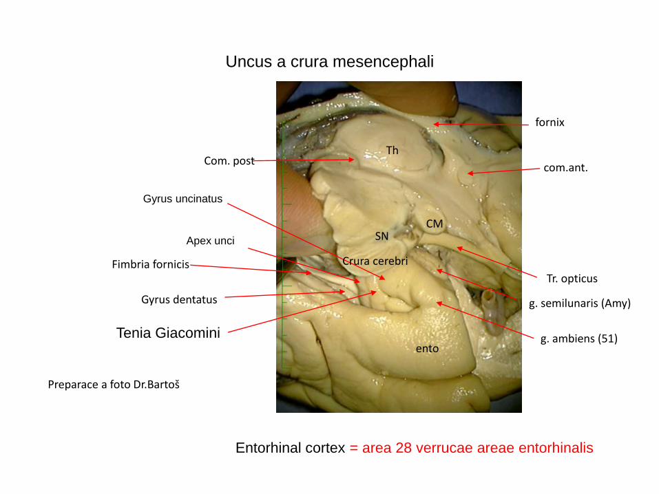
Uncus a crura mesencephali
Preparace a foto Dr.Bartoš
Th
com.ant.
Tr. opticus
ento
Gyrus dentatus
Fimbria fornicis
g. semilunaris (Amy)
g. ambiens (51)
CMSN
Crura cerebri
fornix
Com. post
Entorhinal cortex = area 28 verrucae areae entorhinalis
Tenia Giacomini
Gyrus uncinatus
Apex unci
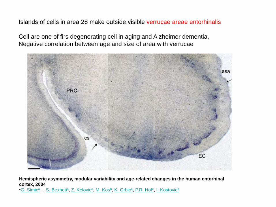
Islands of cells in area 28 make outside visible verrucae areae entorhinalis
Cell are one of firs degenerating cell in aging and Alzheimer dementia,
Negative correlation between age and size of area with verrucae
Hemispheric asymmetry, modular variability and age-related changes in the human entorhinal
cortex, 2004
•G. Simica, , , S. Bexhetia, Z. Kelovica, M. Kosb, K. Grbica, P.R. Hofc, I. Kostovica
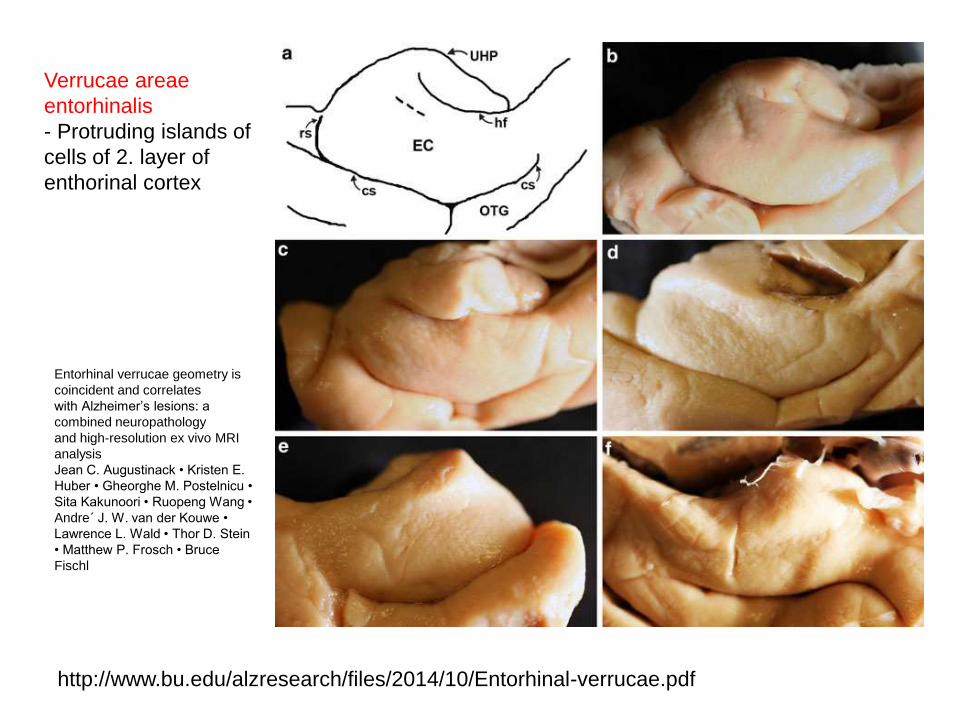
Entorhinal verrucae geometry is
coincident and correlates
with Alzheimer’s lesions: a
combined neuropathology
and high-resolution ex vivo MRI
analysis
Jean C. Augustinack • Kristen E.
Huber • Gheorghe M. Postelnicu •
Sita Kakunoori • Ruopeng Wang •
Andre´ J. W. van der Kouwe •
Lawrence L. Wald • Thor D. Stein
• Matthew P. Frosch • Bruce
Fischl
http://www.bu.edu/alzresearch/files/2014/10/Entorhinal-verrucae.pdf
Verrucae areae
entorhinalis
- Protruding islands of
cells of 2. layer of
enthorinal cortex

1-band of Giacomini ( arrows along the superficial hippocampal sulcus),
2 uncal apex,
3 fimbria.
Structure of posterior uncal segment:
4 alveus covering
the uncal apex (hippocampus inversus),
5 CA3 field, 6 gyrus dentatus,
7 CA1 fi eld, 8 subiculum, 9 uncinate gyrus, 10 intraventricular aspect of
hippocampal head after opening of the ventricular cavity, 11 ambient
gyrus, 12 uncal sulcus
Anterior segment piriform
8 semilunar gyrus, 9 semianular sulcus, 10 ambient gyrus,
11 uncal notch produced by the free edge of the tentorium
cerebelli, 12 entorhinal area and verrucae gyri
hippocampi, 13 rhinal sulcus, 14 parahippocampal gyrus
posterior segment - hippocampus:
1 band of Giacomini ( arrows along the superficial hippocampal
sulcus), 2 medial surface of uncal apex, 3 fimbria, 4 choroid
fissure (the choroid plexuses have been removed), 5 uncinate
gyrus, 6 uncal sulcus
UNCUS
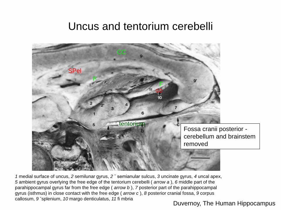
Uncus and tentorium cerebelli
CC
SPel
FF
GD
tentoriumFossa cranii posterior -
cerebellum and brainstem
removed
1 medial surface of uncus, 2 semilunar gyrus, 2 ˘ semianular sulcus, 3 uncinate gyrus, 4 uncal apex,
5 ambient gyrus overlying the free edge of the tentorium cerebelli ( arrow a ), 6 middle part of the
parahippocampal gyrus far from the free edge ( arrow b ), 7 posterior part of the parahippocampal
gyrus (isthmus) in close contact with the free edge ( arrow c ), 8 posterior cranial fossa, 9 corpus
callosum, 9 ˘splenium, 10 margo denticulatus, 11 fi mbriaDuvernoy, The Human Hippocampus
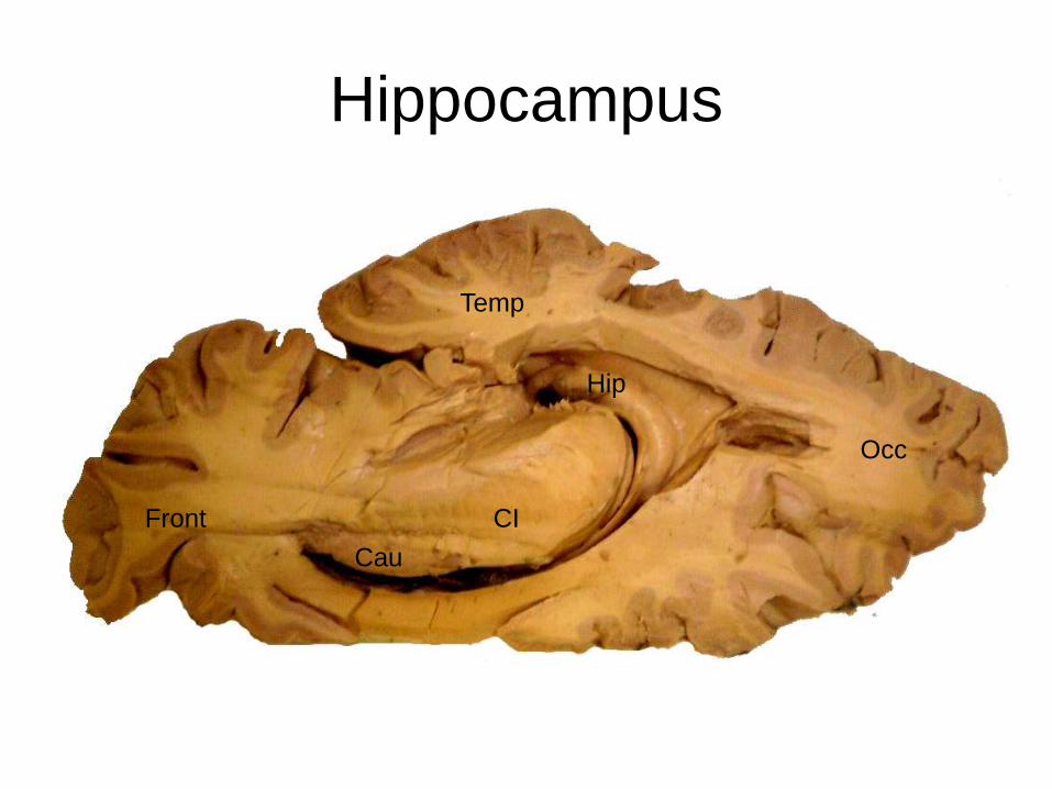
Hippocampus
Cau
Hip
Temp
Occ
CIFront
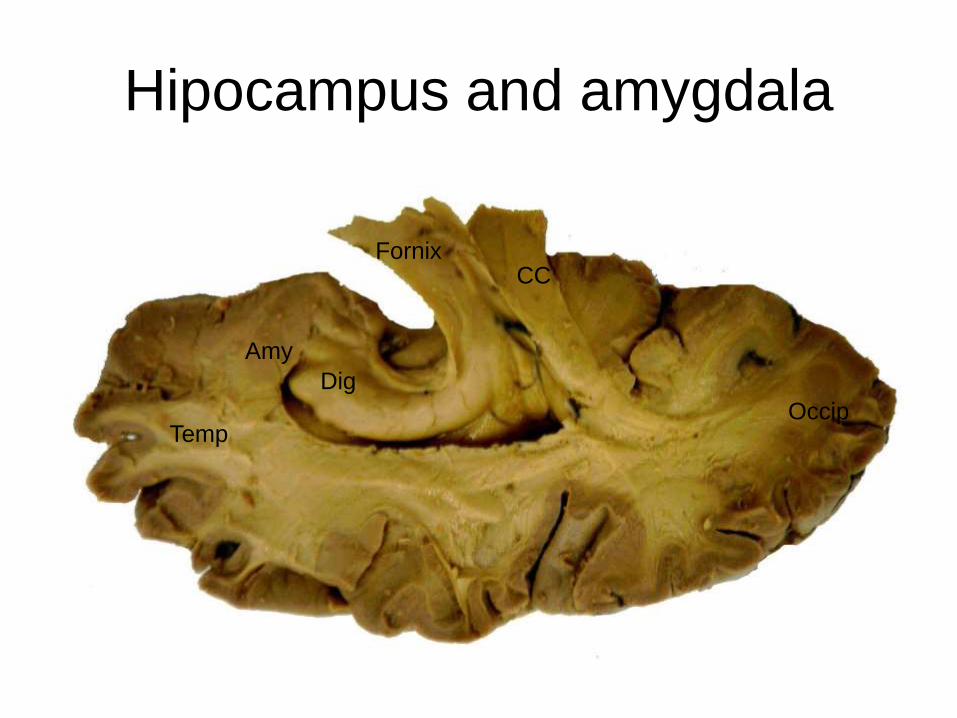
Hipocampus and amygdala
Amy
Dig
FornixCC
TempOccip
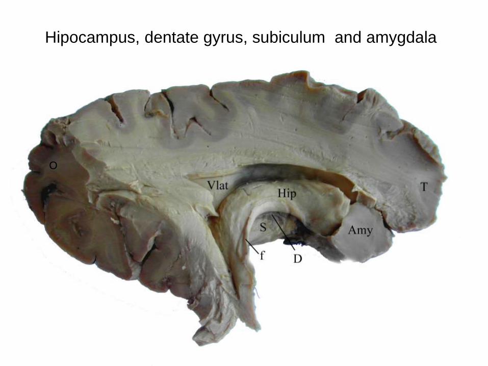
Hipocampus, dentate gyrus, subiculum and amygdala
O
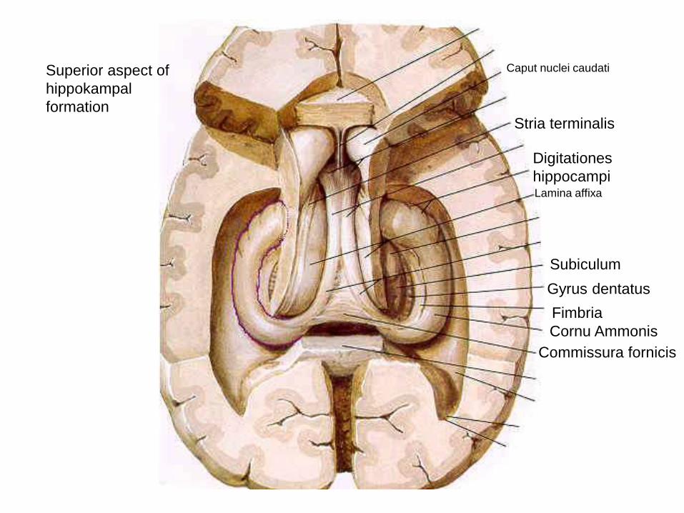
Subiculum
Gyrus dentatus
Fimbria
Cornu Ammonis
Commissura fornicis
Stria terminalis
Superior aspect of
hippokampal
formation
Digitationes
hippocampiLamina affixa
Caput nuclei caudati
Habenula
Stria
medullaris
Stria
terminalis
Fornix
Dent
fissura calcarina
ncl.
interpeduncularismamm-th tr
area subcallosa
septum
verum
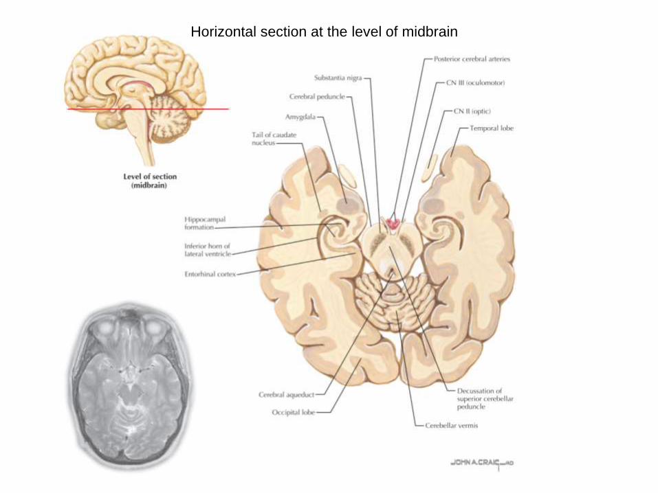
Horizontal section at the level of midbrain
Vývoj kůry telencephalického váčku pozdější v
časném embryonální období ( Petrovický)
Neocortex
Paleocortex
Archicortex
Řezy vyvíjející se
hemisférou
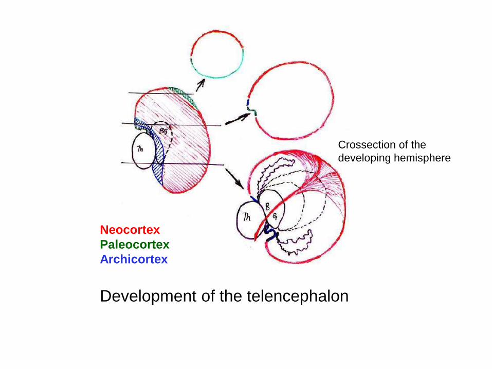
Development of the telencephalon
Crossection of the
developing hemisphere
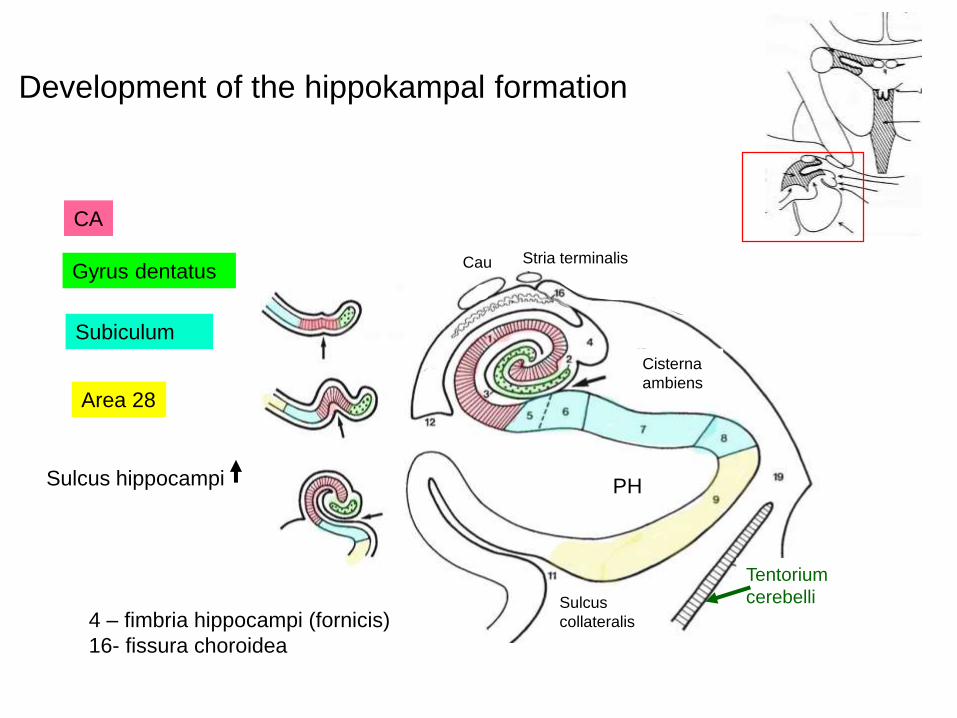
Development of the hippokampal formation
CA
Gyrus dentatus
Subiculum
Area 28
Sulcus hippocampi
Cisterna
ambiens
PH
Tentorium
cerebelli
Cau Stria terminalis
Sulcus
collateralis4 – fimbria hippocampi (fornicis)
16- fissura choroidea
Schema of prof. Petrovický
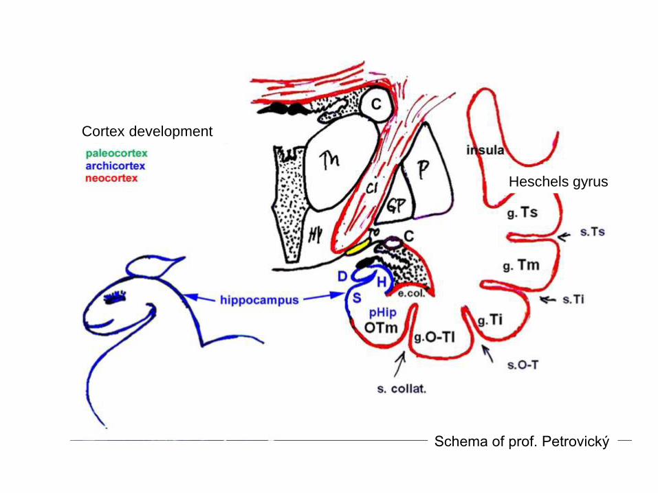
Cortex development
Heschels gyrus
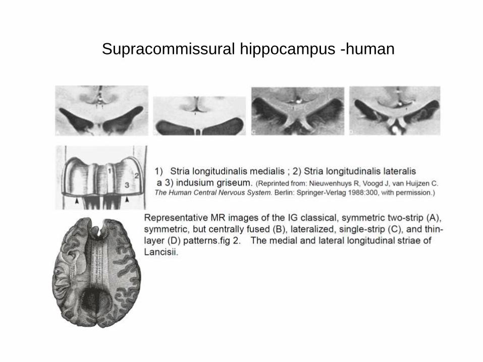
humanSupracommissural hippocampus -human
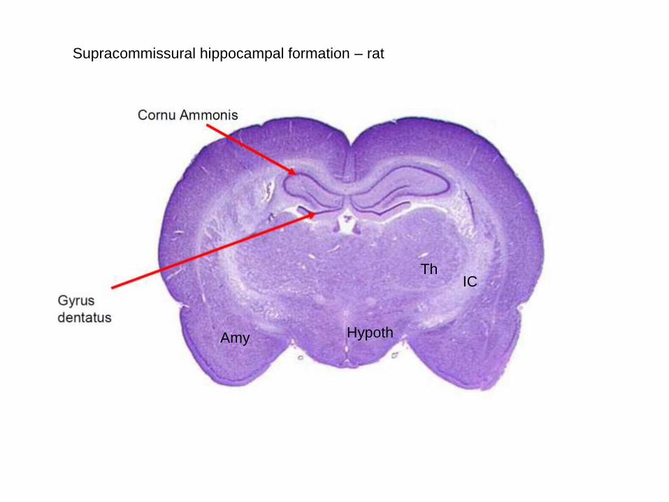
Supracommissural hippocampal formation – rat
Th
Amy Hypoth
IC
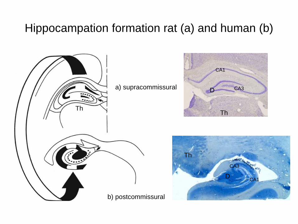
Hippocampation formation rat (a) and human (b)
a) supracommissural
CA1
CA1
CA3
CA3
b) postcommissural
Th
Th
D
D
Th
postcomissural hippocampus
supracomissural hippocampus
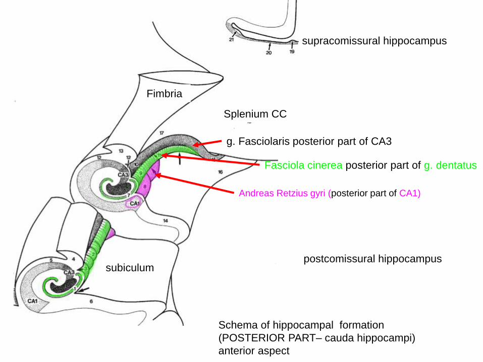
Splenium CC
g. Fasciolaris posterior part of CA3
Fasciola cinerea posterior part of g. dentatus
Andreas Retzius gyri (posterior part of CA1)
subiculum
Fimbria
Schema of hippocampal formation
(POSTERIOR PART– cauda hippocampi)
anterior aspect
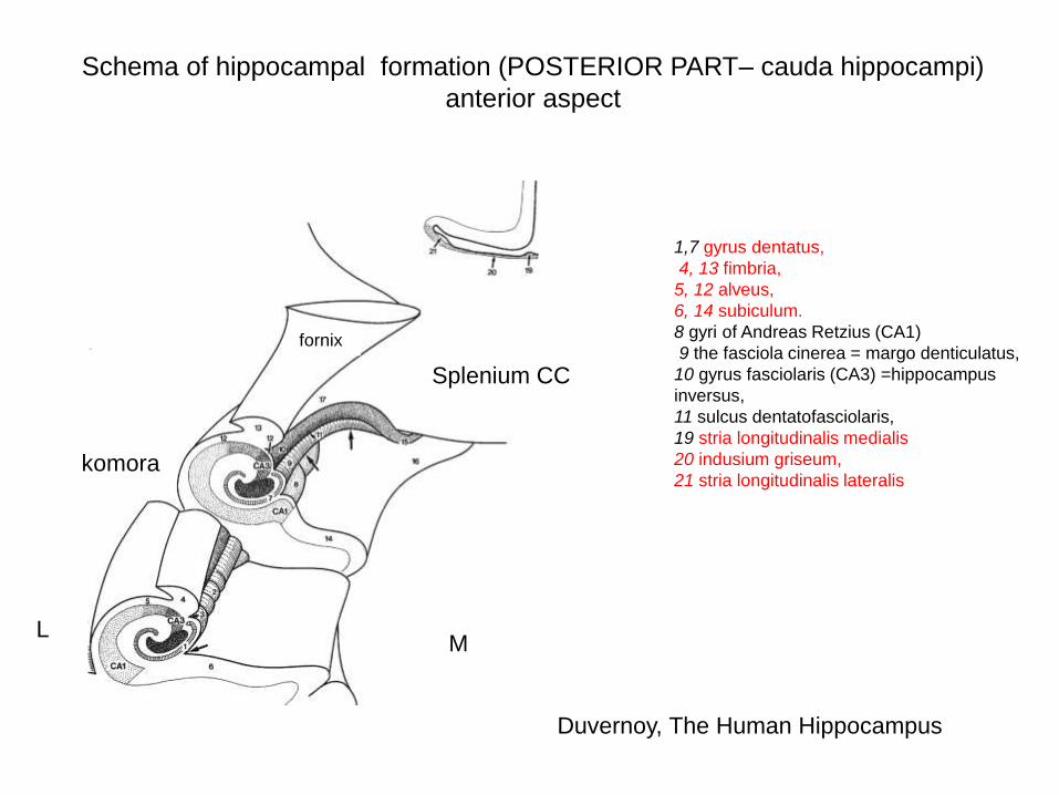
Schema of hippocampal formation (POSTERIOR PART– cauda hippocampi)
anterior aspect
1,7 gyrus dentatus,
4, 13 fimbria,
5, 12 alveus,
6, 14 subiculum.
8 gyri of Andreas Retzius (CA1)
9 the fasciola cinerea = margo denticulatus,
10 gyrus fasciolaris (CA3) =hippocampus
inversus,
11 sulcus dentatofasciolaris,
19 stria longitudinalis medialis
20 indusium griseum,
21 stria longitudinalis lateralis
Splenium CC
fornix
ML
komora
Duvernoy, The Human Hippocampus
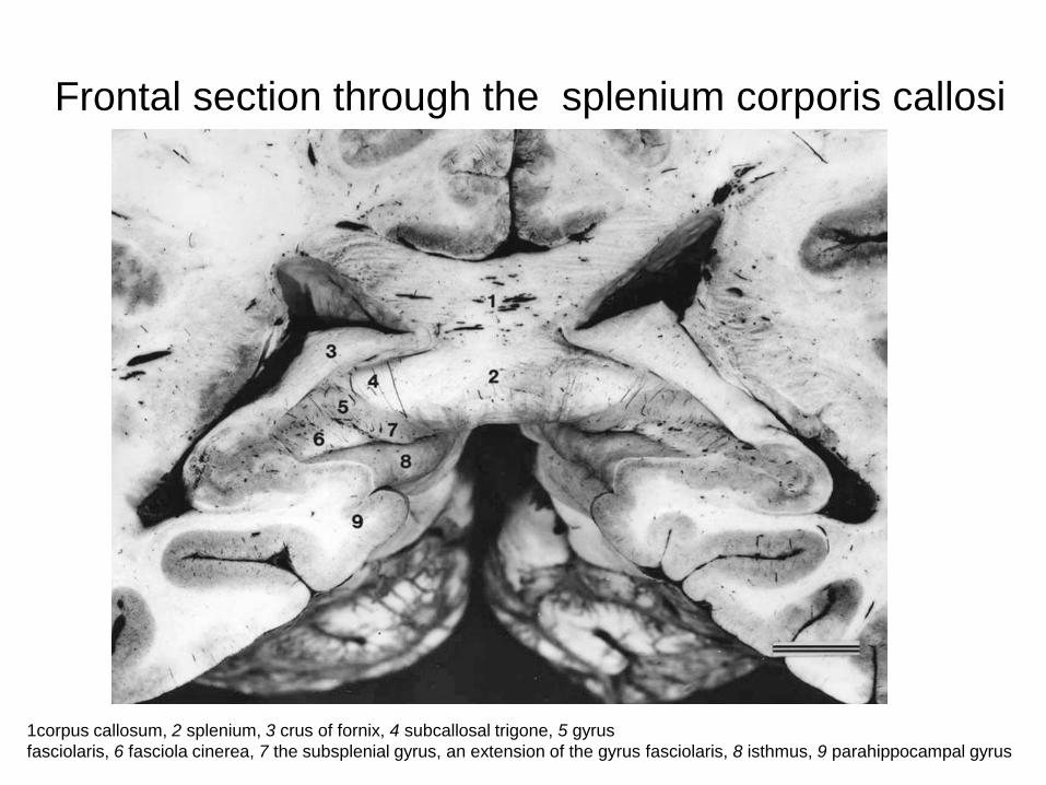
Frontal section through the splenium corporis callosi
1corpus callosum, 2 splenium, 3 crus of fornix, 4 subcallosal trigone, 5 gyrus
fasciolaris, 6 fasciola cinerea, 7 the subsplenial gyrus, an extension of the gyrus fasciolaris, 8 isthmus, 9 parahippocampal gyrus

Limbic structures ventral aspect1-corpus callosum
2- commissura fornicis
3-epiphysis
4-colliculus superior
5-colliculus inferior
6-frenulum veli medullaris sup
7-vellum medullare sup
8-IV.n
9-crura cerebri
10- pulvinar thalami
11- locus coeruleus
12- tegmentum pontis
13-pars basilaris pontis
14-tractus opticus
15-uncus
16-g. parahippocampalis
17-sulcus collateralis
18 –supracommissurální
hippocampus
19- trigonum olfactorium
= area perforata anterior
19
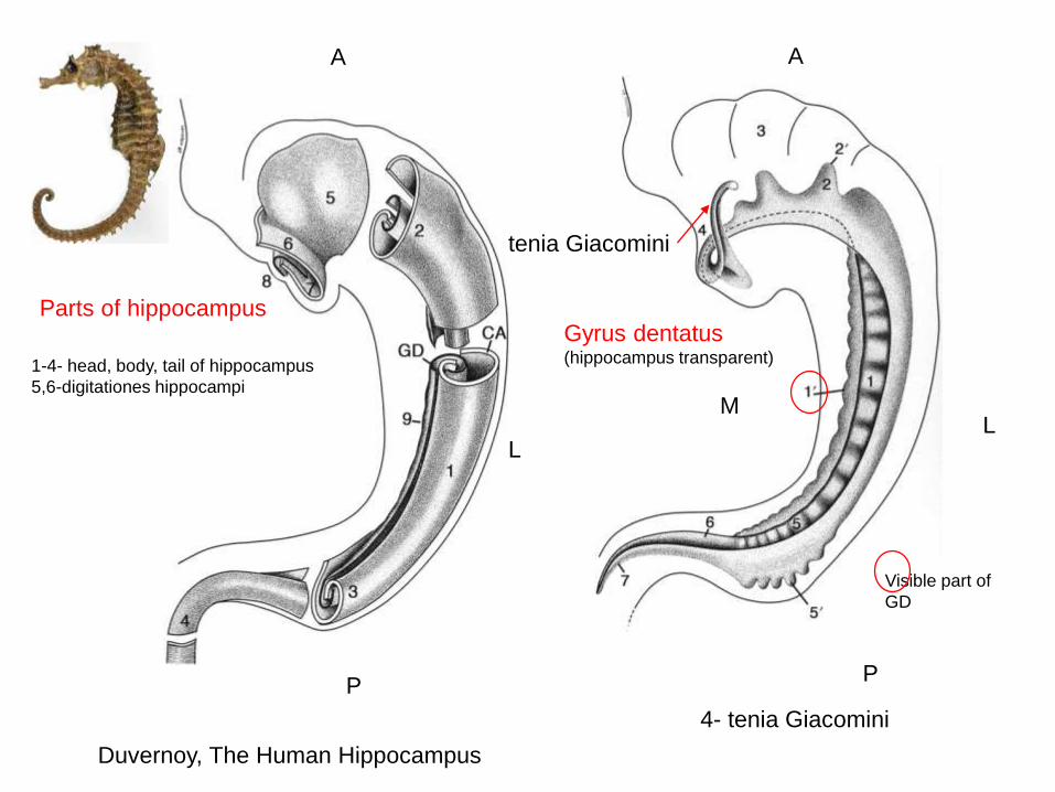
1-4- head, body, tail of hippocampus
5,6-digitationes hippocampi
A
P
Parts of hippocampus
M
LL
A
P
Gyrus dentatus(hippocampus transparent)
4- tenia Giacomini
tenia Giacomini
Duvernoy, The Human Hippocampus
Visible part of
GD
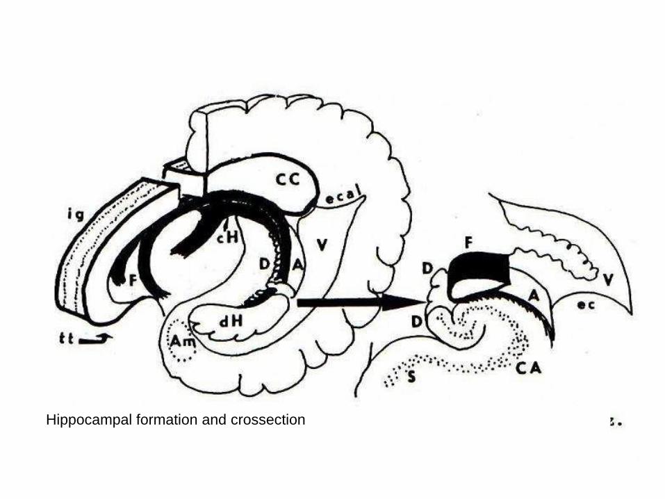
Hippocampal formation and crossection
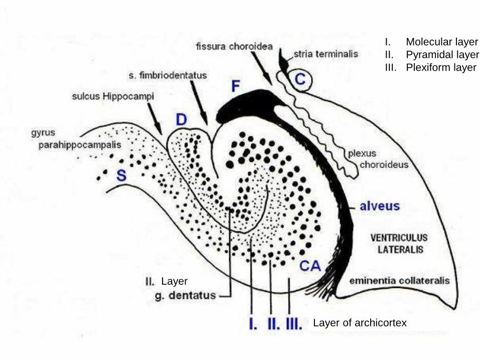
Layer of archicortex
Layer
I. Molecular layer
II. Pyramidal layer
III. Plexiform layer
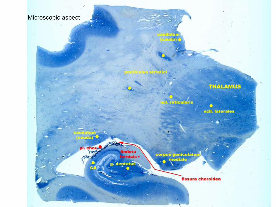
Microscopic aspect
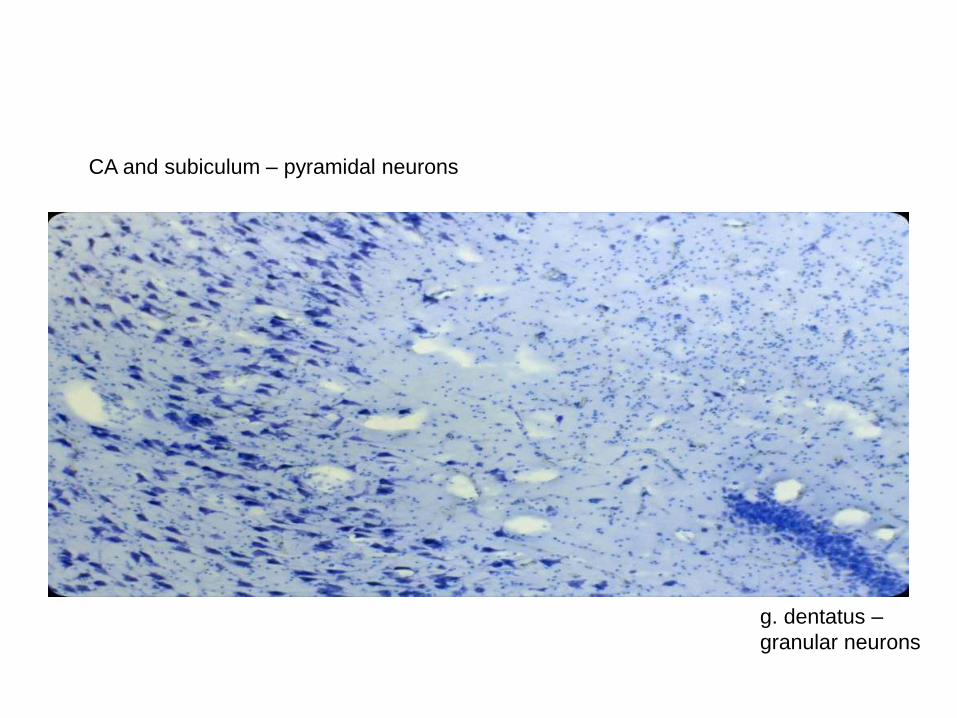
CA and subiculum – pyramidal neurons
g. dentatus –
granular neurons
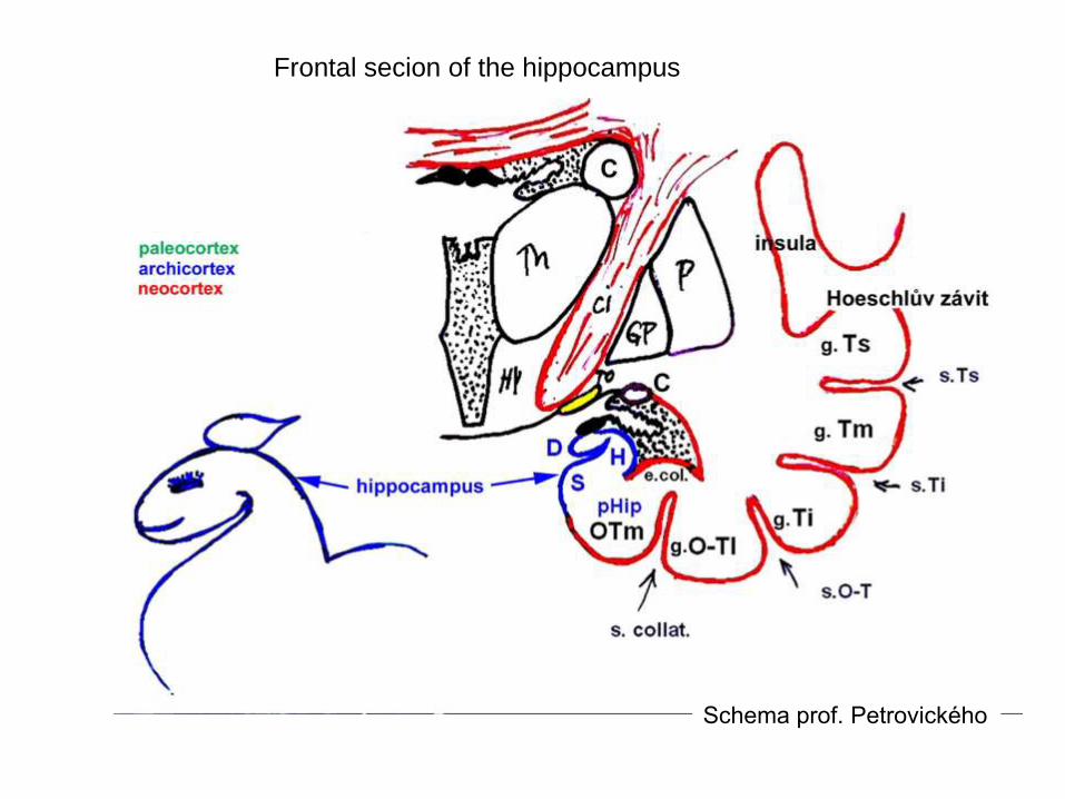
Frontal secion of the hippocampus
Schema prof. Petrovického
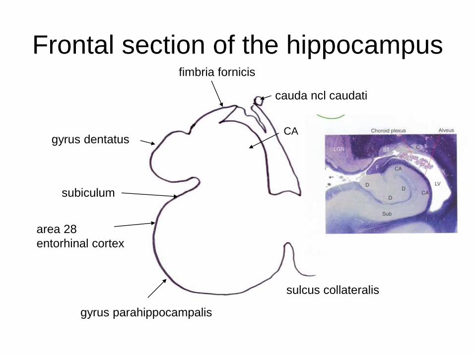
Frontal section of the hippocampus
cauda ncl caudati
fimbria fornicis
gyrus dentatus
subiculum
area 28
entorhinal cortex
gyrus parahippocampalis
sulcus collateralis
CA
Archicortex – hippokampal formation
gyrus dentatus
cornu Ammonis
subiculum
stratum
moleculare
stratum
pyramidale
stratum
polymorfum
• 3 parts 3 layers
CA
D
S
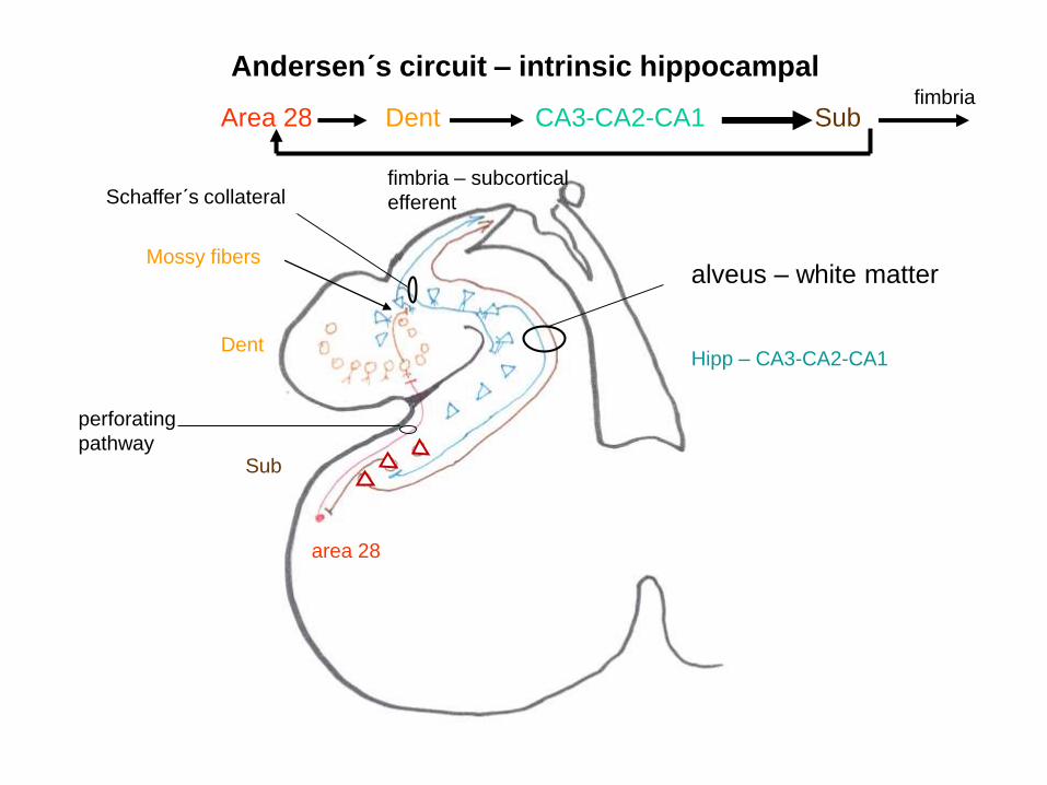
Andersen´s circuit – intrinsic hippocampal
area 28
Sub
DentHipp – CA3-CA2-CA1
fimbria – subcortical
efferent
alveus – white matter
Schaffer´s collateral
perforating
pathway
Area 28 Dent CA3-CA2-CA1 Subfimbria
Mossy fibers
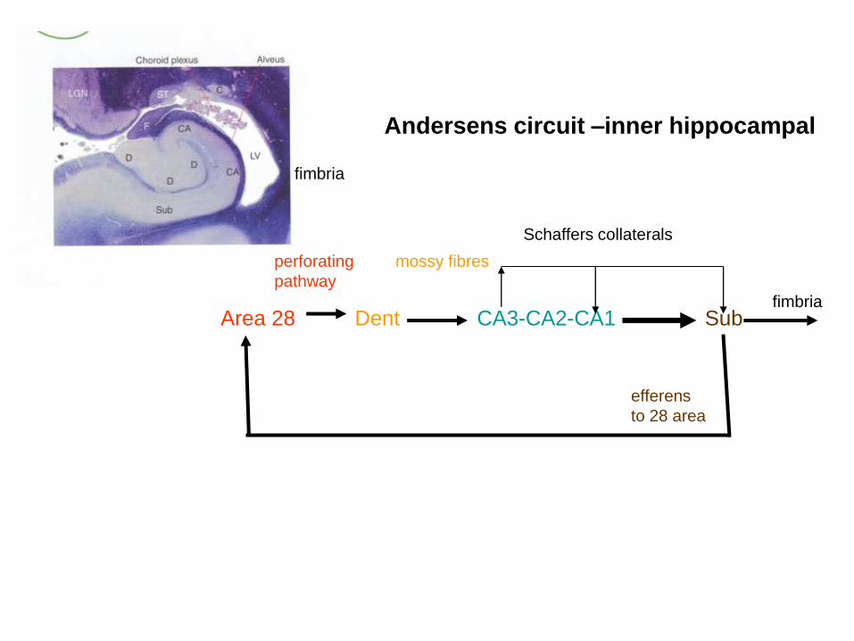
Andersens circuit –inner hippocampal
efferens
to 28 area
fimbria
Schaffers collaterals
perforating
pathway
Area 28 Dent CA3-CA2-CA1 Subfimbria
mossy fibres
???
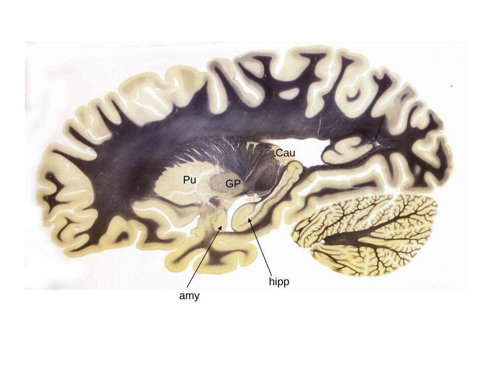
hipp
amy
Pu GP
Cau
Hipp
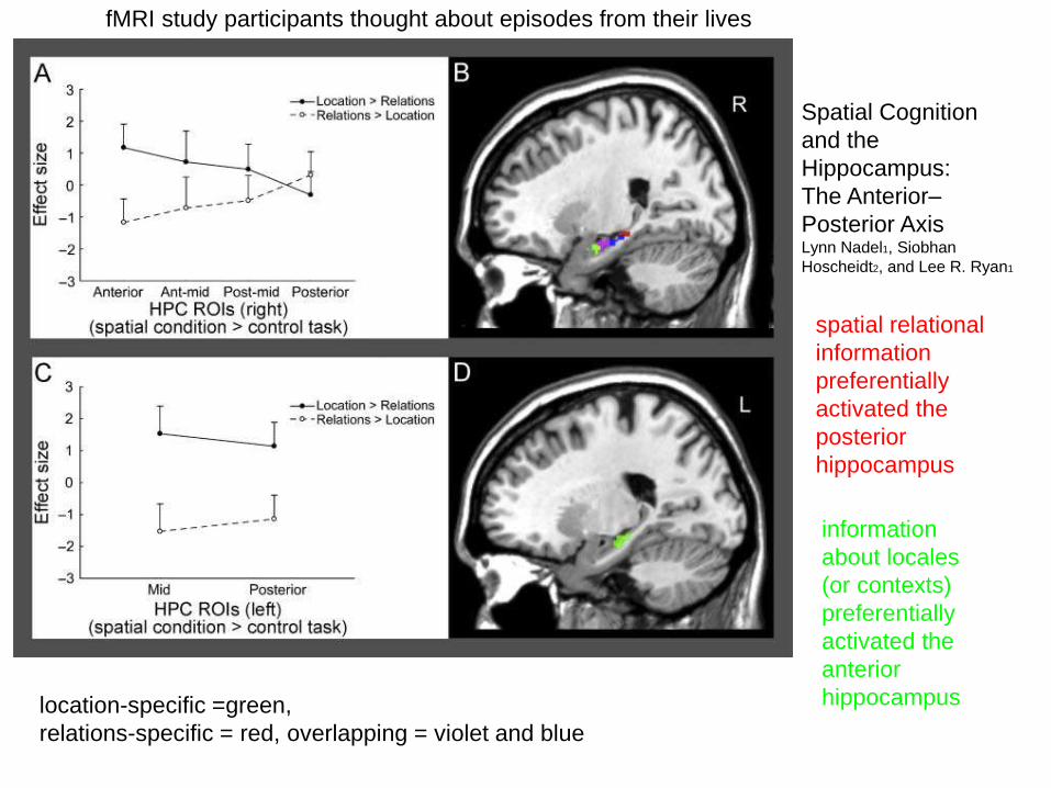
location-specific =green,
relations-specific = red, overlapping = violet and blue
fMRI study participants thought about episodes from their lives
spatial relational
information
preferentially
activated the
posterior
hippocampus
information
about locales
(or contexts)
preferentially
activated the
anterior
hippocampus
Spatial Cognition
and the
Hippocampus:
The Anterior–
Posterior AxisLynn Nadel1, Siobhan
Hoscheidt2, and Lee R. Ryan1
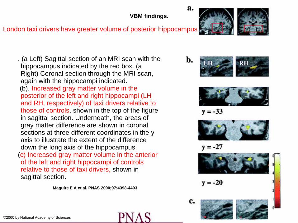
VBM findings.
Maguire E A et al. PNAS 2000;97:4398-4403
©2000 by National Academy of Sciences
. (a Left) Sagittal section of an MRI scan with the hippocampus indicated by the red box. (a Right) Coronal section through the MRI scan, again with the hippocampi indicated.(b). Increased gray matter volume in the posterior of the left and right hippocampi (LH and RH, respectively) of taxi drivers relative to those of controls, shown in the top of the figure in sagittal section. Underneath, the areas of gray matter difference are shown in coronal sections at three different coordinates in the y axis to illustrate the extent of the difference down the long axis of the hippocampus.
(c) Increased gray matter volume in the anterior of the left and right hippocampi of controls relative to those of taxi drivers, shown in sagittal section.
London taxi drivers have greater volume of posterior hippocampus
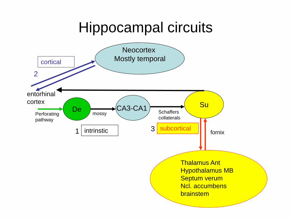
Hippocampal circuits
Neocortex
Mostly temporal
De CA3-CA1Su
entorhinal
cortex
cortical
intrinstic
Thalamus Ant
Hypothalamus MB
Septum verum
Ncl. accumbens
brainstem
1
2
subcortical3fornix
mossyPerforating
pathway
Schaffers
collaterals
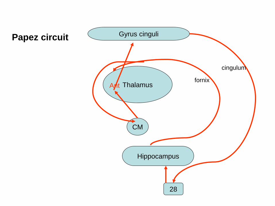
Gyrus cinguli
ThalamusAnt
CM
Hippocampus
28
Papez circuit
fornix
cingulum
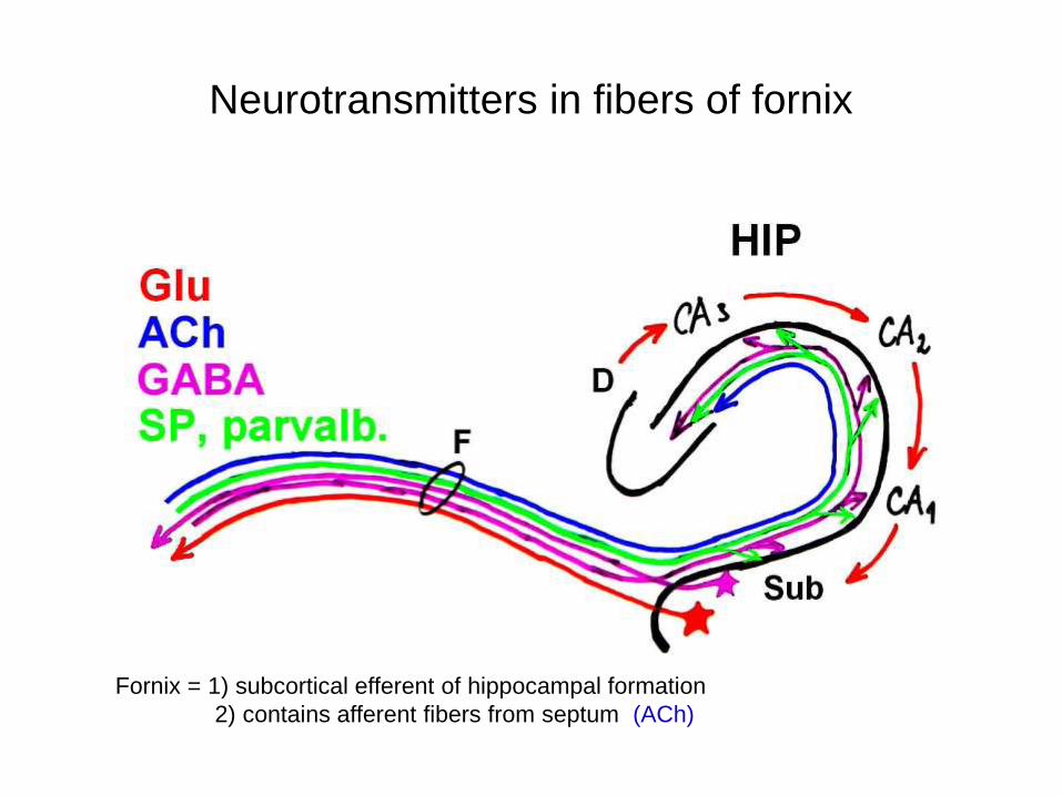
Neurotransmitters in fibers of fornix
Fornix = 1) subcortical efferent of hippocampal formation
2) contains afferent fibers from septum (ACh)
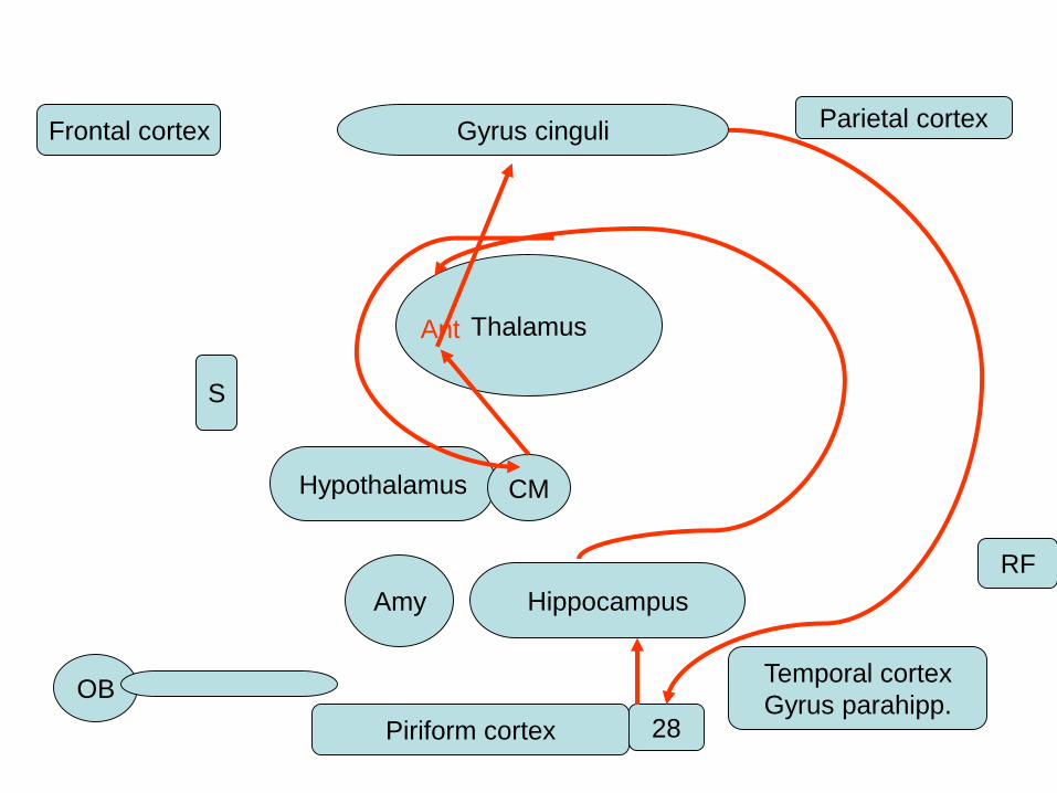
Gyrus cinguliFrontal cortexParietal cortex
ThalamusAnt
Hypothalamus CM
Amy Hippocampus
S
OB
Piriform cortex 28
Temporal cortex
Gyrus parahipp.
RF
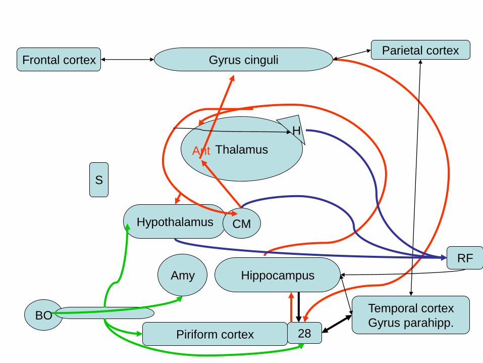
Gyrus cinguliFrontal cortexParietal cortex
ThalamusAnt
Hypothalamus CM
Amy Hippocampus
S
BO
Piriform cortex 28
Temporal cortex
Gyrus parahipp.
RF
H
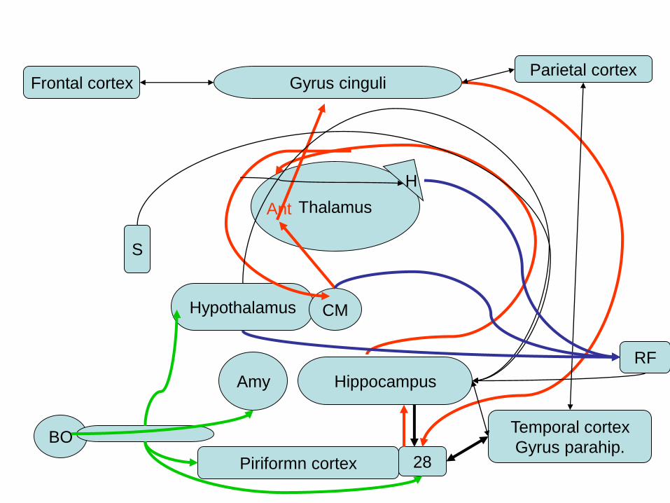
Gyrus cinguliFrontal cortexParietal cortex
ThalamusAnt
Hypothalamus CM
Amy Hippocampus
S
BO
Piriformn cortex 28
Temporal cortex
Gyrus parahip.
RF
H
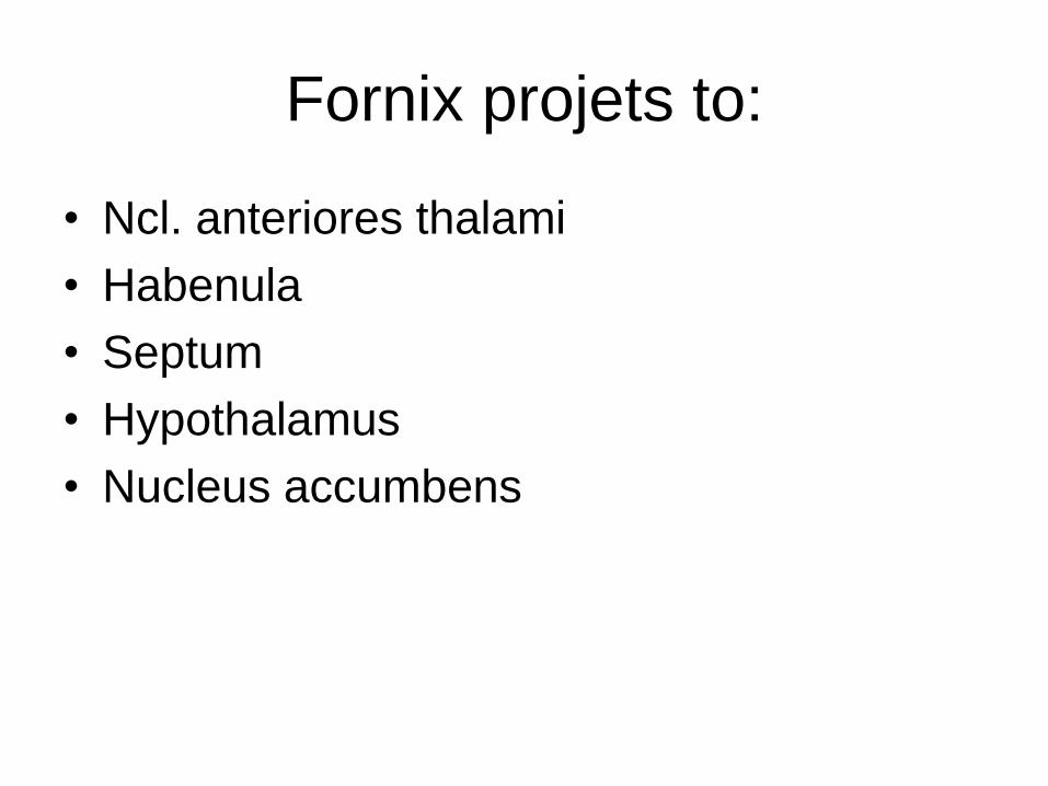
Fornix projets to:
• Ncl. anteriores thalami
• Habenula
• Septum
• Hypothalamus
• Nucleus accumbens
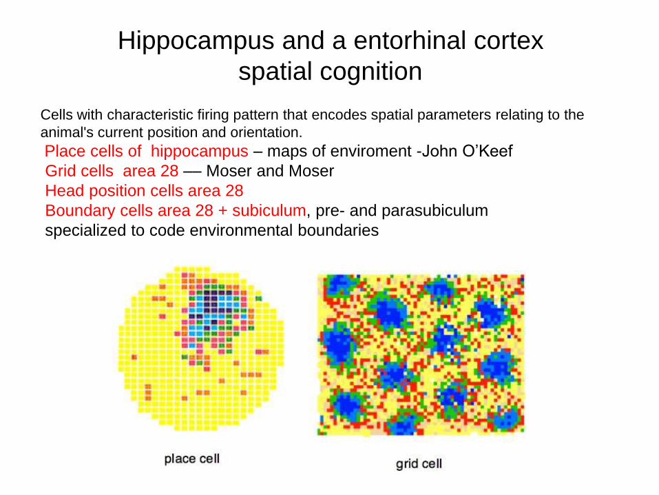
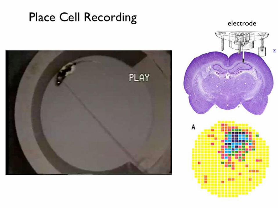
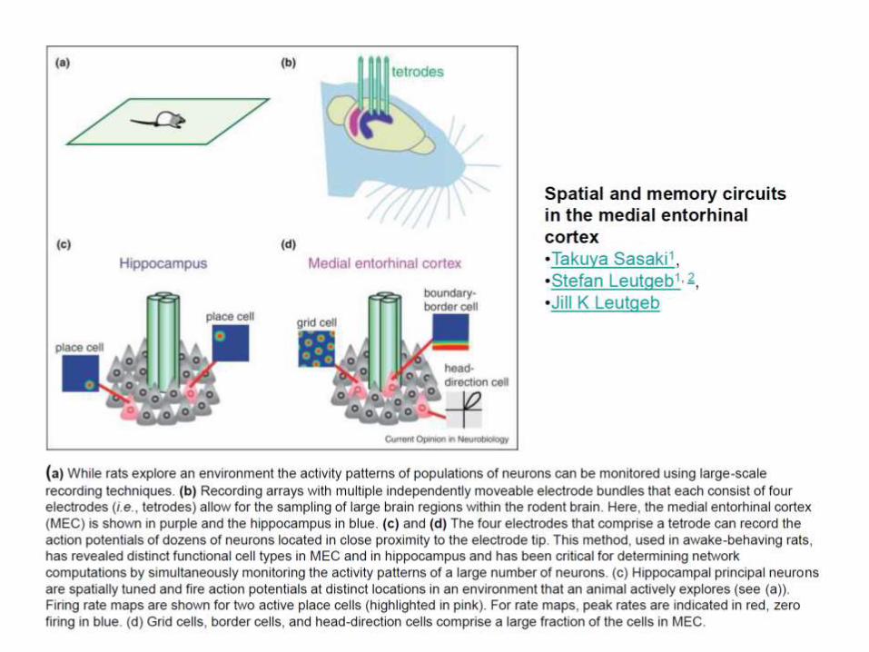
Hippocampus and a entorhinal cortex
spatial cognition
Cells with characteristic firing pattern that encodes spatial parameters relating to the
animal's current position and orientation.
Place cells of hippocampus – maps of enviroment -John O’Keef
Grid cells area 28 –– Moser and Moser
Head position cells area 28
Boundary cells area 28 + subiculum, pre- and parasubiculum
specialized to code environmental boundaries
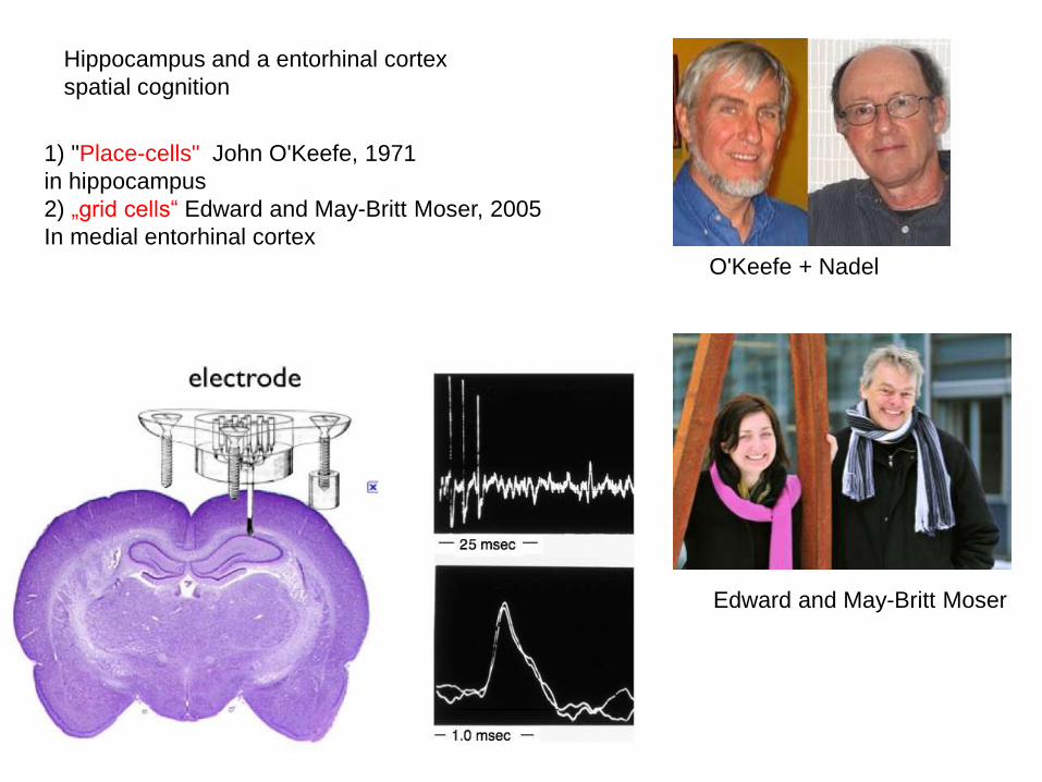
1) "Place-cells" John O'Keefe, 1971
in hippocampus
2) „grid cells“ Edward and May-Britt Moser, 2005
In medial entorhinal cortex
O'Keefe + Nadel
Edward and May-Britt Moser
Hippocampus and a entorhinal cortex
spatial cognition
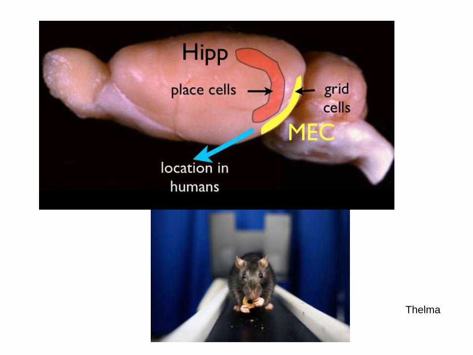
Thelma
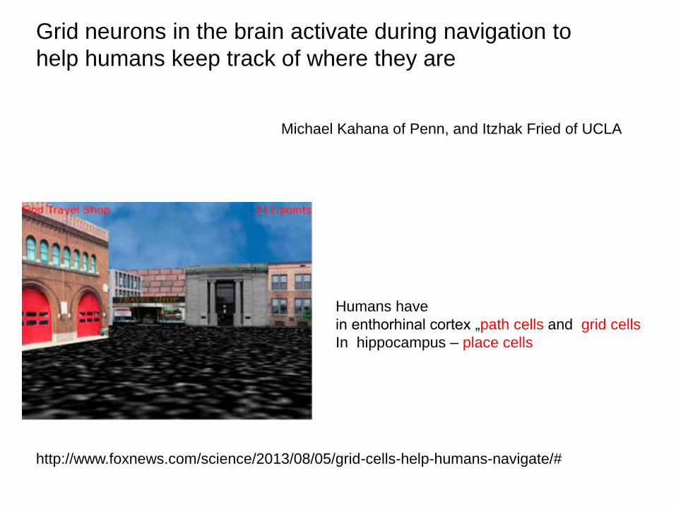
Grid neurons in the brain activate during navigation to
help humans keep track of where they are
http://www.foxnews.com/science/2013/08/05/grid-cells-help-humans-navigate/#
Michael Kahana of Penn, and Itzhak Fried of UCLA
Humans have
in enthorhinal cortex „path cells and grid cells
In hippocampus – place cells
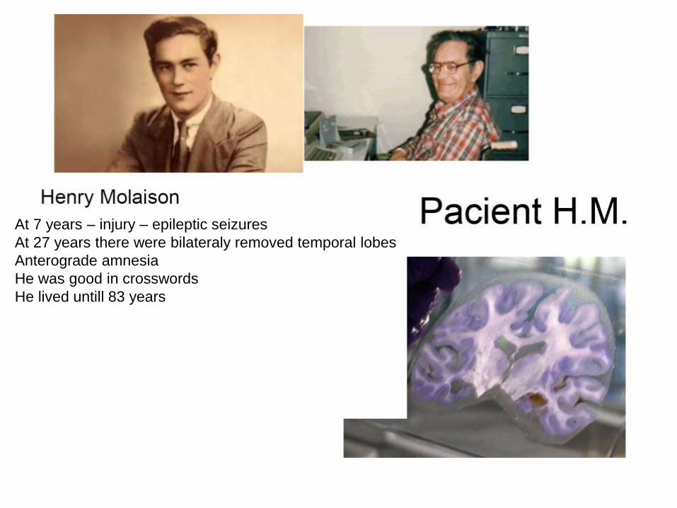
At 7 years – injury – epileptic seizures
At 27 years there were bilateraly removed temporal lobes
Anterograde amnesia
He was good in crosswords
He lived untill 83 years
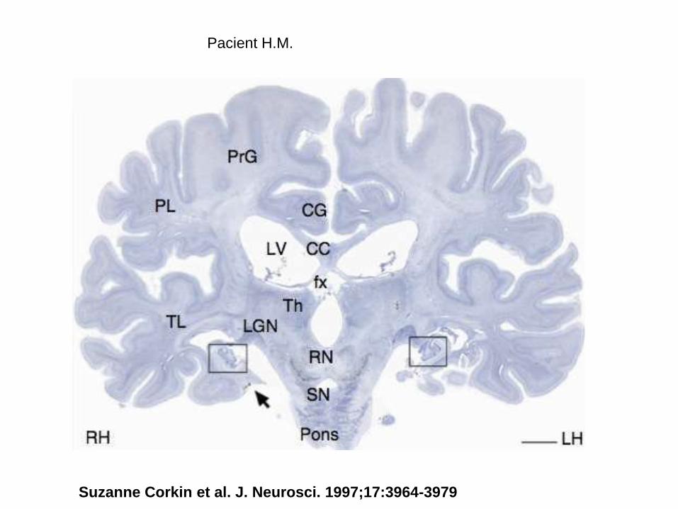
Pacient H.M.
Suzanne Corkin et al. J. Neurosci. 1997;17:3964-3979
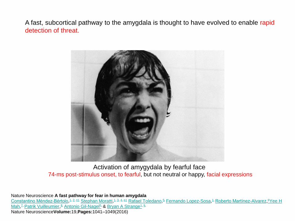
A fast, subcortical pathway to the amygdala is thought to have evolved to enable rapid
detection of threat.
Activation of amygydala by fearful face 74-ms post-stimulus onset, to fearful, but not neutral or happy, facial expressions
Nature Neuroscience A fast pathway for fear in human amygdala
Constantino Méndez-Bértolo,1, 2, n1 Stephan Moratti,1, 3, 4, n1 Rafael Toledano,5, Fernando Lopez-Sosa,1, Roberto Martínez-Alvarez,6Yee H
Mah,7, Patrik Vuilleumier,8, Antonio Gil-Nagel5, & Bryan A Strange1, 9,
Nature NeuroscienceVolume:19,Pages:1041–1049(2016)
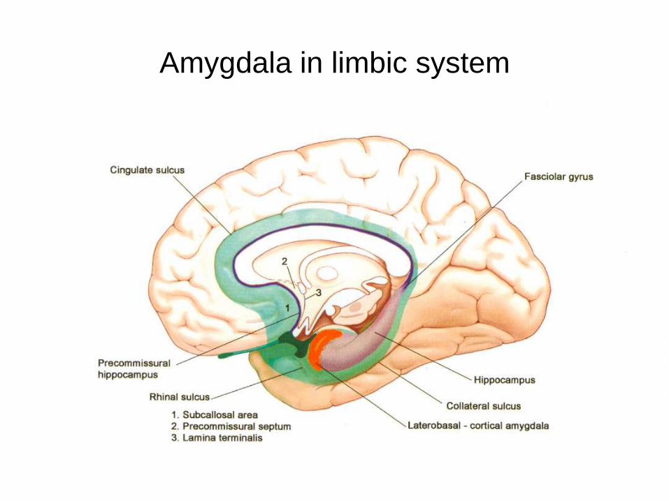
Amygdala in limbic system
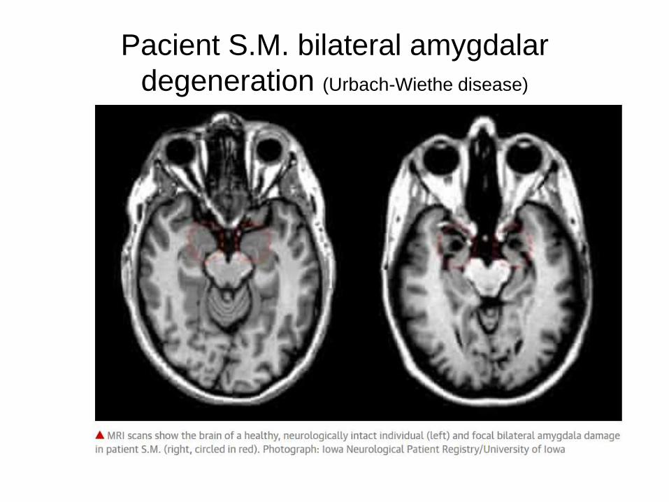
Pacient S.M. bilateral amygdalar
degeneration (Urbach-Wiethe disease)
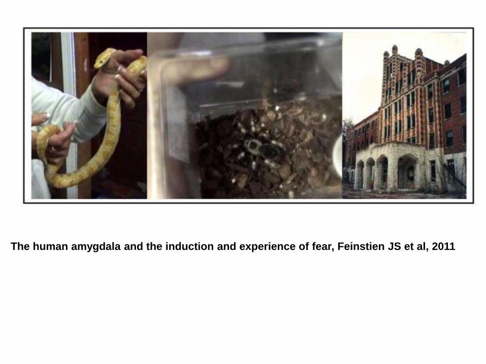
The human amygdala and the induction and experience of fear, Feinstien JS et al, 2011
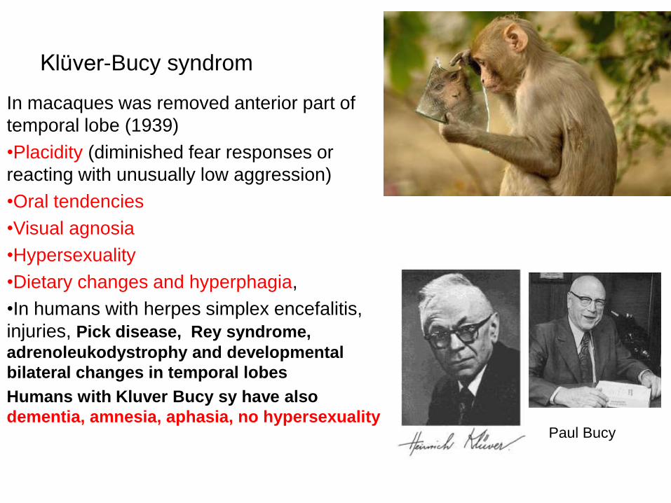
Klüver-Bucy syndrom
In macaques was removed anterior part of
temporal lobe (1939)
•Placidity (diminished fear responses or
reacting with unusually low aggression)
•Oral tendencies
•Visual agnosia
•Hypersexuality
•Dietary changes and hyperphagia,
•In humans with herpes simplex encefalitis,
injuries, Pick disease, Rey syndrome,
adrenoleukodystrophy and developmental
bilateral changes in temporal lobes
Humans with Kluver Bucy sy have also
dementia, amnesia, aphasia, no hypersexualityPaul Bucy
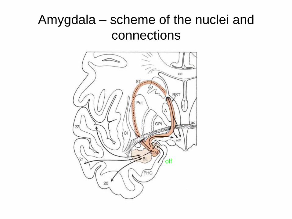
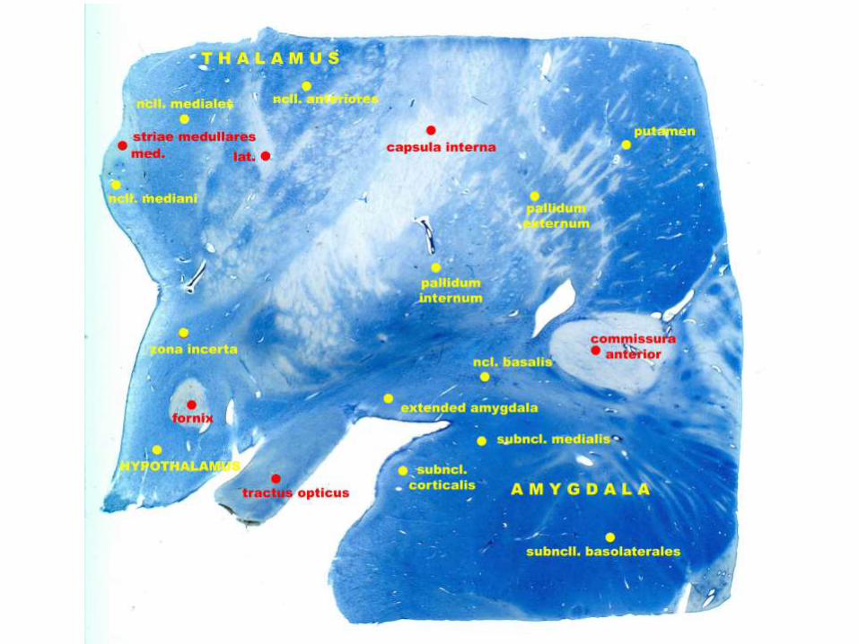
Amygdala – scheme of the nuclei and
connections
olf
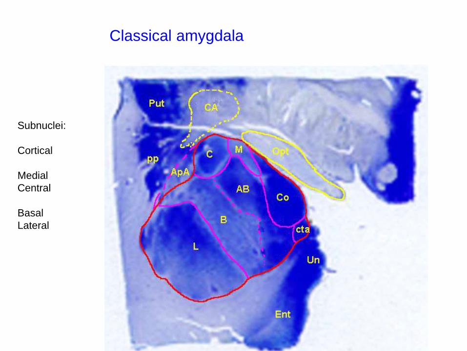
„Klasická“ amygdalaClassical amygdala
Subnuclei:
Cortical
Medial
Central
Basal
Lateral
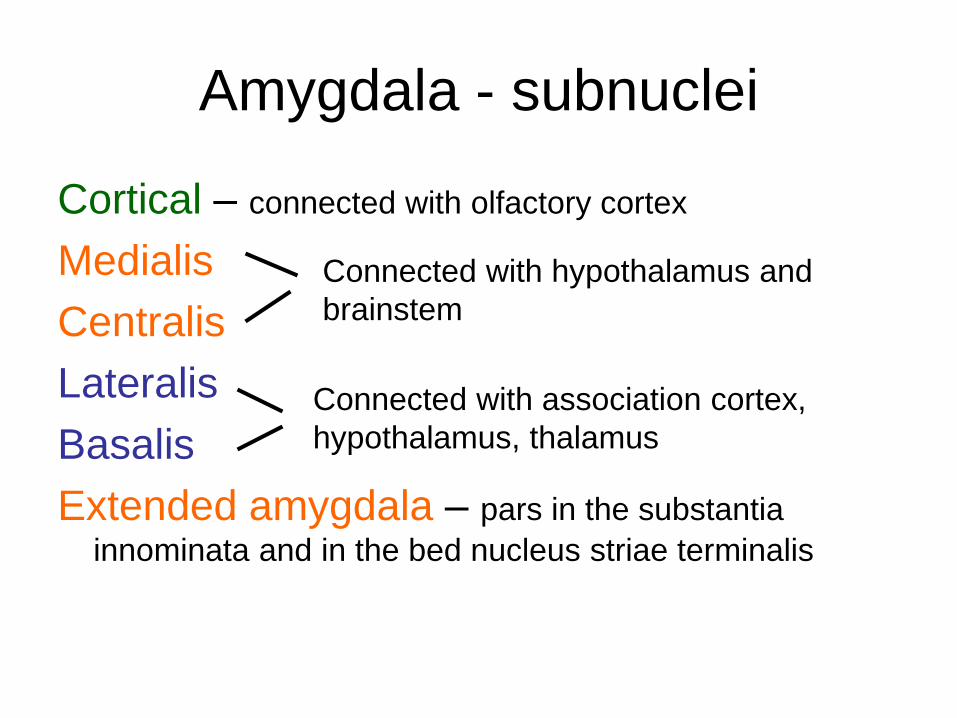
Amygdala - subnuclei
Cortical – connected with olfactory cortex
Medialis
Centralis
Lateralis
Basalis
Extended amygdala – pars in the substantia
innominata and in the bed nucleus striae terminalis
Connected with hypothalamus and
brainstem
Connected with association cortex,
hypothalamus, thalamus
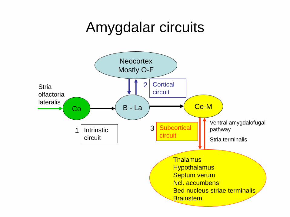
Amygdalar circuits
Neocortex
Mostly O-F
Co B - La Ce-M
Stria
olfactoria
lateralis
Cortical
circuit
Intrinstic
circuit
Thalamus
Hypothalamus
Septum verum
Ncl. accumbens
Bed nucleus striae terminalis
Brainstem
1
2
Subcortical
circuit 3
Ventral amygdalofugal
pathway
Stria terminalis
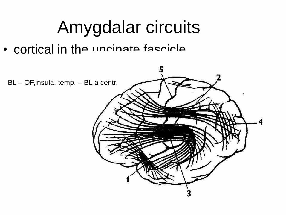
Amygdalar circuits• cortical in the uncinate fascicle
BL – OF,insula, temp. – BL a centr.
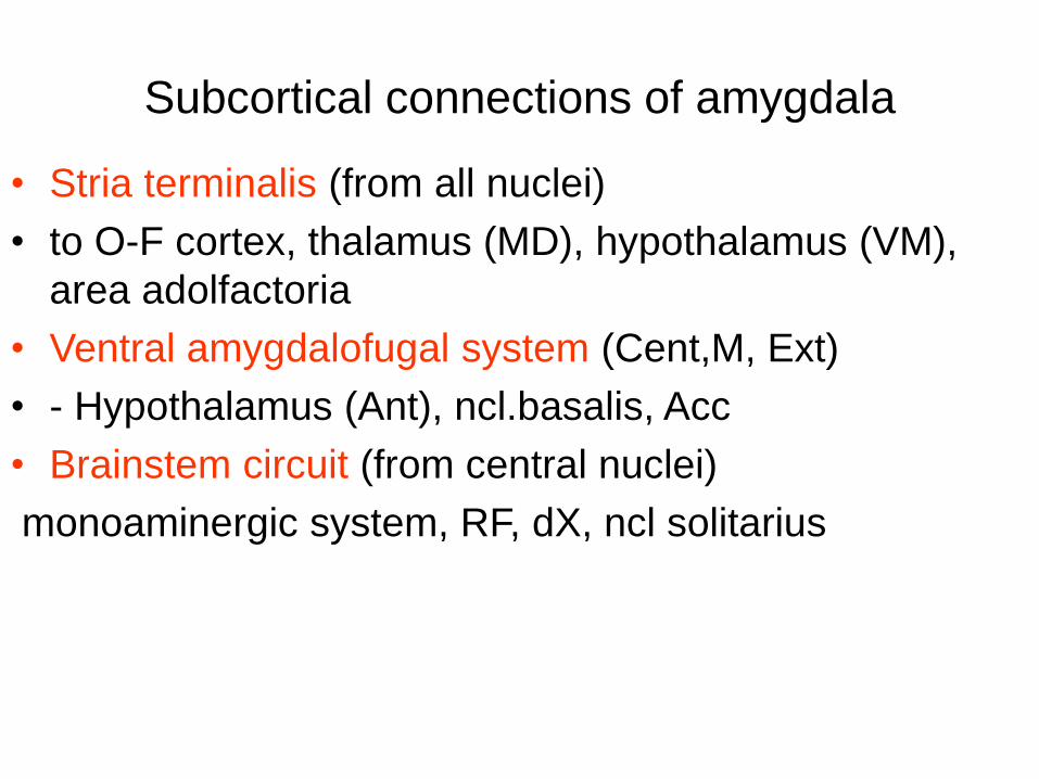
Subcortical connections of amygdala
• Stria terminalis (from all nuclei)
• to O-F cortex, thalamus (MD), hypothalamus (VM),
area adolfactoria
• Ventral amygdalofugal system (Cent,M, Ext)
• - Hypothalamus (Ant), ncl.basalis, Acc
• Brainstem circuit (from central nuclei)
monoaminergic system, RF, dX, ncl solitarius
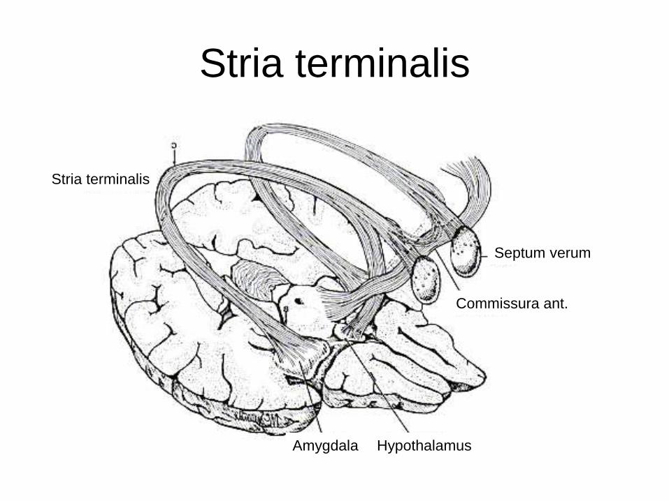
Stria terminalis
Stria terminalis
Commissura ant.
HypothalamusAmygdala
Septum verum

Fornix
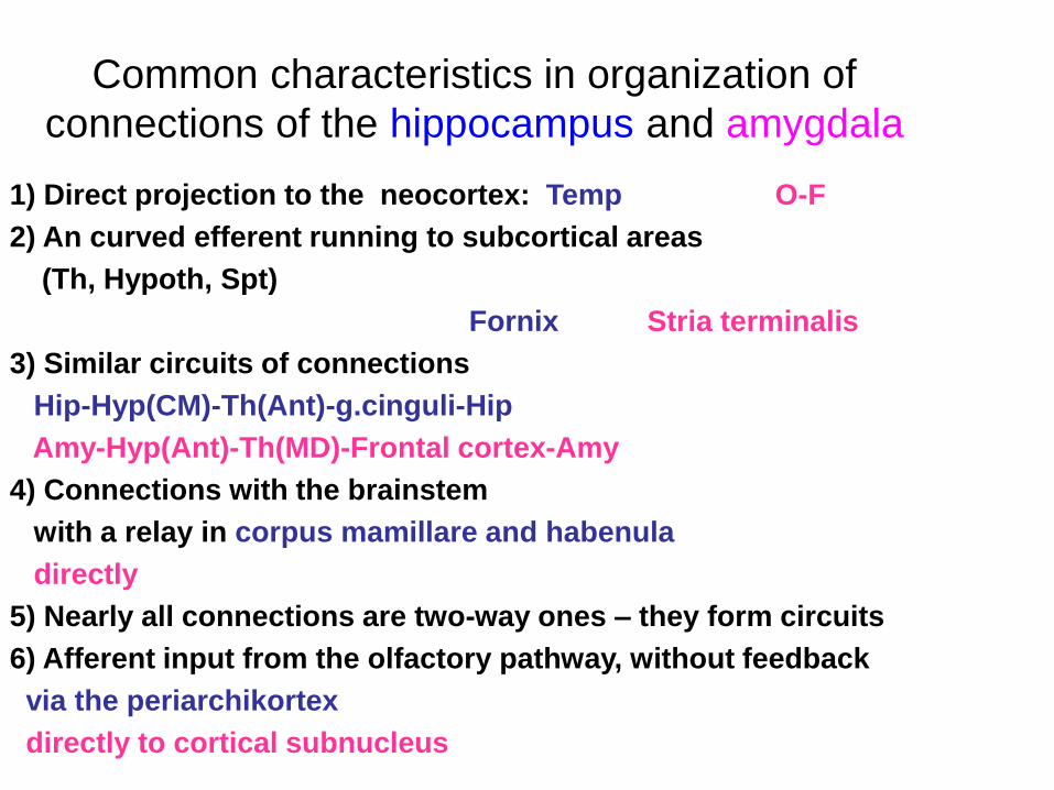
Common characteristics in organization of
connections of the hippocampus and amygdala
1) Direct projection to the neocortex: Temp O-F
2) An curved efferent running to subcortical areas
(Th, Hypoth, Spt)
Fornix Stria terminalis
3) Similar circuits of connections
Hip-Hyp(CM)-Th(Ant)-g.cinguli-Hip
Amy-Hyp(Ant)-Th(MD)-Frontal cortex-Amy
4) Connections with the brainstem
with a relay in corpus mamillare and habenula
directly
5) Nearly all connections are two-way ones – they form circuits
6) Afferent input from the olfactory pathway, without feedback
via the periarchikortex
directly to cortical subnucleus
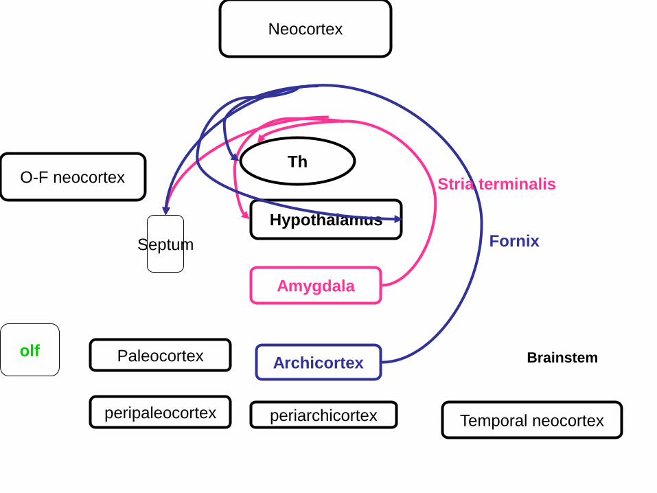
Archicortex
Amygdala
Th
Septum
Neocortex
O-F neocortex
Hypothalamus
periarchicortex Temporal neocortexperipaleocortex
Paleocortexolf
Fornix
Stria terminalis
Brainstem
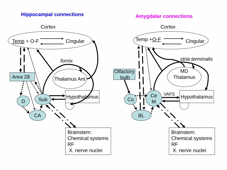
Thalamus Ant
Temp + O-F Cingular Temp +O-F Cingular
SubCe
M
CA
D Co
BL
Hypothalamus
Cortex Cortex
Area 28Olfactory
bulb
Hypothalamus
Brainstem:
Chemical systems
RF
X. nerve nuclei
MD
Thalamus
Brainstem:
Chemical systems
RF
X. nerve nuclei
fornix stria terminalis
Hippocampal connections Amygdalar connections
VAFS
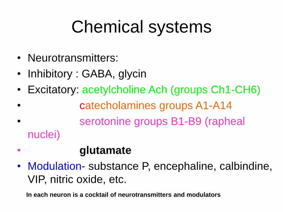
Chemical systems
• Neurotransmitters:
• Inhibitory : GABA, glycin
• Excitatory: acetylcholine Ach (groups Ch1-CH6)
• catecholamines groups A1-A14
• serotonine groups B1-B9 (rapheal
nuclei)
• glutamate
• Modulation- substance P, encephaline, calbindine,
VIP, nitric oxide, etc.
In each neuron is a cocktail of neurotransmitters and modulators
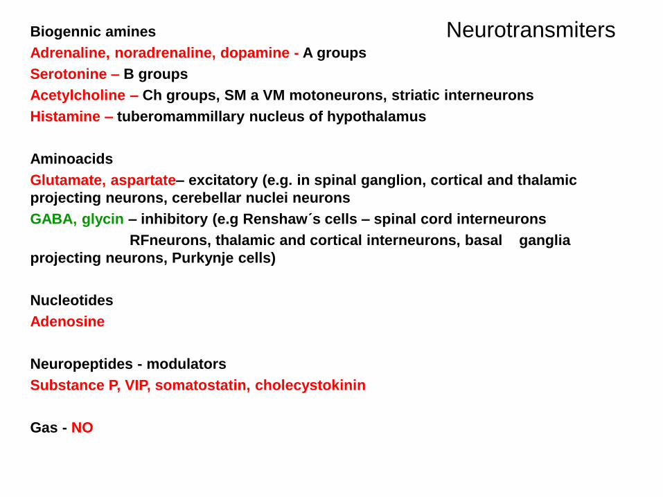
NeurotransmitersBiogennic amines
Adrenaline, noradrenaline, dopamine - A groups
Serotonine – B groups
Acetylcholine – Ch groups, SM a VM motoneurons, striatic interneurons
Histamine – tuberomammillary nucleus of hypothalamus
Aminoacids
Glutamate, aspartate– excitatory (e.g. in spinal ganglion, cortical and thalamic
projecting neurons, cerebellar nuclei neurons
GABA, glycin – inhibitory (e.g Renshaw´s cells – spinal cord interneurons
RFneurons, thalamic and cortical interneurons, basal ganglia
projecting neurons, Purkynje cells)
Nucleotides
Adenosine
Neuropeptides - modulators
Substance P, VIP, somatostatin, cholecystokinin
Gas - NO
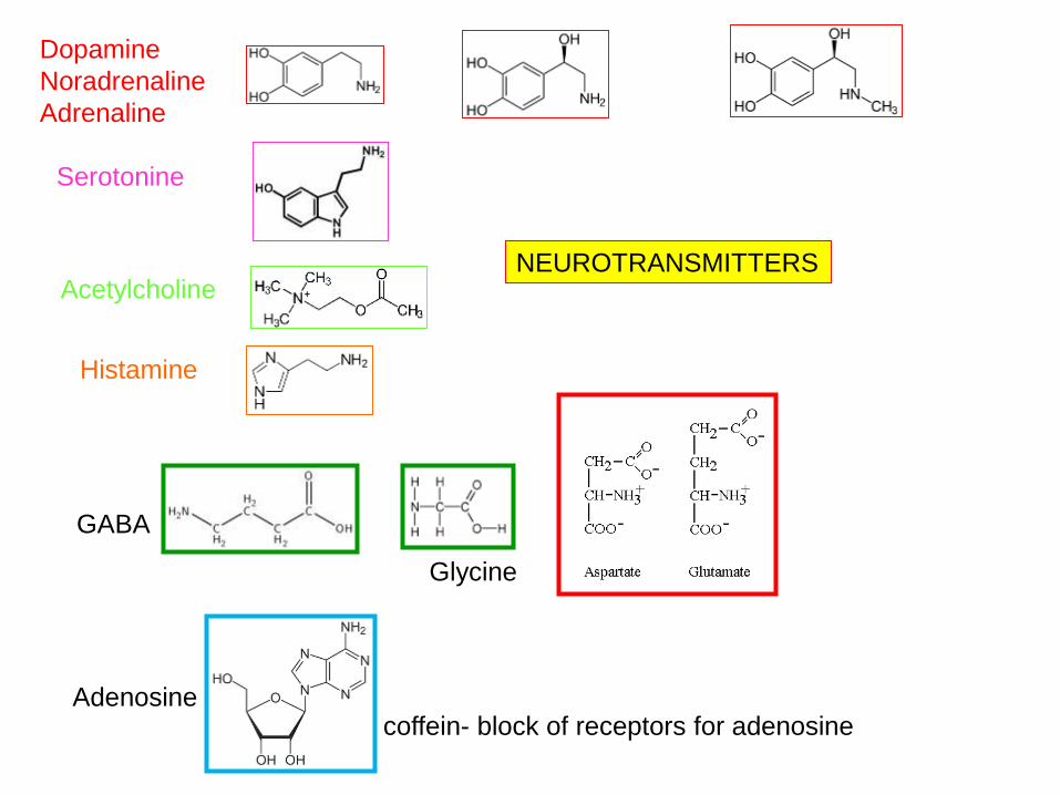
Dopamine
Noradrenaline
Adrenaline
Serotonine
Acetylcholine
Histamine
GABA
Glycine
Adenosinecoffein- block of receptors for adenosine
NEUROTRANSMITTERS
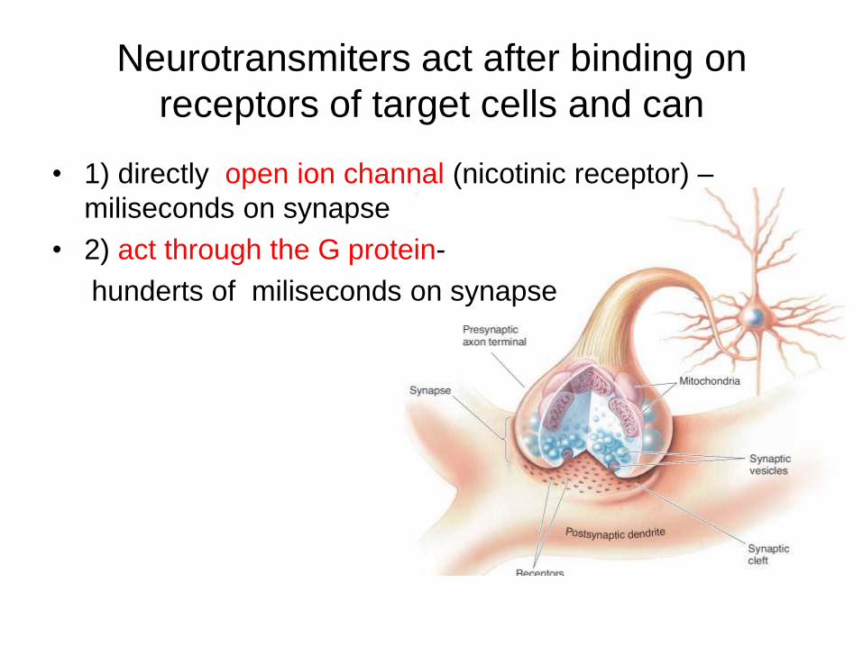
Neurotransmiters act after binding on
receptors of target cells and can
• 1) directly open ion channal (nicotinic receptor) –
miliseconds on synapse
• 2) act through the G protein-
hunderts of miliseconds on synapse
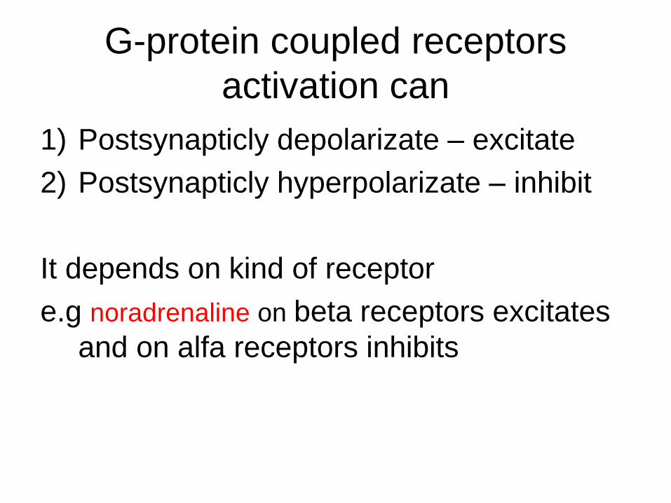
G-protein coupled receptors
activation can
1) Postsynapticly depolarizate – excitate
2) Postsynapticly hyperpolarizate – inhibit
It depends on kind of receptor
e.g noradrenaline on beta receptors excitates
and on alfa receptors inhibits
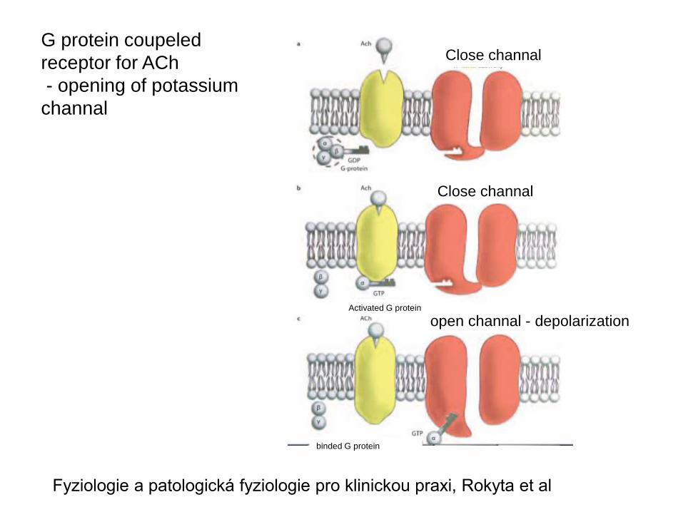
G protein coupeled
receptor for ACh
- opening of potassium
channal
Fyziologie a patologická fyziologie pro klinickou praxi, Rokyta et al
Activated G protein
binded G protein
Close channal
Close channal
open channal - depolarization
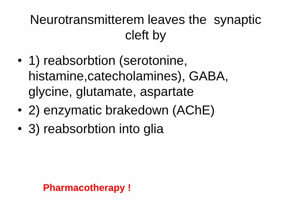
Neurotransmitterem leaves the synaptic
cleft by
• 1) reabsorbtion (serotonine,
histamine,catecholamines), GABA,
glycine, glutamate, aspartate
• 2) enzymatic brakedown (AChE)
• 3) reabsorbtion into glia
Pharmacotherapy !
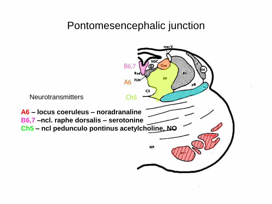
Pontomesencephalic junction
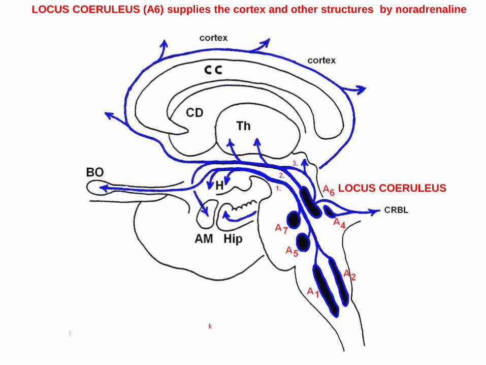
A6 – locus coeruleus – noradranaline
B6,7 –ncl. raphe dorsalis – serotonine
Ch5 – ncl pedunculo pontinus acetylcholine, NO
Neurotransmitters
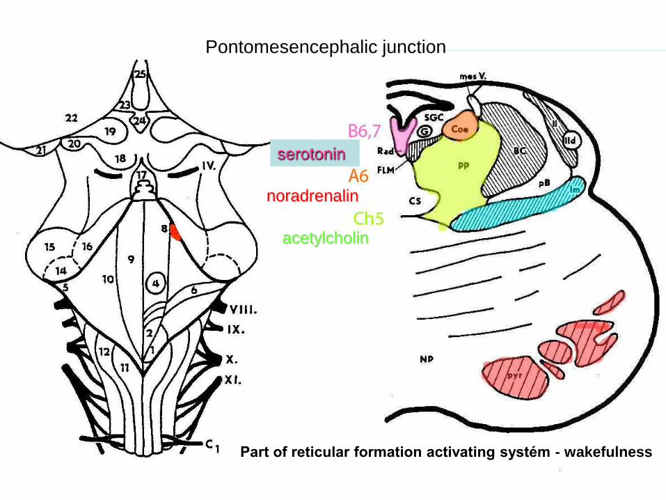
serotonin
noradrenalin
acetylcholin
Pontomesencephalic junction
Part of reticular formation activating systém - wakefulness
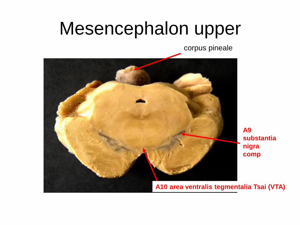
Mesencephalon uppercorpus pineale
A9
substantia
nigra
comp
A10 area ventralis tegmentalia Tsai (VTA)
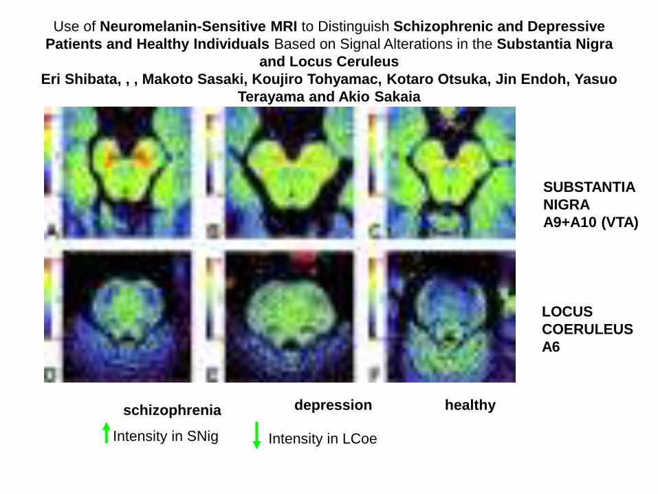
Use of Neuromelanin-Sensitive MRI to Distinguish Schizophrenic and Depressive
Patients and Healthy Individuals Based on Signal Alterations in the Substantia Nigra
and Locus Ceruleus
Eri Shibata, , , Makoto Sasaki, Koujiro Tohyamac, Kotaro Otsuka, Jin Endoh, Yasuo
Terayama and Akio Sakaia
schizophrenia depression healthy
SUBSTANTIA
NIGRA
A9+A10 (VTA)
LOCUS
COERULEUS
A6
Intensity in LCoeIntensity in SNig
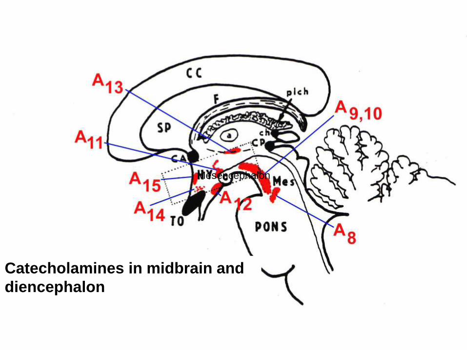
Mesencephalon
Catecholamines in midbrain and
diencephalon
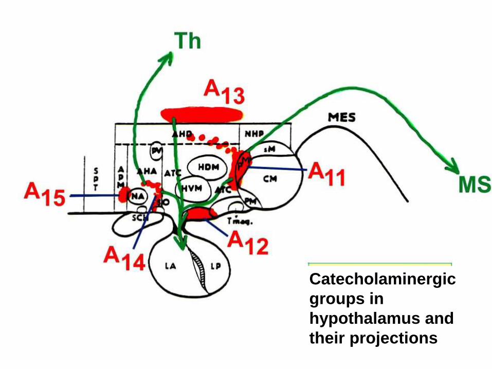
Catecholaminergic
groups in
hypothalamus and
their projections
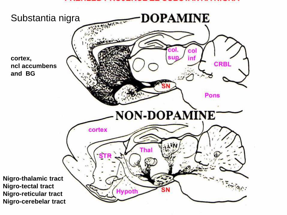
cortex,
ncl accumbens
and BG
Nigro-thalamic tract
Nigro-tectal tract
Nigro-reticular tract
Nigro-cerebelar tract
Substantia nigra
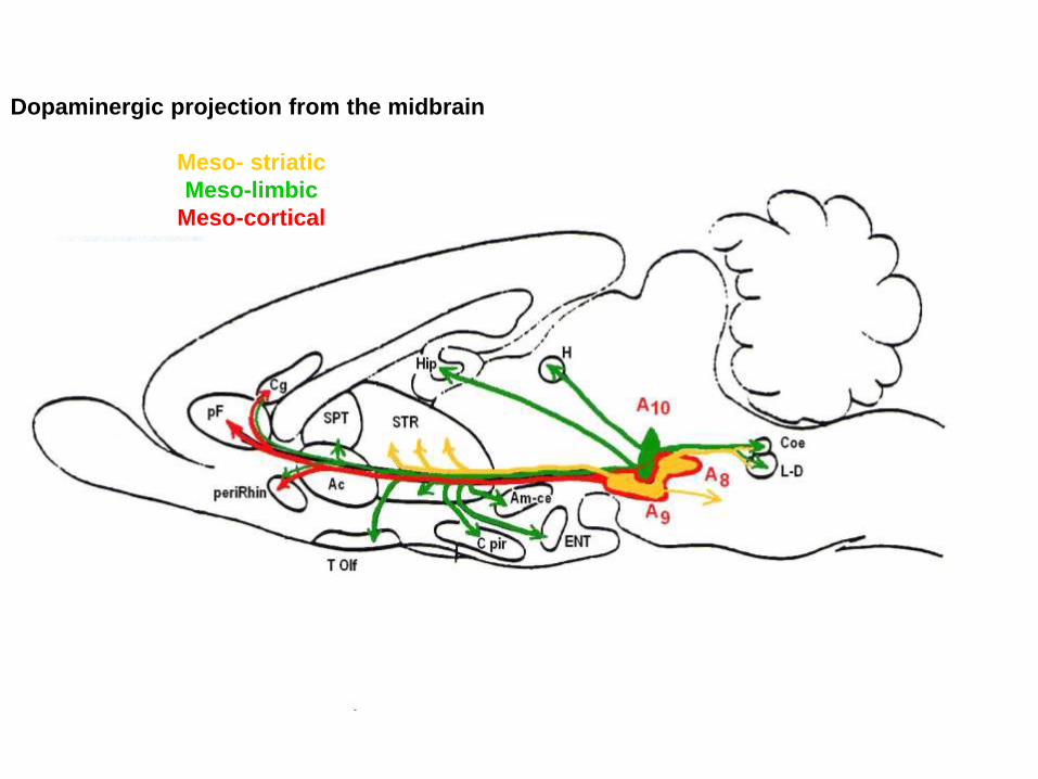
Dopaminergic projection from the midbrain
Meso- striatic
Meso-limbic
Meso-cortical
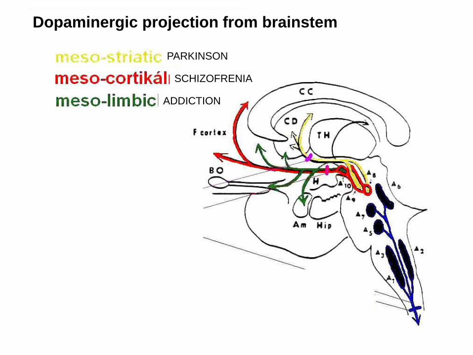
PARKINSON
ADDICTION
SCHIZOFRENIA
Dopaminergic projection from brainstem
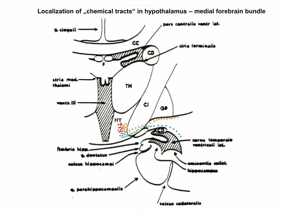
Localization of „chemical tracts“ in hypothalamus – medial forebrain bundle
LOCUS COERULEUS
KATECHOLAMINYLOCUS COERULEUS (A6) supplies the cortex and other structures by noradrenaline
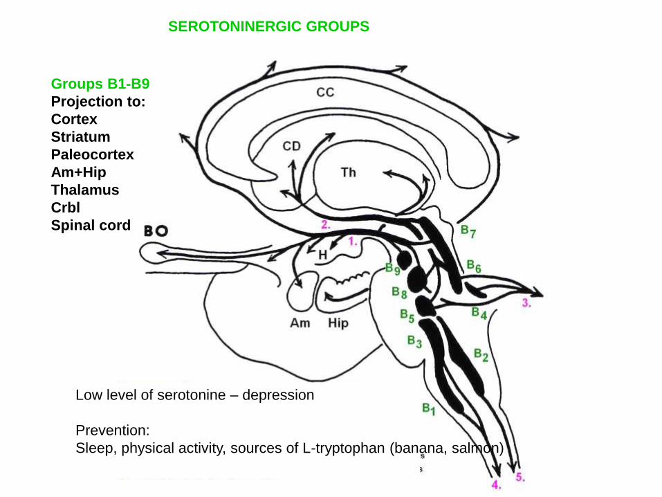
Groups B1-B9
Projection to:
Cortex
Striatum
Paleocortex
Am+Hip
Thalamus
Crbl
Spinal cord
SEROTONINERGIC GROUPS
Low level of serotonine – depression
Prevention:
Sleep, physical activity, sources of L-tryptophan (banana, salmon)
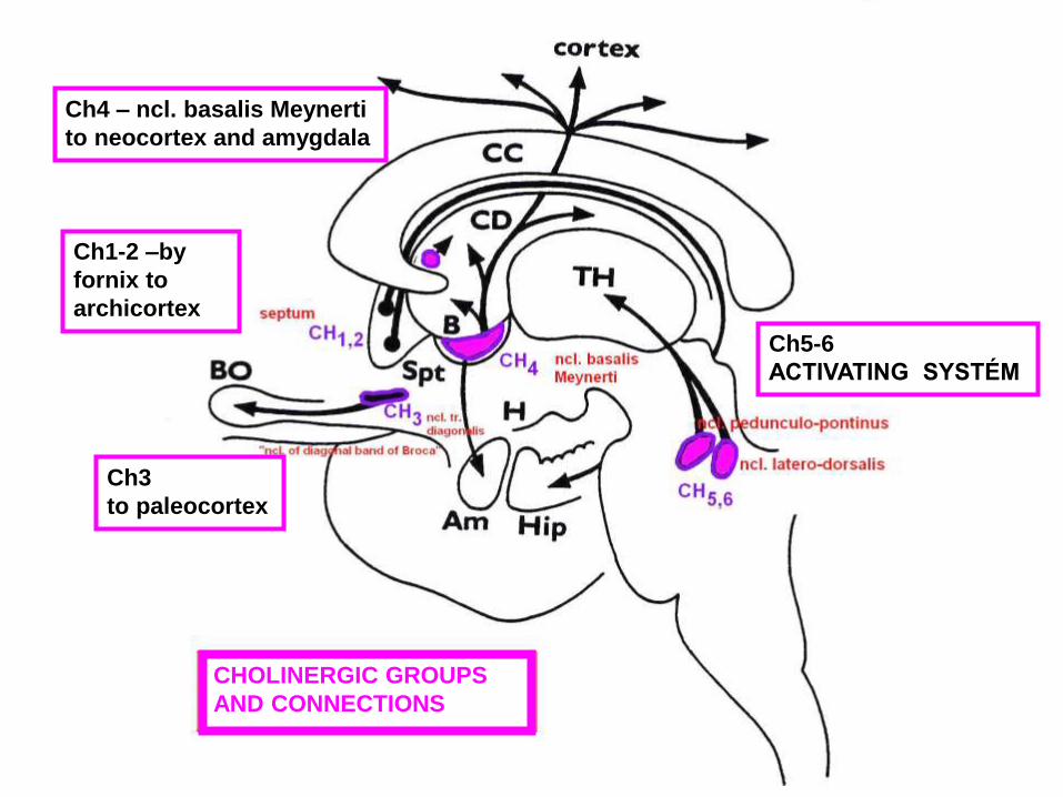
Ch5-6
ACTIVATING SYSTÉM
Ch1-2 –by
fornix to
archicortex
Ch3
to paleocortex
Ch4 – ncl. basalis Meynerti
to neocortex and amygdala
CHOLINERGIC GROUPS
AND CONNECTIONS
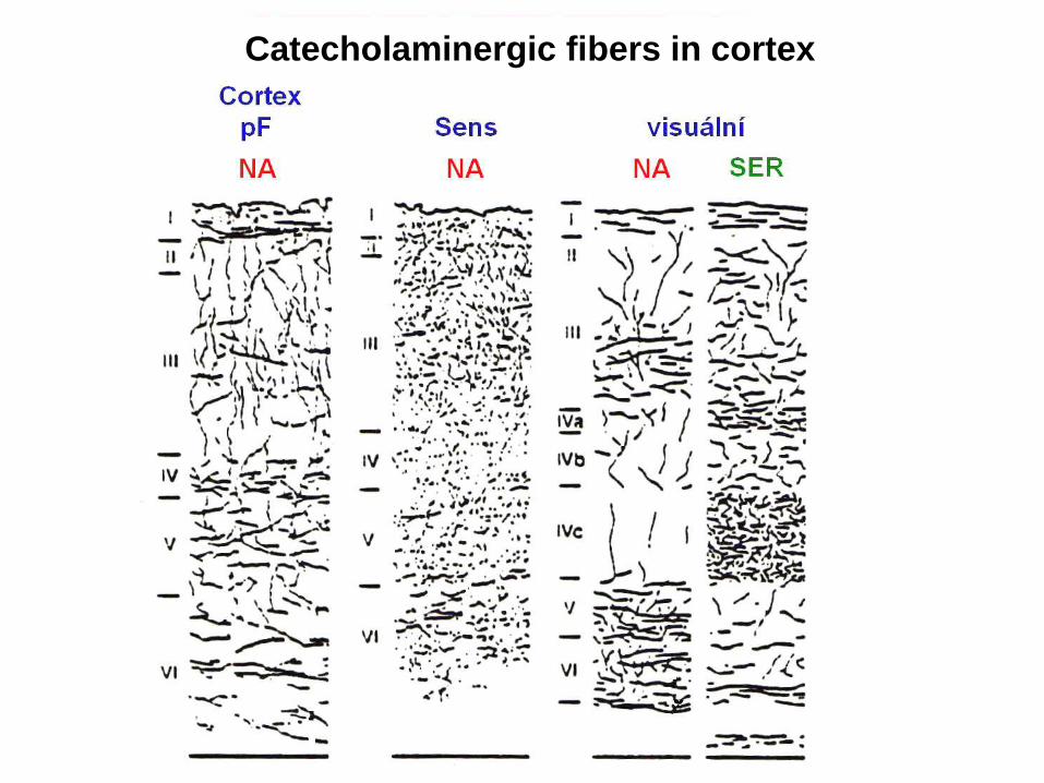
Catecholaminergic fibers in cortex
Sources
• Petrovický, Anatonie III
• Netter
• Nolte: The human brain in photographs
and diagrams
• H-J ten Donkelaar Clinical Neuroanatomy
• Kandel, Principles of Neural Science


