Redakční rada: Ing. Karel Balík, CSc. Tel. +420 2 66009 212 RNDr. František Kolář Ing. Jaroslava Svítilová Ing. Zuzana Weishauptová, CSc. Adresa redakce: RNDr. František Kolář Ústav struktury a mechaniky hornin AV ČR V Holešovičkách 41 182 09 Praha 8 tel.: +420 2 66009 343 fax.: +420 2 6880105 e-mail: [email protected]Spolupracoval: Ing. Tomáš Suchý e-mail: [email protected]Číslo 1/2005 - Obsah strana Oddělení kompozitních a uhlíkových materiálů, kontakt 3 Jan Grégr, Hana Schejbalová Král prvků – historie jeho poznávání a využívání 4 Oddělení kompozitních a uhlíkových materiálů – současný výzkum 9 K. Balík, M. Sochor, T. Suchý, M. Černý, R.Sedláček, V. Pešáková, H. Hulejová Biomechanical properties of porous composites based on glass and polysiloxane 10 Czech – Polish Workshop on Advanced Composites 17 Informace o konferencích 28 Instrukce pro publikování v časopise Acta Montana 31 Ústav struktury a mechaniky hornin AV ČR Oddělení kompozitních a uhlíkových materiálů – kontakt Kontakt ÚSMH AV ČR – oddělení kompozitních a uhlíkových materiálů V Holešovičkách 41 182 09 Praha 8 Česká republika telefon: +420 266 009 212 fax.: +420 268 801 05 Vedoucí: Ing. Karel Balík, CSc. [email protected]Zástupce: Mgr. Petr Glogar, CSc. [email protected]
Transcript
Redakční rada: Ing. Karel Balík, CSc. Tel. +420 2 66009 212 RNDr. František Kolář Ing. Jaroslava Svítilová Ing. Zuzana Weishauptová, CSc. Adresa redakce: RNDr. František Kolář Ústav struktury a mechaniky hornin AV ČR V Holešovičkách 41 182 09 Praha 8
Spolupracoval: Ing. Tomáš Suchý e-mail: [email protected] Číslo 1/2005 - Obsah strana Oddělení kompozitních a uhlíkových materiálů, kontakt 3 Jan Grégr, Hana Schejbalová Král prvků – historie jeho poznávání a využívání
4
Oddělení kompozitních a uhlíkových materiálů – současný výzkum
9
K. Balík, M. Sochor, T. Suchý, M. Černý, R.Sedláček, V. Pešáková, H. Hulejová Biomechanical properties of porous composites based on glass and polysiloxane
10
Czech – Polish Workshop on Advanced Composites 17 Informace o konferencích 28 Instrukce pro publikování v časopise Acta Montana 31
Ústav struktury a mechaniky hornin AV ČR Oddělení kompozitních a uhlíkových materiálů – kontakt
Kontakt ÚSMH AV ČR – oddělení kompozitních a uhlíkových materiálů V Holešovičkách 41 182 09 Praha 8 Česká republika telefon: +420 266 009 212 fax.: +420 268 801 05 Vedoucí: Ing. Karel Balík, CSc. [email protected] Zástupce: Mgr. Petr Glogar, CSc. [email protected]
KRÁL PRVKŮ – HISTORIE JEHO POZNÁNÍ A VYUŽÍVÁNÍ
Grégr Jan, Schejbalová Hana Technická univerzita v Liberci, katedra chemie FP
I když historie objevů a využití chemických prvků v souvislosti s jejich technickým významem i dějinnými událostmi je z hlediska metodologického zajímavá, je jí věnován velmi malý prostor ve výuce přírodních věd na všech stupních vzdělávací soustavy. Fascinující myšlenka, že rozmanitost přírody, která nás obklopuje, je tvořena jen z relativně malého počtu „stavebních kamenů“, se vynořila ve vývoji lidského myšlení poměrně brzy a nahradila tak ještě starší domněnku, že svět vznikl z jediné prahmoty. S touto představou se můžeme setkat již v indických vědách pocházejících zhruba z 12. století před naším letopočtem a v nejstarších písemných pramenech řecké kultury. Jako látka byl uhlík znám již v pravěku (dřevěné uhlí, saze), ale skutečnost , že se jedná o prvek, byla prokázána až v druhé polovině osmnáctého století. Mezinárodní název uhlíku “carbon” je odvozen od latinského carbo, čímž Římané označovali dřevěné uhlí. Uhlík se široce vyskytuje v přírodě, elementární uhlík byl dokázán ve vesmíru: na Slunci, hvězdách, kometách a v atmosféře planet. V porovnání s ostatními prvky má uhlík řadu unikátních vlastností jak fyzikálních (mechanických), tak i chemických, např.:
nejpevnější a nejtvrdší materiál – diamant nejlepší mazadlo (lubrikant) – grafit nejpevnější vlákna nejlepší adsorbent plynů – aktivní uhlí nejlepší héliová bariéra – skelný uhlík nejvíce sloučenin jak anorganických, tak zejména organických nejrozmanitější struktury objevené v posledních desetiletích
jako je molekula fullerenu, nanotrubice, magnetické nanopěny
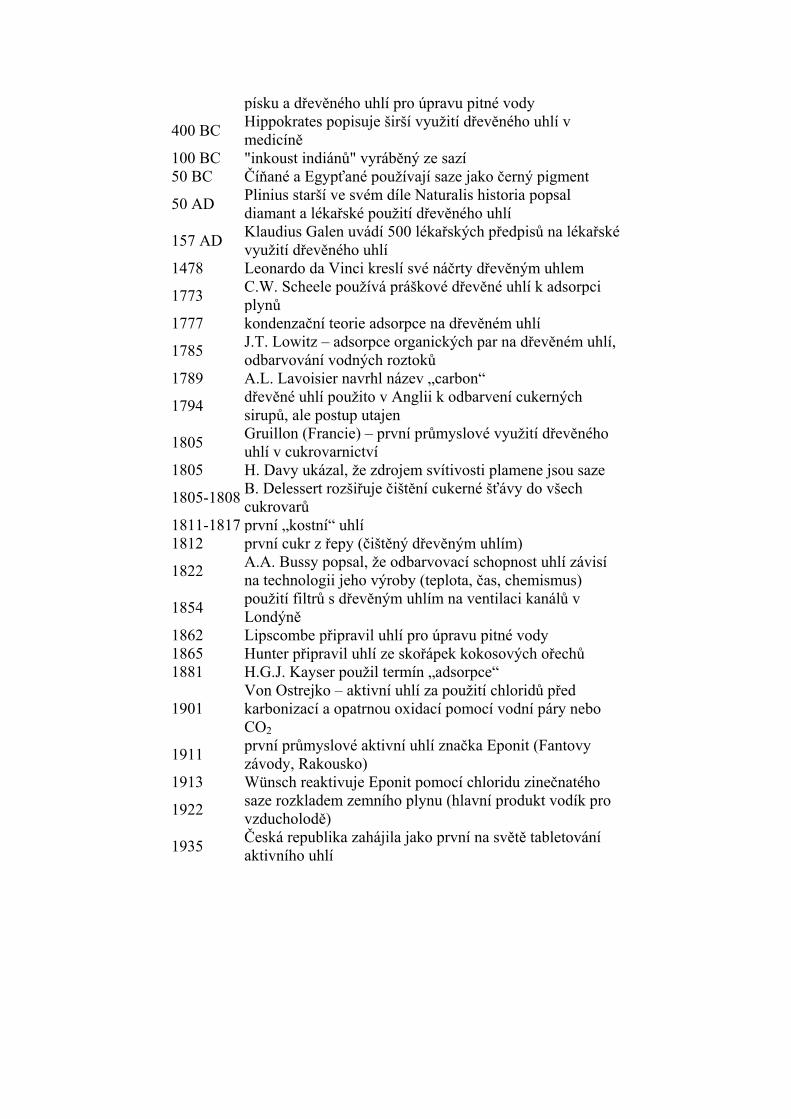
Z výše uvedeného výčtu je zřejmé, že uhlík má mezi prvky zcela výjimečné postavení a můžeme jej korunovat na „krále prvků“. Historie jeho poznávání a využívání je velmi dlouhá a bohatá. Protože však komplexní souhrn informací by byl nepřehledný, dovolili jsme si jej rozdělit podle forem uhlíku, zvlášť tedy základní informace o dřevěném uhlí, aktivním uhlí a sazích, informace o diamantu, grafitu (a uhlíkových vláknech a nanovláknech) a moderních formách uhlíku: Informace o dřevěném uhlí, aktivním uhlí a sazích
velký třeskvznik prvků vodíkovým, heliovým a uhlíkovým hořením – vznik uhlíku ve Vesmíru a jeho přeměna na další prvky
–20 000 letlidstvo poznalo oheň a dřevěné uhlí
3750 BC Egypťané a Sumerové používají dřevěné uhlí k redukci Cu, Sn a Zn z jejich rud
2000 BC Dřevěné uhlí se používá jako domácí bezdýmé palivo 1500 BC Egypťané použili dřevěné uhlí v medicíně (trávicí potíže)
450 BC Féničané uskladňují pitnou vodu v sudech pokrytých zevnitř dřevěným uhlím, na Hindu byly použity filtry z
písku a dřevěného uhlí pro úpravu pitné vody
400 BC Hippokrates popisuje širší využití dřevěného uhlí v medicíně
100 BC "inkoust indiánů" vyráběný ze sazí 50 BC Číňané a Egypťané používají saze jako černý pigment
50 AD Plinius starší ve svém díle Naturalis historia popsal diamant a lékařské použití dřevěného uhlí
157 AD Klaudius Galen uvádí 500 lékařských předpisů na lékařské využití dřevěného uhlí
1478 Leonardo da Vinci kreslí své náčrty dřevěným uhlem
1773 C.W. Scheele používá práškové dřevěné uhlí k adsorpci plynů
1777 kondenzační teorie adsorpce na dřevěném uhlí
1785 J.T. Lowitz – adsorpce organických par na dřevěném uhlí, odbarvování vodných roztoků
1789 A.L. Lavoisier navrhl název „carbon“
1794 dřevěné uhlí použito v Anglii k odbarvení cukerných sirupů, ale postup utajen
1805 Gruillon (Francie) – první průmyslové využití dřevěného uhlí v cukrovarnictví
1805 H. Davy ukázal, že zdrojem svítivosti plamene jsou saze
1805-1808B. Delessert rozšiřuje čištění cukerné šťávy do všech cukrovarů
1811-1817 první „kostní“ uhlí 1812 první cukr z řepy (čištěný dřevěným uhlím)
1822 A.A. Bussy popsal, že odbarvovací schopnost uhlí závisí na technologii jeho výroby (teplota, čas, chemismus)
1854 použití filtrů s dřevěným uhlím na ventilaci kanálů v Londýně
1862 Lipscombe připravil uhlí pro úpravu pitné vody 1865 Hunter připravil uhlí ze skořápek kokosových ořechů 1881 H.G.J. Kayser použil termín „adsorpce“
1901 Von Ostrejko – aktivní uhlí za použití chloridů před karbonizací a opatrnou oxidací pomocí vodní páry nebo CO2
1911 první průmyslové aktivní uhlí značka Eponit (Fantovy závody, Rakousko)
1913 Wünsch reaktivuje Eponit pomocí chloridu zinečnatého
1922 saze rozkladem zemního plynu (hlavní produkt vodík pro vzducholodě)
1935 Česká republika zahájila jako první na světě tabletování aktivního uhlí
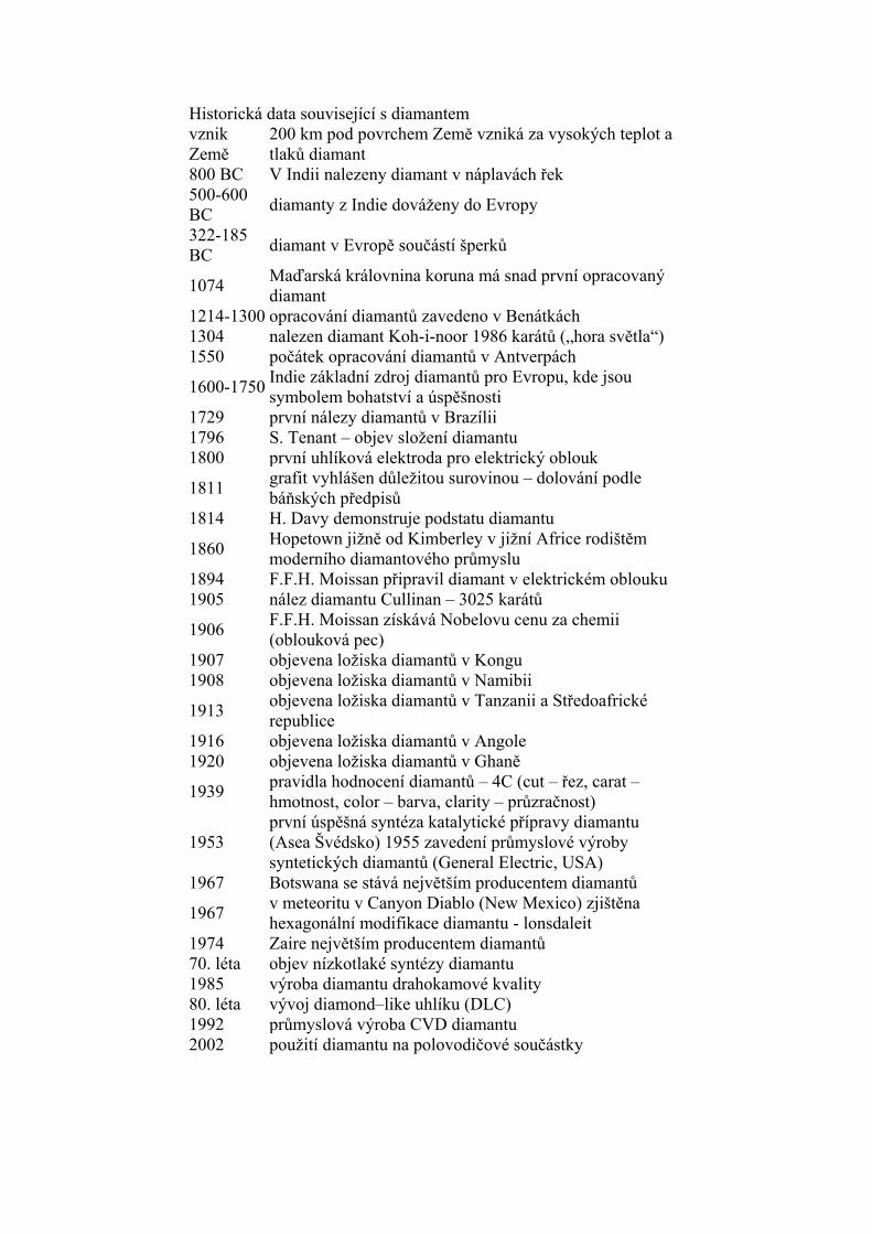
Historická data související s diamantem vznik Země
200 km pod povrchem Země vzniká za vysokých teplot a tlaků diamant
800 BC V Indii nalezeny diamant v náplavách řek 500-600 BC
diamanty z Indie dováženy do Evropy
322-185 BC
diamant v Evropě součástí šperků
1074 Maďarská královnina koruna má snad první opracovaný diamant
1214-1300 opracování diamantů zavedeno v Benátkách 1304 nalezen diamant Koh-i-noor 1986 karátů („hora světla“) 1550 počátek opracování diamantů v Antverpách
1600-1750Indie základní zdroj diamantů pro Evropu, kde jsou symbolem bohatství a úspěšnosti
1729 první nálezy diamantů v Brazílii 1796 S. Tenant – objev složení diamantu 1800 první uhlíková elektroda pro elektrický oblouk
1811 grafit vyhlášen důležitou surovinou – dolování podle báňských předpisů
1814 H. Davy demonstruje podstatu diamantu
1860 Hopetown jižně od Kimberley v jižní Africe rodištěm moderního diamantového průmyslu
1906 F.F.H. Moissan získává Nobelovu cenu za chemii (oblouková pec)
1907 objevena ložiska diamantů v Kongu 1908 objevena ložiska diamantů v Namibii
1913 objevena ložiska diamantů v Tanzanii a Středoafrické republice
1916 objevena ložiska diamantů v Angole 1920 objevena ložiska diamantů v Ghaně
1939 pravidla hodnocení diamantů – 4C (cut – řez, carat – hmotnost, color – barva, clarity – průzračnost)
1953 první úspěšná syntéza katalytické přípravy diamantu (Asea Švédsko) 1955 zavedení průmyslové výroby syntetických diamantů (General Electric, USA)
1967 Botswana se stává největším producentem diamantů
1967 v meteoritu v Canyon Diablo (New Mexico) zjištěna hexagonální modifikace diamantu - lonsdaleit
1974 Zaire největším producentem diamantů 70. léta objev nízkotlaké syntézy diamantu 1985 výroba diamantu drahokamové kvality 80. léta vývoj diamond–like uhlíku (DLC) 1992 průmyslová výroba CVD diamantu 2002 použití diamantu na polovodičové součástky
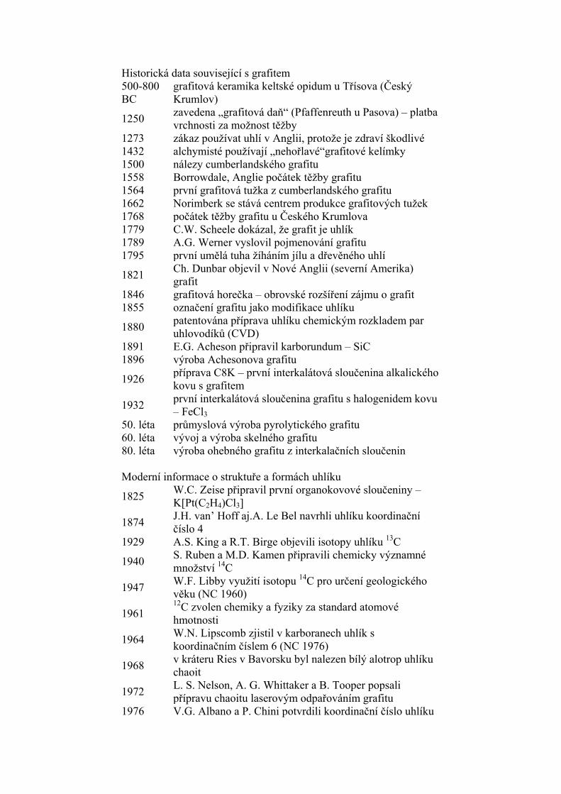
Historická data související s grafitem 500-800 BC
grafitová keramika keltské opidum u Třísova (Český Krumlov)
1250 zavedena „grafitová daň“ (Pfaffenreuth u Pasova) – platba vrchnosti za možnost těžby
1273 zákaz používat uhlí v Anglii, protože je zdraví škodlivé 1432 alchymisté používají „nehořlavé“grafitové kelímky 1500 nálezy cumberlandského grafitu 1558 Borrowdale, Anglie počátek těžby grafitu 1564 první grafitová tužka z cumberlandského grafitu 1662 Norimberk se stává centrem produkce grafitových tužek 1768 počátek těžby grafitu u Českého Krumlova 1779 C.W. Scheele dokázal, že grafit je uhlík 1789 A.G. Werner vyslovil pojmenování grafitu 1795 první umělá tuha žíháním jílu a dřevěného uhlí
1821 Ch. Dunbar objevil v Nové Anglii (severní Amerika) grafit
1846 grafitová horečka – obrovské rozšíření zájmu o grafit 1855 označení grafitu jako modifikace uhlíku
1880 patentována příprava uhlíku chemickým rozkladem par uhlovodíků (CVD)
1891 E.G. Acheson připravil karborundum – SiC 1896 výroba Achesonova grafitu
1926 příprava C8K – první interkalátová sloučenina alkalického kovu s grafitem
1932 první interkalátová sloučenina grafitu s halogenidem kovu – FeCl3
50. léta průmyslová výroba pyrolytického grafitu 60. léta vývoj a výroba skelného grafitu 80. léta výroba ohebného grafitu z interkalačních sloučenin
Moderní informace o struktuře a formách uhlíku
1825 W.C. Zeise připravil první organokovové sloučeniny – K[Pt(C2H4)Cl3]
1874 J.H. van’ Hoff aj.A. Le Bel navrhli uhlíku koordinační číslo 4
1929 A.S. King a R.T. Birge objevili isotopy uhlíku 13C
1940 S. Ruben a M.D. Kamen připravili chemicky významné množství 14C
1947 W.F. Libby využití isotopu 14C pro určení geologického věku (NC 1960)
1961 12C zvolen chemiky a fyziky za standard atomové hmotnosti
1964 W.N. Lipscomb zjistil v karboranech uhlík s koordinačním číslem 6 (NC 1976)
1968 v kráteru Ries v Bavorsku byl nalezen bílý alotrop uhlíku chaoit
1972 L. S. Nelson, A. G. Whittaker a B. Tooper popsali přípravu chaoitu laserovým odpařováním grafitu
1976 V.G. Albano a P. Chini potvrdili koordinační číslo uhlíku
8 v [Co8C(CO)18]
–2 1980 J. Jansta a F.P. Dousek popsali syntézu sp-uhlíku z teflonu 1985 objev molekuly fullerenu
1989 uhlíkové aerogely z resorcin-formaldehydového sol-gelu (Pekala R.W.)
1991 první informace o nanotrubicích (Iijima) 1993 výroba nanotrubic ve velkém (Bethme) 1995 uhlíkové anody pro lithiové dobíjecí články
1996 H.W. Kroto, R.F. Curl a R.E. Smalley získali Nobelovu cenu za objev fullerenu
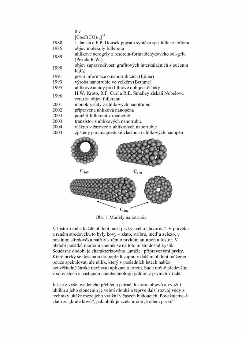
2001 monokrystaly z uhlíkových nanotrubic 2002 připravena uhlíková nanopěna 2003 použití fullerenů v medicíně 2003 tranzistor z uhlíkových nanotrubic 2004 vlákno v žárovce z uhlíkových nanotrubic 2004 zjištěny paramagnetické vlastnosti uhlíkových nanopěn
Obr. 1 Modely nanotrubic V historii měla každé období mezi prvky svého „favorita“. V pravěku a raném středověku to byly kovy – zlato, stříbro, měď a železo, v pozdním středověku patřily k těmto prvkům antimon a fosfor. V období počátků moderní chemie se na toto místo dostal kyslík. Současné období je charakterizováno „uměle“ připravenými prvky. Které prvky se dostanou do popředí zájmu v dalším období můžeme pouze spekulovat, ale uhlík, který v posledních letech nabízí neuvěřitelně široké možnosti aplikací a forem, bude určitě především v souvislosti s nástupem nanotechnologií jedním z prvních v řadě. Jak je z výše uvedeného přehledu patrné, historie objevů a využití uhlíku a jeho sloučenin je velmi dlouhá a teprve další rozvoj vědy a techniky ukáže meze jeho využití v časech budoucích. Považujeme–li zlato za „krále kovů“, pak uhlík je zcela určitě „králem prvků“.
ODDĚLENÍ KOMPOZITNÍCH A UHLÍKOVÝCH
MATERIÁLŮ SOUČASNÝ VÝZKUM
Výzkumná činnost je zaměřena na studium moderních vláknových kompozitů (biokompatibilních kompozitů uhlík – uhlík a sklo-siloxan a tepelně odolných kompozitů s keramickou matricí) připravovaných pyrolýzou kompozitů (prekurzorů) s polymerní matricí. Věnuje se optimalizaci procesů přípravy, vztahům mezi strukturou a vlastnostmi studovaných materiálů, a hledání možných aplikací. Jako vláknová výztuž kompozitů jsou používána uhlíková vlákna nebo tkaniny, keramická vlákna (SiC nebo Al2O3) nebo vlákna a tkaniny z E-skla, R-skla či taveného čediče. Pravidelné vrstevnaté uspořádání vláken smočených polymerní matricí vytváří strukturu budoucího kompozitu a určuje anizotropii jeho vlastností. Druh použitého polymeru předurčuje typ matrice vzniklé pyrolýzou (tj. tepelným rozkladem v inertní atmosféře): při použití fenolické pryskyřice vzniká kompozit s matricí uhlíkovou, zatímco z polysiloxanové pryskyřice vzniká tepelně odolná keramická matrice obsahující skupiny SiOC. Procesy přípravy zahrnují také opakovanou impregnaci kompozitů pryskyřicí s cílem zvýšit jejich hustotu a snížit výskyt dutin, vysokoteplotní zpracování (až 2500°C pro kompozit uhlík-uhlík) pro zlepšení mechanických vlastností a infiltraci pyrolytickým uhlíkem či křemíkem z plynné fáze pro zlepšení povrchových vlastností. Mikrostruktura připravených kompozitů je sledována optickou mikroskopií a kvantitativně hodnocena metodou obrazové analýzy. Mechanické vlastnosti (modul pružnosti a mez pevnosti) jsou studovány pomocí univerzálního testovacího stroje do teploty 1400°C. Dynamické moduly v tahu a ve smyku jsou měřeny metodou rezonančních frekvencí při laboratorní teplotě. Poznatky slouží nejen k posouzení sladění mechanických vlastností kompozitů s vlastnostmi kostí v případě biomateriálů, ale též k hodnocení odolnosti (či degradace) kompozitů v nepříznivých podmínkách a k optimalizaci volby výchozích složek a procesů tepelné přípravy. Biokompatibilita připravených kompozitů, tj. zejména testy „ in vitro“a „in vivo“ a jejich možné využití v chirurgických implantátech jsou zkoumány ve spolupráci s externími pracovišti. Otiskujeme poslední publikovaný článek z 12. mezinárodní konference biomedicínského inženýrství (The 12th International Conference on Biomedical Engineering 7 - 10 December 2005, Suntec Singapore International Convention and Exhibition Centre, SINGAPORE).
BIOMECHANICAL PROPERTIES OF POROUS
COMPOSITES BASED ON GLASS AND POLYSILOXANE K. Balik*, M. Sochor**, T. Suchy*,**, M. Cerny*, R. Sedlacek**, H.
Hulejova*** and V. Pesakova*** * Institute of Rock Structure and Mechanics, Academy of Sciences of
the Czech Republic, Prague, Czech Republic ** Faculty of Mechanical Engineering, Department of Mechanics,
Czech Technical University, Prague, Czech Republic *** Institute of Rheumatism, Prague, Czech Republic
Abstract Fiber composites, based on glass fibers and polysiloxanes, were prepared. On their surface, pores, having sizes ranging 200-400, 400-600 and larger than 600 m, were made, and, into the matrices of selected composites, powdered hydroxyapatite was added. The materials were then tested mechanically: their flexural strength, Young’s modulus of elasticity in tension and modulus of elasticity in shear and strength in compression were measured. Also the wettability of the composites was measured, and in-vitro and in-vivo tests were carried out. The composites displayed good mechanical properties, comparable with those of the human bone, and a satisfactory bio-tolerance. INTRODUCTION Efforts to produce artificial replacements of parts of the human body have a very long history. In ancient civilizations, whether ancient Indians, Egyptians or Chinese, this concerned primarily replacements of facial parts: ears, noses, teeth, etc. With regard to the bone surgery, natural replacements, which can be obtained by operating on the patient, are now frequently used, which, however, entail some disadvantages: an additional operation, possible infection and loss of blood. To prevent them, artificial replacements are used instead. The best known ones are metal implants. Their problem consists in their higher rigidity being unnatural for the bone, which can lead to the spongialization of the bone [1]. On the other hand, disadvantages of synthetic polymers lie in their low mechanical strength. Ceramic and glass materials are also frequently used. Some of them show a very good bioactivity, which enables a good osseo-integration. However, their frequent disadvantages are either a low mechanical strength, or, when having a higher strength, a considerable brittleness. Composite materials, to be used in the bone surgery, have been studied as well. Carbon-polymer composites have outstanding mechanical properties, but when exposed for a longer period of time in the body, the polymer tends to degrade to a monomer, which is no longer biotolerant. Carbon-carbon composites also show satisfactory mechanical properties, they are bio-inert, but their preparation is very demanding and expensive, and, when exposed to the live organism, they often release carbon particles [2, 3, 4, 5, 6].
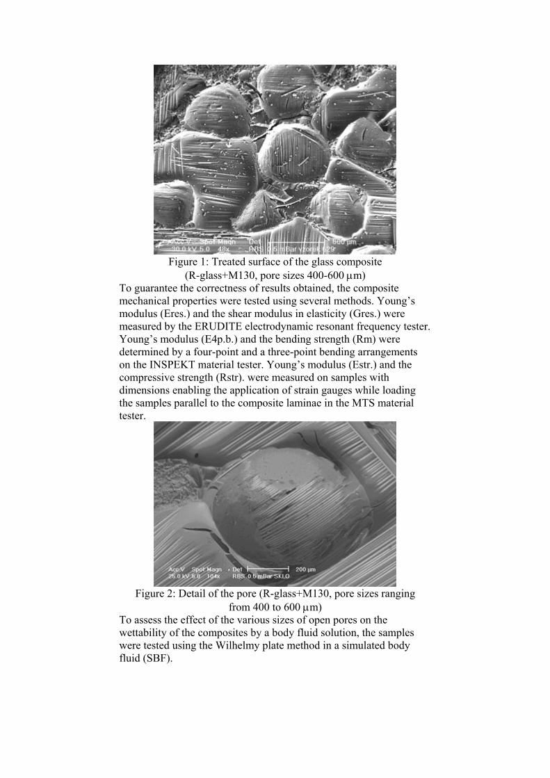
The purpose of this study was to suggest a simple method of preparing composite materials, which would display close mechanical properties to the human bones, with optimum size of open pores, which would enable the down-growth of osteoblasts, and containing additional bioactive components in the matrix. Materials and Methods The composites were prepared from a plain-woven V240 cloth (E-glass, VETROTEX, Litomysl, Czech Republic) and from 21055 satin-woven fabric (R-glass, VETROTEX, Saint Gobain, France) and polysiloxanes LUKOSIL 901 (L901) and LUKOSIL M130 (M130) (Lucebni zavody Kolin, Czech Republic) as the matrix precursor. The precursor used enables curing at temperatures of 200-350°C in a nitrogen atmosphere. The composite formed of R-glass + L901 was excluded from further tests, because it displayed excessive delamination. The percentages by fiber volume (Vf) in the composites with woven material from E-glass were 52%, and in the sample with R-glass 65%. For further treatment, aiming at enhancing the material ability to stimulate the down-growth of bone tissue, we chose the R-glass+M130 composite type, which displayed the most suitable mechanical properties (see Results) as compared with the properties of the human cortical bone and, at the same time, satisfied the high demands imposed on the biotolerance of implants. With regard to the treatment of the surface structure, a method of pressing separated fractions of special salt into the surface of the uncured composite and their subsequent elution after curing was applied (see Figures 1 and 2). In this way, three types of surfaces were obtained with different open pore sizes: 200-400, 400-600 and over 600 m. The porous samples, inclusive of the samples with untreated surface, were then subjected to in vivo and in vitro tests. Another group of samples consisted of materials with the same surface treatment, but simultaneously modified by adding matrix with an admixture of powder hydroxyapatite (HAp), particle size 5 m.
Figure 1: Treated surface of the glass composite (R-glass+M130, pore sizes 400-600 m)
To guarantee the correctness of results obtained, the composite mechanical properties were tested using several methods. Young’s modulus (Eres.) and the shear modulus in elasticity (Gres.) were measured by the ERUDITE electrodynamic resonant frequency tester. Young’s modulus (E4p.b.) and the bending strength (Rm) were determined by a four-point and a three-point bending arrangements on the INSPEKT material tester. Young’s modulus (Estr.) and the compressive strength (Rstr). were measured on samples with dimensions enabling the application of strain gauges while loading the samples parallel to the composite laminae in the MTS material tester.
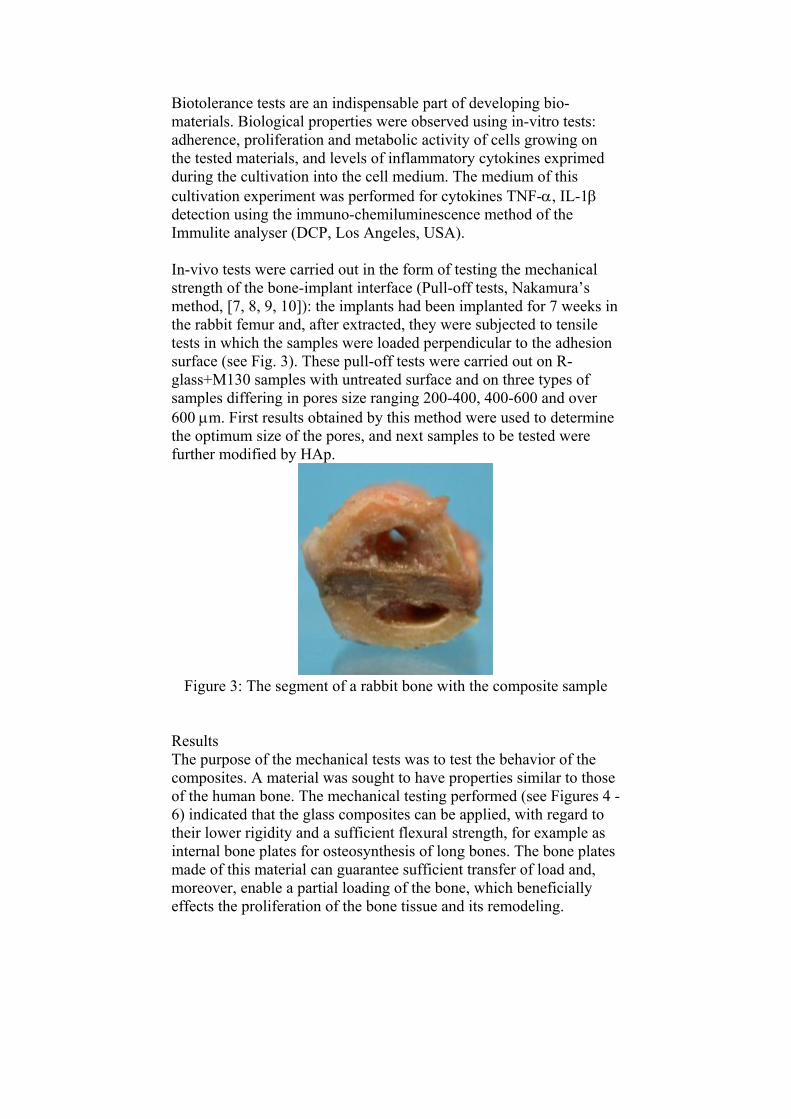
Figure 2: Detail of the pore (R-glass+M130, pore sizes ranging from 400 to 600 m)
To assess the effect of the various sizes of open pores on the wettability of the composites by a body fluid solution, the samples were tested using the Wilhelmy plate method in a simulated body fluid (SBF).
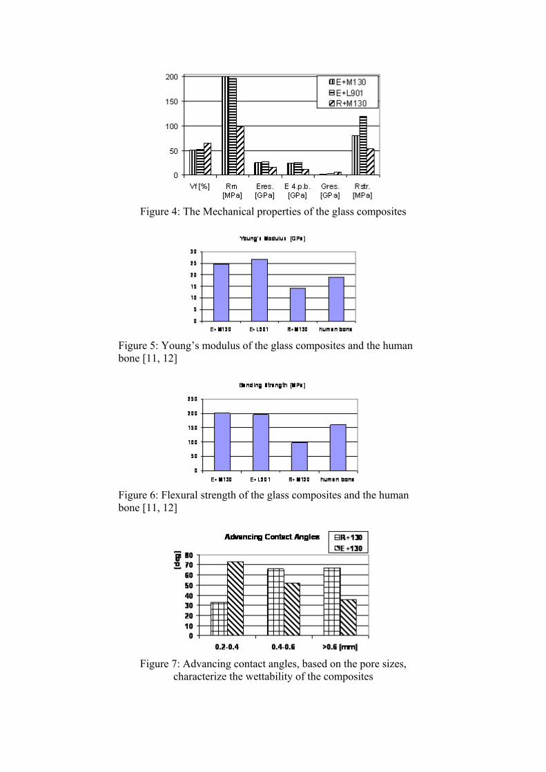
Biotolerance tests are an indispensable part of developing bio-materials. Biological properties were observed using in-vitro tests: adherence, proliferation and metabolic activity of cells growing on the tested materials, and levels of inflammatory cytokines exprimed during the cultivation into the cell medium. The medium of this cultivation experiment was performed for cytokines TNF-, IL-1 detection using the immuno-chemiluminescence method of the Immulite analyser (DCP, Los Angeles, USA). In-vivo tests were carried out in the form of testing the mechanical strength of the bone-implant interface (Pull-off tests, Nakamura’s method, [7, 8, 9, 10]): the implants had been implanted for 7 weeks in the rabbit femur and, after extracted, they were subjected to tensile tests in which the samples were loaded perpendicular to the adhesion surface (see Fig. 3). These pull-off tests were carried out on R-glass+M130 samples with untreated surface and on three types of samples differing in pores size ranging 200-400, 400-600 and over 600 m. First results obtained by this method were used to determine the optimum size of the pores, and next samples to be tested were further modified by HAp.
Figure 3: The segment of a rabbit bone with the composite sample Results
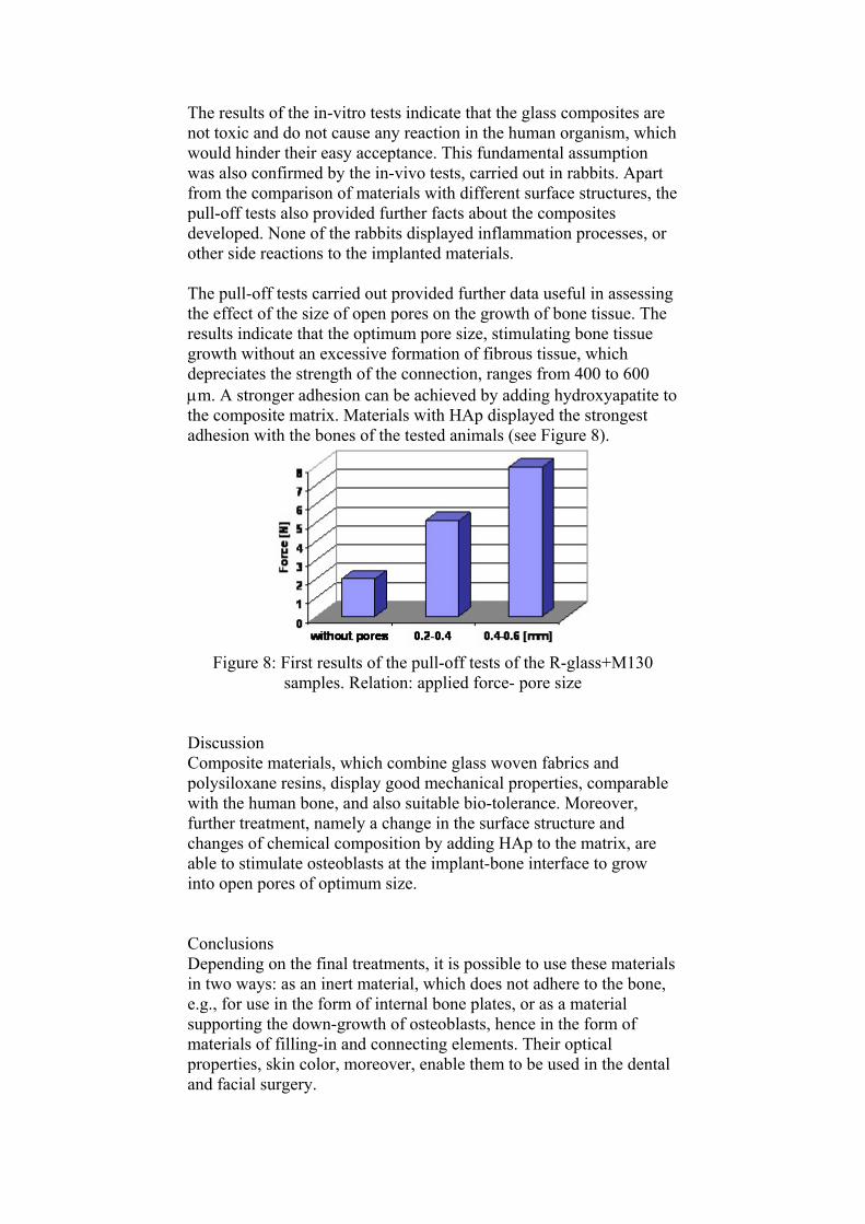
The purpose of the mechanical tests was to test the behavior of the composites. A material was sought to have properties similar to those of the human bone. The mechanical testing performed (see Figures 4 - 6) indicated that the glass composites can be applied, with regard to their lower rigidity and a sufficient flexural strength, for example as internal bone plates for osteosynthesis of long bones. The bone plates made of this material can guarantee sufficient transfer of load and, moreover, enable a partial loading of the bone, which beneficially effects the proliferation of the bone tissue and its remodeling.
Figure 4: The Mechanical properties of the glass composites
Figure 5: Young’s modulus of the glass composites and the human bone [11, 12]
Figure 6: Flexural strength of the glass composites and the human bone [11, 12]
Figure 7: Advancing contact angles, based on the pore sizes, characterize the wettability of the composites
The results of the in-vitro tests indicate that the glass composites are not toxic and do not cause any reaction in the human organism, which would hinder their easy acceptance. This fundamental assumption was also confirmed by the in-vivo tests, carried out in rabbits. Apart from the comparison of materials with different surface structures, the pull-off tests also provided further facts about the composites developed. None of the rabbits displayed inflammation processes, or other side reactions to the implanted materials. The pull-off tests carried out provided further data useful in assessing the effect of the size of open pores on the growth of bone tissue. The results indicate that the optimum pore size, stimulating bone tissue growth without an excessive formation of fibrous tissue, which depreciates the strength of the connection, ranges from 400 to 600 m. A stronger adhesion can be achieved by adding hydroxyapatite to the composite matrix. Materials with HAp displayed the strongest adhesion with the bones of the tested animals (see Figure 8).
Figure 8: First results of the pull-off tests of the R-glass+M130 samples. Relation: applied force- pore size
Discussion Composite materials, which combine glass woven fabrics and polysiloxane resins, display good mechanical properties, comparable with the human bone, and also suitable bio-tolerance. Moreover, further treatment, namely a change in the surface structure and changes of chemical composition by adding HAp to the matrix, are able to stimulate osteoblasts at the implant-bone interface to grow into open pores of optimum size. Conclusions Depending on the final treatments, it is possible to use these materials in two ways: as an inert material, which does not adhere to the bone, e.g., for use in the form of internal bone plates, or as a material supporting the down-growth of osteoblasts, hence in the form of materials of filling-in and connecting elements. Their optical properties, skin color, moreover, enable them to be used in the dental and facial surgery.
Acknowledgements This study was supported by the Grant Agency of the Czech Republic, GACR, under the project No. 106/03/1167 and by the Ministry of Education project: Transdisciplinary research in Biomedical Engineering II., No. MSM 6840770012. References
1. VALENTA J. et al. (1993): ‘Biomechanics’, (Academia, Prague, Co-published with Elsevier Science Publishers, Amsterdam)
2. RAMAKRISHNA S., MAYER J., WINTERMANTEL E. and LEONG KAM W. (2001): ‘Biomedical applications of polymer-composite materials: a review’, Composite Science and Technology, 61, pp. 1189-1224
3. PESAKOVA V., KLEZL Z., BALIK K., ADAM M. (2000): ‘Biomechanical and biological properties of the implant material carbon-carbon composite covered with pyrolytic carbon’. J. Mater. Sci. Mater.Med., 11, pp. 793-798
4. PESAKOVA V., SMETANA JR. K., BALIK K., HRUSKA J., PETRTYL M., HULEJOVA H. and ADAM M. (2003): ‘Biological and biomechanical properties of the carbon composite and polyethylene implant materials’, Journal of Materials Science: Materials in Medicine, 14, pp. 531-537
5. PESAKOVA V., SMETANA K., SOCHOR M., HULEJOVA H. and BALIK K. (2005): ‘Biological properties of the intervertebral cages made of titatium and containing a carbon-carbon composite covered with different copolymers’, Journal of Material Science:Materials in Medicine, 16, pp. 143-148
6. SUCHANEK W., YOSHIMURA M. (1998): ‘Processing and properties of hydroxyapatite-based biomaterials for use as hard tissue replacement implants’, Journal of Materials Research, 13, pp. 94-117
7. NAKAMURA ET AL. (1985): ‘A new glass-ceramic for bone replacement: Evaluation of its bonding to bone tissue’, Journal of Biomedical Materials Research, 19, pp. 94-96
8. KITSUGI T., NAKAMURA T., OKA M. ET AL. (1996): ‘Bone-bonding behavior of plasma-aprazed coatings of Bioglass, AW-glass ceramic, and tricalcium phosphate on titanium alloy’, J. Biomed. Mater. Res., 30, p. 261
9. TAKATSUKA K., ZAMAMURO T., NAKAMURA T. and KOKUBO T. (1995): ‘Bone-bonding behavior of titanium alloy evaluated mechanically with detaching failure load’, J. Biomed. Mater. Res., 29, p. 157
10. TAMURA J., KITSUGI T., IIDA H. ET AL. (1997): ‘Bone bonding ability of bioactive bone cements’, Clin. Orthop., 343, p. 183
11. FUNG Y.C. (1993): ‘Mechanical Properties of Living Tissues’, in FUNG Y.C. (Ed.): ‘Biomechanics’, (Springer – Verlag Inc., New York), p. 500
12. CURREY J.D. (1983): ‘Handbook of Composites’, in KELLY A. MILEIKO S.T. (Ed.): ‘Handbook of Composites’, (Elsevier Science Publishers B.V.), p. 501
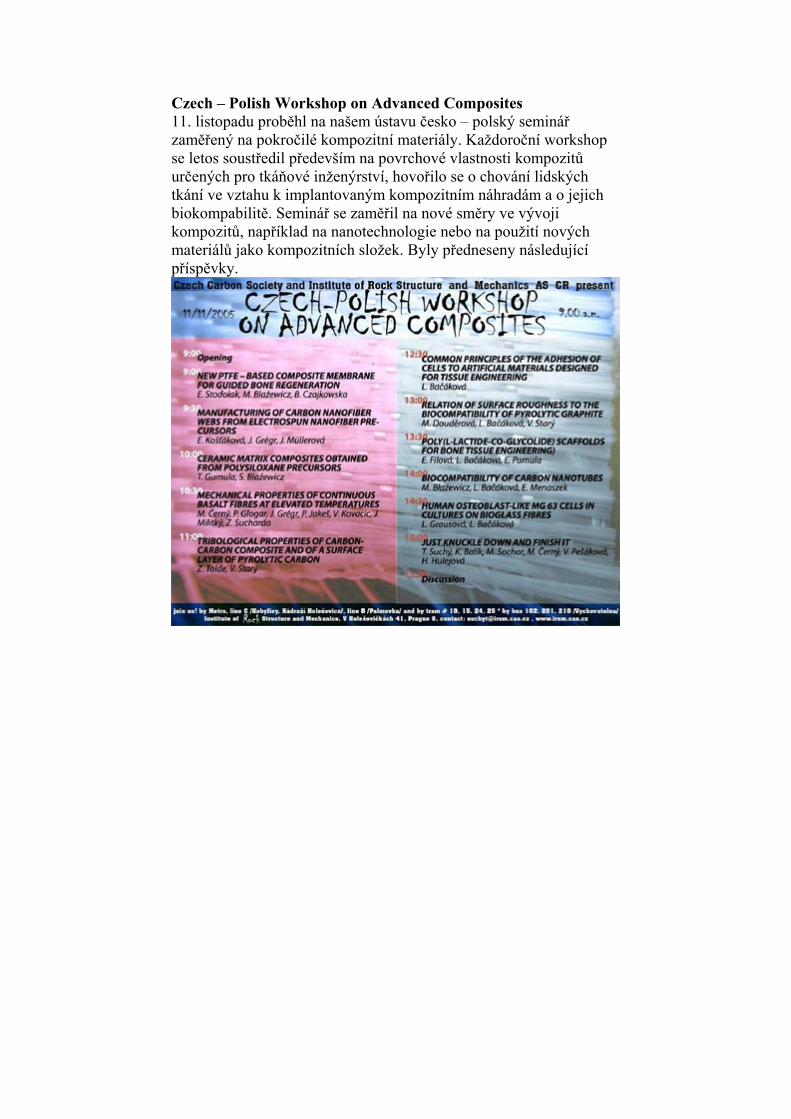
Czech – Polish Workshop on Advanced Composites 11. listopadu proběhl na našem ústavu česko – polský seminář zaměřený na pokročilé kompozitní materiály. Každoroční workshop se letos soustředil především na povrchové vlastnosti kompozitů určených pro tkáňové inženýrství, hovořilo se o chování lidských tkání ve vztahu k implantovaným kompozitním náhradám a o jejich biokompabilitě. Seminář se zaměřil na nové směry ve vývoji kompozitů, například na nanotechnologie nebo na použití nových materiálů jako kompozitních složek. Byly předneseny následující příspěvky.
NEW PTFE – BASED COMPOSITE MEMBRANE FOR GUIDED BONE REGENERATION
E. Stodolak*, M. Blazewicz*, B. Czajkowska** * Department of Biomaterials, Faculty of Materials Science and Ceramics, AGH University Science and Technology, Krakow, Poland ** Department of Immunology, Collegium Medicum, Jagiellonian University, Krakow, Poland GBR technique allows to rebuild the osseous structure in predictably way. It can be made by separating and isolating competing tissues from a healing defect by the means of specific type of material barrier. For such technique an implant material in the form of membrane is frequently used. Besides obvious biocompatibility, such material ought to be capable of inhibiting the connective tissue migration into the diseased site. It has to provide a specific stiffness and sufficient strength and make a suitable space over the healing site. An alternative for pure polymers membrane are composites consisting of biostable polymers reinforced with biocompatible fibrous components. The goal of this work was to manufacture new composite membrane for guided bone regeneration. The composite was made of three components, namely PTFE matrix modified with carbon fibers as reinforcement possessing proven biocompatibility. As a third alginate compounds biopolymer was used. The composite samples in the form of thin membranes were prepared. [email protected]
MANUFACTURING OF CARBON NANOFIBER WEBS FROM ELECTROSPUN NANOFIBER PRECURSORS
E. Košťáková*, J. Grégr**, J. Müllerová** *Department of Nonwovens, Faculty of Textile Engineering, Technical University of Liberec, Hálkova 6, 46117 Liberec, Czech Republic **Department of Chemistry, Faculty of Education, Technical University of Liberec, Hálkova 6, 46117 Liberec, Czech Republic Introduction Nanofibers are generally fibers of diameter smaller than 1 m. This article is focused on nanofibers produced during electrospinning process (Forhams, 1934). Modification of the method with higher efficiency of nanofiber production has been developed recently at TUL and patented (Jirsák et al., 2003 b). First experiments of carbonization of electrospun nanofibers have been realized in last years. Electrospun nanofiber materials made from non-aqueous solutions are used as a precursor for carbonization in majority of articles: e.g. polyacrylonitril (Wang et al., 2003 a, Wang et al., 2003 b), polybenzimidazol (Kim and Kim, 2004, Kim et al., 2004). Only one article being engaged in carbonization water-soluble polymer – polyvinyl alcohol nanofibers was published on web (Fong, 2004). Polyvinyl alcohol unfix physically fixed water in the course at heating, a decomposition reactions occur at the temperature over 200 °C (Smolinski, 2003). Primarily polyens arise, thus water peels by degradation of hydroxyl (OH) groups and hydrogen from carbon chain of macromolecule. This reaction manifests itself by gradual yellowing, browning and then blackening of fibers. If there is faster heating, it leads to decomposition to acetaldehyde and crotonaldehyde and whole original structure of chains can be damaged (destroyed). Polyen chains can their self each other join by means of Diels-Alder addition at careful heating and eventual catalysis, or their take place to intramolecular cycling. Created unsaturated cyclic compounds are already considerably more stable against additional increasing of temperature. An aromatization of these cycles comes into being at temperature above 450 °C, thus a basic graphite structure arises. Remains of water in the structure accelerate decomposition of structure to volatile aldehydes, accordingly usage of flame retardant catalysts is convenient for acquirement of the biggest possible carbonization gain.
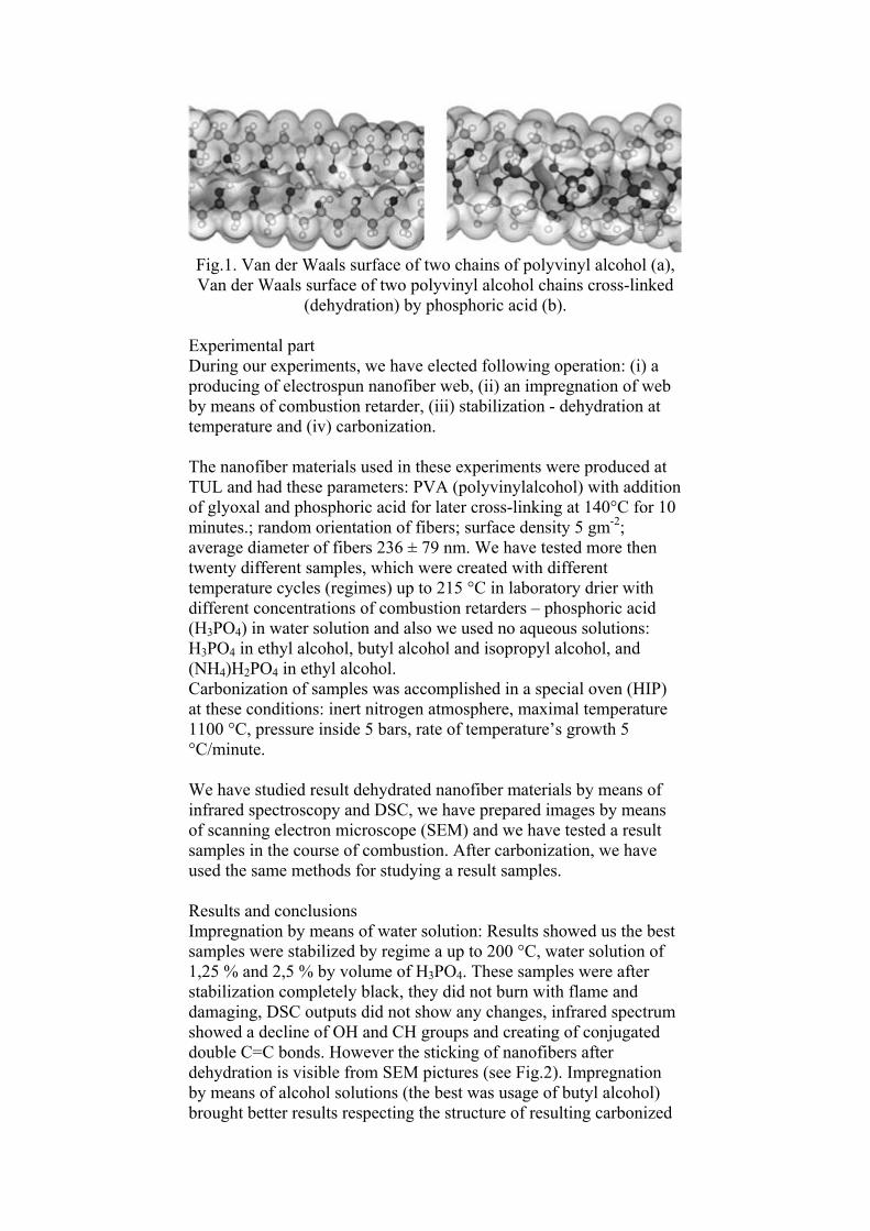
Fig.1. Van der Waals surface of two chains of polyvinyl alcohol (a), Van der Waals surface of two polyvinyl alcohol chains cross-linked
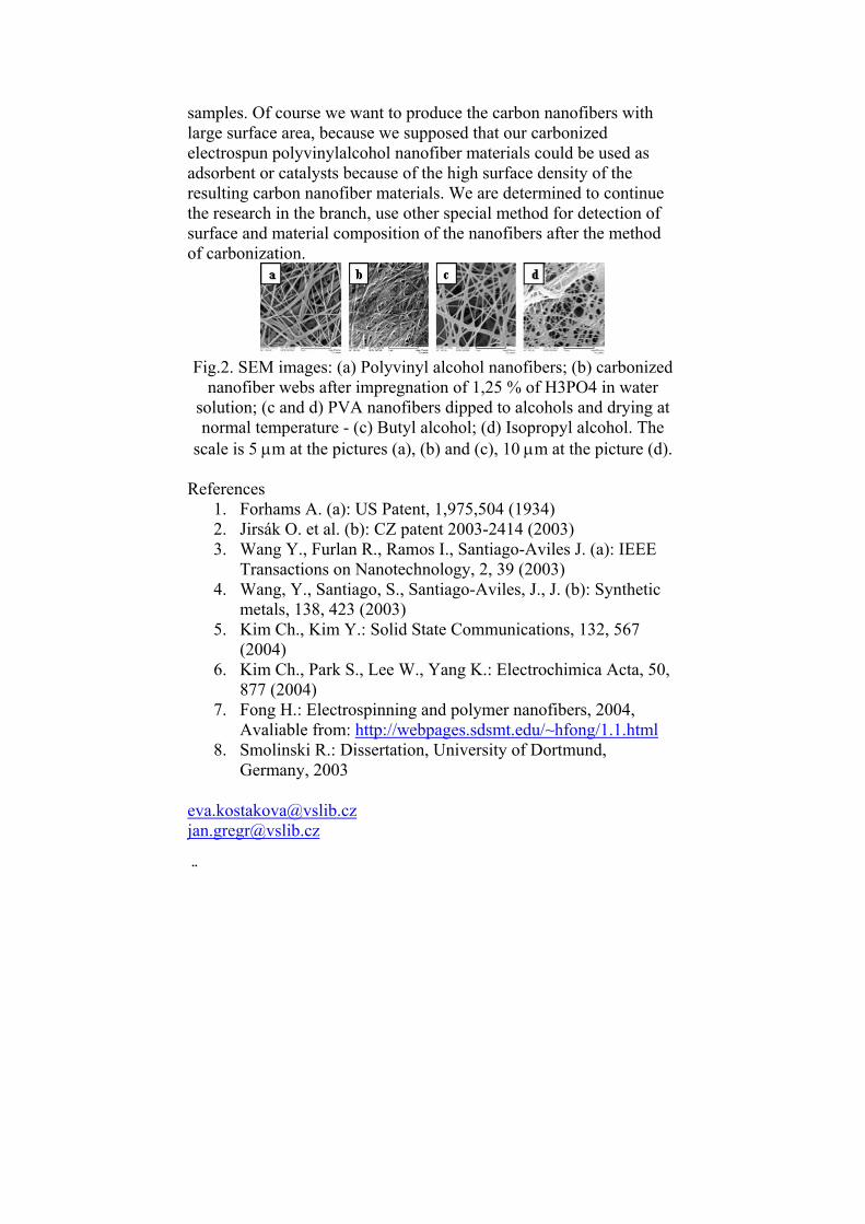
(dehydration) by phosphoric acid (b). Experimental part During our experiments, we have elected following operation: (i) a producing of electrospun nanofiber web, (ii) an impregnation of web by means of combustion retarder, (iii) stabilization - dehydration at temperature and (iv) carbonization. The nanofiber materials used in these experiments were produced at TUL and had these parameters: PVA (polyvinylalcohol) with addition of glyoxal and phosphoric acid for later cross-linking at 140°C for 10 minutes.; random orientation of fibers; surface density 5 gm-2; average diameter of fibers 236 ± 79 nm. We have tested more then twenty different samples, which were created with different temperature cycles (regimes) up to 215 °C in laboratory drier with different concentrations of combustion retarders – phosphoric acid (H3PO4) in water solution and also we used no aqueous solutions: H3PO4 in ethyl alcohol, butyl alcohol and isopropyl alcohol, and (NH4)H2PO4 in ethyl alcohol. Carbonization of samples was accomplished in a special oven (HIP) at these conditions: inert nitrogen atmosphere, maximal temperature 1100 °C, pressure inside 5 bars, rate of temperature’s growth 5 °C/minute. We have studied result dehydrated nanofiber materials by means of infrared spectroscopy and DSC, we have prepared images by means of scanning electron microscope (SEM) and we have tested a result samples in the course of combustion. After carbonization, we have used the same methods for studying a result samples. Results and conclusions Impregnation by means of water solution: Results showed us the best samples were stabilized by regime a up to 200 °C, water solution of 1,25 % and 2,5 % by volume of H3PO4. These samples were after stabilization completely black, they did not burn with flame and damaging, DSC outputs did not show any changes, infrared spectrum showed a decline of OH and CH groups and creating of conjugated double C=C bonds. However the sticking of nanofibers after dehydration is visible from SEM pictures (see Fig.2). Impregnation by means of alcohol solutions (the best was usage of butyl alcohol) brought better results respecting the structure of resulting carbonized
samples. Of course we want to produce the carbon nanofibers with large surface area, because we supposed that our carbonized electrospun polyvinylalcohol nanofiber materials could be used as adsorbent or catalysts because of the high surface density of the resulting carbon nanofiber materials. We are determined to continue the research in the branch, use other special method for detection of surface and material composition of the nanofibers after the method of carbonization.
Fig.2. SEM images: (a) Polyvinyl alcohol nanofibers; (b) carbonized nanofiber webs after impregnation of 1,25 % of H3PO4 in water
solution; (c and d) PVA nanofibers dipped to alcohols and drying at normal temperature - (c) Butyl alcohol; (d) Isopropyl alcohol. The
scale is 5 m at the pictures (a), (b) and (c), 10 m at the picture (d). References
1. Forhams A. (a): US Patent, 1,975,504 (1934) 2. Jirsák O. et al. (b): CZ patent 2003-2414 (2003) 3. Wang Y., Furlan R., Ramos I., Santiago-Aviles J. (a): IEEE
Transactions on Nanotechnology, 2, 39 (2003) 4. Wang, Y., Santiago, S., Santiago-Aviles, J., J. (b): Synthetic
metals, 138, 423 (2003) 5. Kim Ch., Kim Y.: Solid State Communications, 132, 567
(2004) 6. Kim Ch., Park S., Lee W., Yang K.: Electrochimica Acta, 50,
877 (2004) 7. Fong H.: Electrospinning and polymer nanofibers, 2004,
Avaliable from: http://webpages.sdsmt.edu/~hfong/1.1.html 8. Smolinski R.: Dissertation, University of Dortmund,
AGH University of Science and Technology,Faculty of Materials Science and Ceramics, Department of Biomaterials, Krakow, Poland The aim of this work was to obtain new ceramic matrix composites reinforced with carbon fibres by pyrolytic conversion of organosilicon polymer. Four types of polysiloxane resins differing in carbon to silicon molar ratio and oxygen concentration were used. The conversion mechanism from pure polysiloxane resins to carbide phase and conversion mechanism of these resins to ceramic phase in the presence of carbon fibres were investigated. The experiments were led in three temperature ranges, corresponding to composite manufacturing stages, namely moulding and curing, heat treatment up to 1000oC and final heat treatment in the temperature ranges from 1000 to 1700oC. The study on composites revealed that thermal decomposition mechanism of pure resins in the presence of carbon fibres up to 1000oC is similar as without fibres. Above 1000oC thermal decomposition of the matrices in the presence of fibres is more intense - the process occurs both in solid and in gas phases. The presence of carbon fibres results in developing of matrix surface area and produces higher mass losses and higher porosity of composite. As it results from the XRD analysis, at the temperature of 1700oC composite matrices contain nanosized silicon carbide. SEM and EDS analysis show that silicon carbide protective layer onto the fibre-matrix interface is created. Moreover, nanosized silicon carbide fibres crystallize in composite pores. Owing to the presence of the protective silicon carbide layer created from gas phase in the fibre-matrix interface, highly porous C/SiC composites represent significantly high oxidation resistance.
The process of repeated densification of porous matrix with polysiloxane polymer and additional heat treatment lead to further improvement of mechanical properties and oxidation resistance of composites. [email protected]
MECHANICAL PROPERTIES OF CONTINUOUS BASALT FIBRES AT ELEVATED TEMPERATURES
M. Černý*, P. Glogar*, J. Grégr**, P. Jakeš**, V. Kovacic***, J. Militký***, Z. Sucharda*
*Institute of Rock Structure and Mechanics, V Holesovickach 41, CZ-18209 Prague 8, Czech Republic **MDI Technologies, Ohradní 61, Prague 4, Czech Republic, ***Technical University of Liberec, Czech Republic Continuous basalt fibers (CBF) - a novel class of man made mineral fibers - possess good thermal and electric insulating properties. CBF can also be used – in form of filaments or fabrics – as reinforcement in composites with concrete or thermosetting resin matrix. Good thermal stability of CBF allows even utilising their reinforcing function in fibrous composites manufactured with help of an additional heat treatment. It is therefore desirable to investigate thermomechanical properties of CBF at elevated temperature because they can play a significant role in forming the microstructure and the resulting mechanical properties of the composites. In the present study, two commercially available basalt fiber types are examined and their properties are compared to those of conventional glass fibers. A trial basalt fiber prepared in the MDI Technologies laboratory using microwave melting and drawing the fiber through ceramic bushings was also included in the study. [email protected]
TRIBOLOGICAL PROPERTIES OF CARBON-CARBON
COMPOSITE AND OF A SURFACE LAYER OF PYROLYTIC CARBON
Z. Tolde, V. Starý Dept. of Materials Engineering, Fac. of Mech. Engn., CTU in Prague, Karlovo nám. 13, CZ-121 35 Prague 2, Czech Republic We measured the coefficient of friction and wear resistance of 2D carbon-carbon composite and of a surface layer of pyrolytic carbon (graphite). To explain these measurements we also measured the surface roughness and microhardness of both the composite and the layer. Due to its exclusive properties, this material system is often used in biomedical applications (bone and joint implants), machinery (friction-bearing parts) and in the aircraft industry (parts of the braking system). We studied the samples both with native (as prepared) surfaces and also with surfaces prepared by grinding and polishing, to obtain samples with various roughnesses. The measurements demonstrate the excellent tribological properties of the surfaces, especially the very low friction coefficient and the very good wear resistance of the surface of a pyrolytic carbon layer on the polished 2D C-C composite. [email protected]
COMMON PRINCIPLES OF THE ADHESION OF CELLS TO ARTIFICIAL MATERIALS DESIGNED FOR TISSUE
ENGINEERING L. Bačáková
Dept. of Growth and Differentiation of Cell Populations, Institute of Physiology, Academy of Sciences of the Czech Republic, Vídeňská 1083, CZ-142 20 Praha 4, Czech Republic Artificial and nature-derived materials, such as synthetic polymers, carbon materials, ceramics, metals and various composites of all these materials are of increasing importance in medicine and various biotechnologies. A new interdisciplinary scientific field called Tissue Engineering aims at construction of so-called bioartificial replacements of damaged tissues or organs, i.e. structures containing artificial material mimicking the natural extracellular matrix (ECM) and differentiated well functioning cells. For this purpose, the artificial materials should actively control the cell behavior, such as extent and strength of cell adhesion, migration and growth activity of cells, starting differentiation program in cells, secretion of various molecules by cells etc. The design of bioactive materials is based on the knowledge of the molecular mechanisms of cell-material interaction. On conventionally used artificial materials (i.e. materials not endowed with ligands for cell adhesion receptors), the cells adhere through ECM molecules (mainly fibronectin, vitronectin, collagen, laminin) adsorbed to the material surface from body fluids or the serum of the culture media. Cells bind specific sites on the adsorbed ECM molecules, e.g. certain amino acid sequences of the adsorbed proteins, by their adhesion receptors (integrins or non-integrin adhesion molecules, e.g. proteoglycans). Adhesion and further behavior of cells is strongly dependent on the species of the adsorbed molecules (e.g., cell adhesion-mediating fibronectin or vitronectin versus cell non-adhesive albumin), absolute amount of these molecules, and particularly on their spatial conformation, flexibility and accessibility of specific amino acid sequences or other cell-binding domains by cell adhesion receptors. This character of protein adsorption can be, at least partly, controlled by physicochemical properties of the material surface, such as presence of certain chemical functional groups (-OH, =O, -COOH, -NH2), polarity, wettability, electrical charge, surface compliance (i.e., stiffness vs. elasticity), and surface topography, i.e. the size, shape and distribution of the surface irregularities. Nanostructured surfaces of advanced biomaterials promote cell adhesion by adsorption of ECM molecules in conformation very close to that in the natural ECM. On the other hand, the adsorption of entire protein molecules is less controllable and associated with the risk of immune reaction and pathogen transfer. Thus there is an effort to functionalize the materials only with cell-binding domains of the ECM molecules, such as amino acid sequences containing RGD, REDV, KQAGDV, YIGSR, IKVAV, KRSR and other motifs. These oligopeptides are attached against bioinert background, not allowing aberrant protein adsorption and cell adhesion, in defined concentration and spatial
distribution which can contribute to a more precise control of cell behavior. Some of these oligopeptides bind preferentially a certain cell type (e.g. REDV endothelial cells, KQAGDV vascular smooth muscle cells, YIGSR and IKVAV neurons, KRSR osteoblasts) which could be utilized for regionally-selective cell adhesion (e.g. endothelial cells on the luminal surface of bioartificial vessels, osteoblast on bone implants instead competitive cell types, mainly fibroblasts etc.). After binding adhesion ligands, adhesion receptors are recruited into specific nano- or microdomains on the cell membrane, i.e. focal adhesion plaques, where they communicate with many structural and signaling molecules (e.g. talin, vinculin, focal adhesion kinase etc.), and through actin cytoskeleton also with various enzymes, cellular organelles and nucleus. By this way, the signal from extracelular environments, represented by an artificial material, is delivered to the cellular genome, and can influence its expression and proteosynthesis. The results presented in this review were supported by the Grant Agency of the Acad. Sci. CR (grants No. A5011301, A4050202 and 1P05QC012) and the Ministry of Education, Youth and Sports of CR (COST, Action 527.130, grant No. 1P05OC012). [email protected]
RELATION OF SURFACE ROUGHNESS TO THE BIOCOMPATIBILITY OF PYROLYTIC GRAPHITE
M. Douděrová, L. Bačáková, V. Starý Dept. of Materials Engineering, Fac. of Mech. Engn., CTU in Prague, Institute of Physiology, Academy of Sciences of the Czech Republic The material which should come into the contact with cells has to be biocompatible. It means, that the material must not have a negative influence on to cells and simultaneously cells must not degrade the material. For the material are during in vitro examinations important especially chemical and morphological properties of the surface. An important morphological property of the surface is e.g. its roughness. Dependence of surface roughness parameters on cell areas was evaluated in this study. [email protected]
POLY(L-LACTIDE-CO-GLYCOLIDE) SCAFFOLDS FOR BONE TISSUE ENGINEERING
E. Filová*, L. Bačáková*, E. Pamula** *Institute of Physiology, Academy of Sciences of the Czech Republic, Vídeňská 1083, 142 40 Prague 4 - Krč, Czech Republic **AGH University of Science and Technology, Faculty of Materials Science and Ceramics, Department of Biomaterials, 30 Mickiewicza Ave., 30-059 Kraków, Poland Degradable copolymer of L-lactide and glycolide (PLG) was synthesized by ring opening polymerization using zirconium acetylacetonate [Zr(acac)4] as a biocompatible initiator. The structure of the copolymer was studied by nuclear magnetic resonance spectroscopy (NMR) and gel permeation chromatography (GPC). The porous scaffolds of defined microstructure were prepared by solvent casting / salt particulate leaching which resulted in creation of three types of scaffolds with the same porosity (87% ± 1%) but different diameters of pores (600 m, 200 m and 40 m). The potential of the scaffolds for cell colonization was tested in a conventional static cell culture system using human osteoblast-like MG 63 cells. The morphology of cells, their number and presence inside the pores were evaluated on days 5 and 7 after seeding. For this evaluation, conventional fluorescence microscopy of cells stained with propidium iodide, laser confocal microscopy as well as counting of trypsinized cells in a Bürker hemocytometer were used. The highest number of cells was found on the scaffolds of the largest pore size (more than 120,000 cells/sample on day 7), whereas on the scaffolds with the medium and smallest pore diameter, the cell number was almost three times lower and similar for both pore sizes. The cells on the scaffolds of large or medium pore size infiltrated the inside part of the material, whereas on the scaffolds of small pore size, the cells were able to bridge the pore entrances and form a monolayer only on the material surface. These results suggest that the PLG scaffolds with the largest pore diameter (600 m) are the most suitable for colonization with osteogenic cells. Supported by the Ministry of Education, Youth and Sports of the Czech Republic (project COST, Action 527.130, grant No. 1P05OC012). [email protected][email protected]
BIOCOMPATIBILITY OF CARBON NANOTUBES M. Błażewicz*, L. Bačáková**, E. Menaszek***
*AGH University of Science and Technology, Faculty of Materials Science and Ceramics, Department of Biomaterials **Institte of Physiology, Academy of Science of the Czech Republic, Praha ***Department of Cytobiology and Histochemistry, Collegium Medicum, Jagiellonian University, Krakow, Poland The work presents selected examples of application of carbon nanotubes and nanofibers in cell culture and tissue engineering. Some aspects of biocompatibility in vivo and in vitro are considered. The response of living cells to carbon nanoparticles and bulk surface functionalized with nanosized carbon particles is performed. Histological and histochemical analyses of possible mechanism of migration of carbon nanoparticles in the form of single and multi wall nanotubes in living organism is shown. [email protected] HUMAN OSTEOBLAST-LIKE MG 63 CELLS IN CULTURES
ON BIOGLASS FIBERS Ľ. Grausová, L. Bačáková
Institute of Physiology, Academy of Sciences of the Czech Republic, Vídeňská 1083, 142 40 Prague 4 - Krč, Czech Republic The adhesion and proliferation of human osteoblast-like MG63 cells on bioglass fibers was studied. Four types of fibers were studied, different in their thickness and amount of SiO2: (1) fibers containing 20% of SiO2 and produced at the speed of 800 m/min; (2) fibers containing 20% of SiO2 and produced at the speed of 1600 m/min; (3) fibers containing 30% of SiO2 produced at the speed of 800 m/min; (4) fibers containing 30% of SiO2 produced at the speed of 1600 m/min. The diameter of the fibers produced at lower speed was 26 m, whereas in the fibers manufactured at the higher speed, it was twice a lower. The fibers were sterilized by H2O2–plasma method (Sterrad), placed in 24-well polystyrene multidishes (TPP, Switzerland; diameter of 1.5 cm) and seeded with MG 63 cells. Each well contained 500 fibers of the length of 1 cm, 30 000 cells and 1.5 ml of the Dulbecco-modified Eagle minimum essential medium supplemented with 10 % of fetal bovine serum. As control samples, the polystyrene culture dishes as well as microscopic glass coverslips were used. On day 1, 3 and 7 after seeding, the cells on some samples were visualized by staining with propidium iodide and their morphology was evaluated in fluorescence microscope. From the other samples, the cells were detached by trypsinization and counted in Bϋrker haemocytometer. On day 1 after seeding the number of cell initially adhering on bioglass fibers ranged in average from 950 to 2,090 cells/cm2, which was significantly less than on the polystyrene dishes (18560 cells/cm2) or glass coverslips (15000 cells/cm2). Similar trend also persisted in both 3- and 7-day-old cultures. On day 7 after seeding, the final cell population densities on the polystyrene
and glass coverslips reached in average 98500 and 82100 cells/cm2, respectively, whereas on the bioglass fibers, the density ranged only from 20150 to 23970 cells/cm2 (no significant differences among the different groups of fibers were found). Nevertheless, the cells on the glass fibers were viable, capable of proliferation and relatively well spread (i.e., spindle-shaped with the long axis oriented in parallel with fibers), which suggests that their lower number was due to the relatively small diameter and surface curvature of the fibers, less appropriate for a higher degree of cell spreading, rather that to a possible cytotoxicity of the material. The bioglass fibers could be used e.g. for reinforcement of polymeric materials for bone tissue engineering. Supported by the Ministry of Education, Youth and Sports of the Czech Republic (project COST, Action 527.130, grant No. 1P05OC012). [email protected]
JUST KNUCKLE DOWN AND FINISH IT GLASS FIBERS AND POLYSILOXANE BASED
COMPOSITES FOR BIOMEDICAL APPLICATIONS T. Suchý*, **, K. Balík**, M. Sochor*, M. Černý**, V. Pešáková***,
H. Hulejová*** *Faculty of Mechanical Engineering/Department of Mechanics, Czech Technical University in Prague, Prague, Czech Republic **Institute of Rock Structure and Mechanics/Department of Carbon and Composites, Czech Academy of Sciences, Prague, Czech Republic ***Institute of Rheumatism, Prague, Czech Republic The study presented deals with development of advanced composite materials to be used in the bone tissue engineering, based on the polysiloxane matrix and glass fibres, and endowed with 3D porous architecture. An extended experimental investigation was necessary for obtaining physical and chemical properties of the composite materials, namely their mechanical properties, porosity, void fraction, surface roughness. All these parameters had to be modified and studied, using both in vitro and in vivo testing, for achieving an optimum bone tissue ingrowth and maturation. [email protected]
Informace o konferencích
26-30 March 2006 The American Chemical Society, Fuel Division Symposium on Chemistry of Carbon Materials and Nanomaterials to be held in Atlanta. The symposium features research on the synthesis, properties, and applications of carbon materials and their novel nanoforms. Emphasis is on the common chemistry that underlies carbon science for both new and traditional materials. Topics include, but are not limited to: Molecular Engineering in Carbon Material Synthesis Carbon-Based Catalysts and Sorbents Novel Carbon Forms and Their Application Fields Carbon Nanoparticles and Their Derivatives Premium Carbon Products from Coal Carbon Composites and Nanocomposites Activated Carbon for Environment Applications Surface Chemistry and Functionalization http://www.anl.govIPCS/acsfuellpreprintinfo.html http://oasys.acs.orgloasys.htm
1-5 May 2006 33rd International Conference on Metallurgical Coatings and Thin Films will be held at the Town and Country Hotel in San Diego, Ca. Sponsored by the Applied Surface Science Division of the AVS, the meeting draws more than 600 attendees to participate over 50 technical sessions, including a symposium dedicated to carbon and nitride materials. Other symposia include: Coatings for Use at High Temperature Hard Coatings and Vapor Deposition Technology Optical Thin Films Tribology and Mechanical Behavior of Coatings and Thin Films Advanced Materials Characterization Applications, Manufacturing and Equipment New Horizons in Coatings and Thin Filins Coatings for Automotive Applications Coatings for Aerospace Applications Coatings for Fuel Cells
Specific carbon-related sessions include: Carbon Nitride, Boron Nitride and Group-III (AI, Ga, In) Nitride Materials Diamond and Diamond-like Carbon Materials Special forum on Carbon Nanotubes and Related Nano-structures Frontier Devices for Bio-, Energy- and Optoelectronic-Applications Based on Carbon and Nitride Materials http://www2.avs.org/conferences/icmctf/call/default.asp www.icmctf.org [email protected]
16-21 July 2006 The Carbon conference series comprises leading international meetings dedicated to the science and technology of carbon materials. Carbon 2006 will be hosted by the British Carbon Group and will be held at The Robert Gordon University in the historic Scottish city of Aberdeen. In addition, this year the conference will be preceded by an optional 'Nanoporous Carbons' summer school, and the main conference will be incorporating NanoteC06. C2006 conference sessions include, but are not limited to: Adsorption Fundamentals Applications Activated carbon Biological applications Carbon Fibres Carbonization, Mesophase, Pyrolysis and Thermal processes C-C composite materials Diamond Environmental Applications Fullerenes, nanotubes and nanoforms Fundamentals Applications Theory Electrical, battery and related applications Industrial & Energy Products Intercalation Nuclear graphite Physical & Chemical properties Fundamentals Reactivity and Catalysis Special forms of carbon www.carbon2006.org [email protected]
20-23 February 2007 Second International Conference on Recent Advances in Composite Materials will be held in New Delhi, India. The meeting will attempt to discuss all the critical aspects of metal-matrix composites, fibre composites, and ceramic composites. http://www.bhu.ac.in/notification/ICRACM%202007_r_.pdf [email protected]
Informace pro publikování v Časopise Acta Montana Acta Montana je multidisciplinární časopis s náplní:
Vybrané aspekty geodynamiky (řada A) Paliva, uhlíkové materiály a úpravnictví (řada B) Disertace, výsledky grantových projektů vypracovaných v
ÚSMH (řada AB)
Základní informace naleznete na internetové adrese: http://www.irsm.cas.cz/Newweb/ActaMont.htm Každá řada časopisu Acta Montana vychází 3 až 4 krát ročně. Cena jednoho výtisku je 100Kč + poštovné. Bankovní spojení: Česká národní banka, Praha 1, Na Příkopě 28, 115 03, č. účtu 635-081/0710, Variabilní symbol 468888 Adresa redakce: J. Netrestová IRSM AS CR V Holešovičkách 41 182 09 Prague 8, Czech Republic E-mail: [email protected] Tel. +420 266 009 318 INSTRUKCE PRO ČESKÉ AUTORY Časopis Acta Montana přijímá původní příspěvky splňující podmínky výše zmíněných disciplin. Rukopis zašlete e-mailem, resp. 3.5" disketě, ZIP nebo CD-ROM (MS Word libovolné verze), příp. spolu s dalšími soubory (obrázky, tabulky apod.). Dále je třeba dodat vytištěný příspěvek, ve třech vyhotoveních. Redakčbí rada rozhodne o přijetí příspěvku na základě recenze dvou oponentů. První autor obdrží jeden výtisk Acta Montana a 20 reprintů svých článků zdarma. Článek musí obsahovat: Název, celá jména autorů s adresami, včetně telefonních čísel, faxu, příp. e-mailu, abstrakt, klíčová slova, vlastní sdělení (vše v angličtině). Dále může obsahovat: Tabulky max. velikosti 24x16 cm a min. velikosti písma 9 b., v textu nebo na zvláštních stránkách. Tabulky musí být psány jako skutečné tabulky – ve sloupcích, nikoli jako text v řádcích. Záhlaví všech tabulek musí být na zvláštní stránce. Ilustrace: Přijímají se černobílé fotografie, kresby a mapy v dobré kvalitě (min. 600 dpi). Grafy se přijímají černobílé, velikosti bodů, tloušťka čar a popisy os musí být dostatečně velké kvůli možnému zmenšení. U všech grafů v článku je třeba dodržet stejný styl popisu. Ilustrace mohou být umístěny v textu nebo v samostatných souborech. Přípustné formáty těchto souborů
jsou (*.doc, *.xls, *.ptt, *.bmp, *.pcx, *.tif, *.jpg, *.gif). Ilustrace jsou číslovány v pořadí tak, jak je o nich referováno. Popis nesmí být umístěn přímo v obrázku, soupis všech popisů musí být na samostatné stránce. U popisu obrázku je třeba uvést jeho požadovanou velikost v textu. Redakce přijímá rovněž barevné ilustrace (min. 1200 dpi, max. velikost vytištěného obrázku je 24x16 cm). Barevné ilustrace jsou tištěny jednostranně a umístěny na konci článku jako příloha. Náklady na tisk těchto barevných příloh hradí autor. (2000 Kč za stranu). Proměnné, konstanty aj. symboly v matematických rovnicích a odkazy na ně v textu je třeba psát výhradně v editorech rovnic MS Equation Editor nebo MathType. Všechny použité symboly musí být vysvětleny v textu nebo v seznamu symbolů. Odkazy na literaturu v textu musí být ve formě (autor, rok), např., (Balik, 2001), (Rudajev et al., 2002). Soupis všech odkazů v abecedním pořadí musí být na samotné stránce ve tvaru: Příjmení, iniciály prvního autora, (příjmení a iniciály dalších autorů, kromě posledního) and (příjmení a iniciály posledního autora): rok, název článku, název časopisu, číslo, stránky od-do např. Mierzejewski, M., Korzak, F. and Kaczalek, M.: 2002, Geodynamic research of recent movements in the Karkonosze Mts, Acta Montana ser.A., 15(126), 56-78 Příspěvek musí být zaslán v konečné verzi. V autorském náhledu je možno opravovat pouze překlepy, jiné zásahy do textu jsou nepřípustné.
Acta Montana is a multidisciplinary journal concerned with:
Selected aspects of the geodynamics (series A) Fuel, carbon and mineral processing (series B) Extended essays, PhD dissertations or results of grant projects
elaborated in IRSM (series AB)
Topical information on internet address: http://www.irsm.cas.cz/Newweb/ActaMont.htm
Acta Montana issues periodically 4 times per annum. Aim: The main purpose of Acta Montana enables a possibility to publish the results of scientific works and contribute to information exchange among scientists, engineers and others professionals. Issues, price list: Acta Montana appears about three times annually for each series. The price of one issue is 12 EUR plus carriage charges. Back issues can be obtaining on request. Bank connection: Czech National Bank, Prague 1, Na Příkopě street 28, 115 03, account no. 635-081/0710 Variable Symbol 468888 Subscription office: J. Netrestová IRSM AS CR V Holešovičkách 41 182 09 Prague 8, Czech Republic E-mail: [email protected] Tel. +420 266 009 318 INSTRUCTIONS FOR AUTHORS Submissions: Acta Montana accepts original papers in English concerning all aspects of mentioned disciplines. Authors should submit three hard copies of their contribution and identical text file in MS Word (any version) and in case of need other files (figures, tables etc.) by e-mail, on 3.5" floppy disc, ZIP or CD-ROM. The Editorial Board on the basis of reviews of at least two referees makes the decision upon their publication. Author first named will receive one volume of Acta Montana and twenty reprints free of charge. The manuscript must contain: Title, full names of all authors, their affiliations and addresses including phone number, fax number and e-mail address, abstract, keywords and main body of paper (all in English). It can be included: Tables at max. size 24x16 cm and min. font size 9 pt, in text or on separate pages. Tables must be written really as tables (in columns), not as text (in rows). Captions of all tables must be on separate page.
Figures: Black and white photographs, drawing or maps in good quality (min. 600 dpi) are acceptable. Charts and diagrams must be in black and white, description of axes must be at sufficient size in due to possibility of reducing. Figures may be placed in text or as separate files. Only permitted formats of these files are (*.doc, *.xls, *.ptt, *.bmp, *.pcx, *.tif, *.jpg, *.gif). Figures are to be referred as Fig. Arabic numeral and should be numbered consecutively, according to their sequence in the text. Caption must no be in placed in the figure area. List of captions of all figures must be on the separate page. Required size of the pictures in the text must be denoted at the list of captions. Color illustrations are tolerable (min. 1200 dpi), but at author's expense 75 EUR per 1 page A4). These pages, (pressed on one side) will be placed on end of the paper as appendix. Variables, constants and other symbols in mathematical functions and also in the text are accepted written exclusively at MS Equation Editor or MS MathType. All used symbols must be explained in text or in the List of symbols. References quoted in the text must be in form (author, year), e.g., (Balik, 2001), (Rudajev et al., 2002). All references should be listed together at the end of the paper in alphabetic order as: First name, signatures of surnames, (names of other authors except last) and (First name, signatures of surnames of last author): year, title, journal, number, pages e.g. Mierzejewski, M., Korzak, F. and Kaczalek, M.: 2002, Geodynamic research of recent movements in the Karkonosze Mts, Acta Montana ser.A., 15(126), 56-78 Paper must be supplied as final version. At press-proof it is possible to correct only typing errors, other changes of text are not acceptable.