UNIVERSITY OF SOUTH BOHEMIA IN ČESKÉ BUDĚJOVICE FACULTY OF AGRICULTURE Study programme: N4101 Agriculture Engineering Branch of study: Agricultural Biotechnology Department: Department of Special Plant Production Head of Department: prof. Ing. Vladislav Čurn, Ph.D. DIPLOMA THESIS Diversity and distribution study of viruses in the entomopathogenic fungus Beauveria bassiana in the Czech Republic Studium diversity a rozšíření virů entomopatogenní houby Beauveria bassiana v České republice Thesis Supervisor: Noemí Herrero Asensio, Ph.D. Institute of Entomology, Biology Centre of the CAS Co‐Supervisor: prof. Ing. Vladislav Čurn, Ph.D. Faculty of Agriculture, University of South Bohemia Author: Bc. Petr Vaněček České Budějovice, April 2015
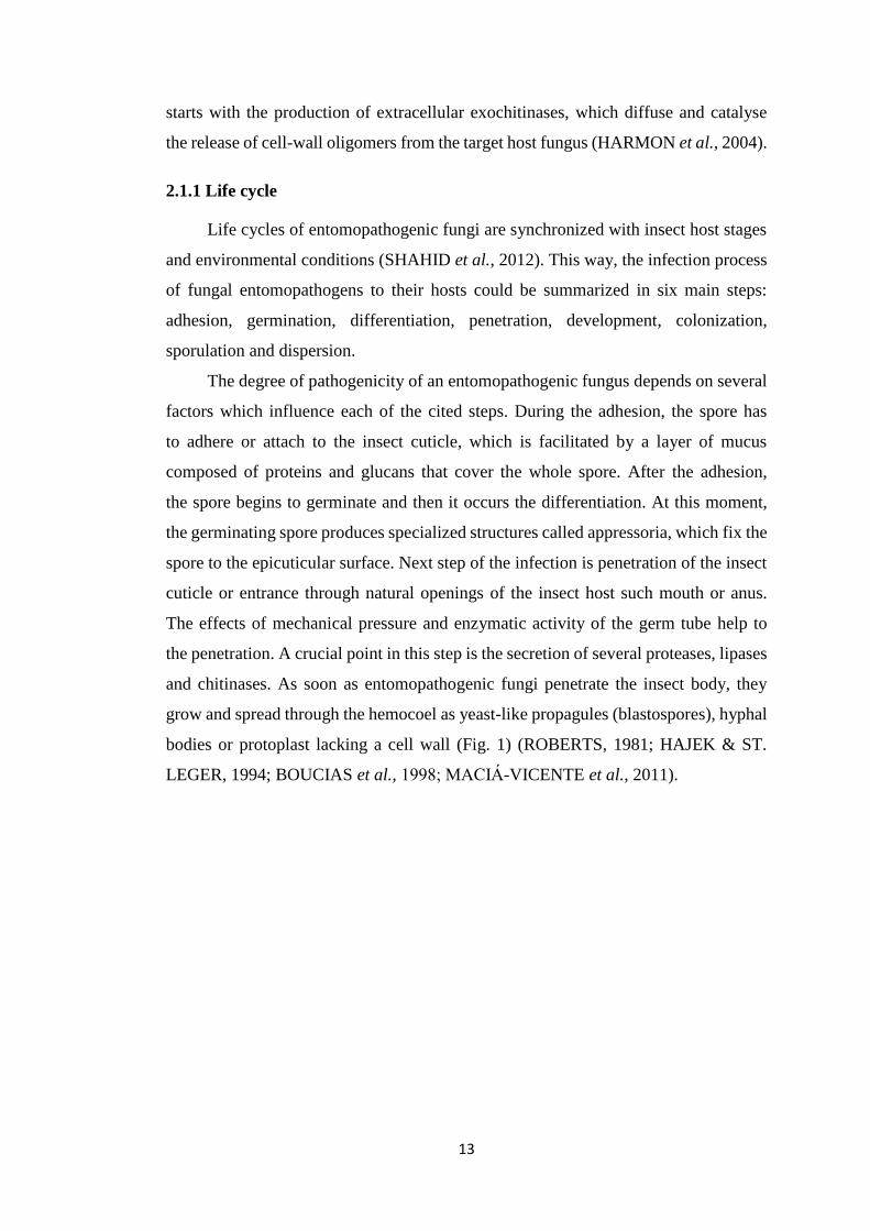
Transcript
UNIVERSITY OF SOUTH BOHEMIA IN ČESKÉ BUDĚJOVICE
FACULTY OF AGRICULTURE
Study programme: N4101 Agriculture Engineering
Branch of study: Agricultural Biotechnology
Department: Department of Special Plant Production
Head of Department: prof. Ing. Vladislav Čurn, Ph.D.
DIPLOMA THESIS
Diversity and distribution study of viruses
in the entomopathogenic fungus Beauveria bassiana
in the Czech Republic Studium diversity a rozšíření virů entomopatogenní houby
Beauveria bassiana v České republice
Thesis Supervisor: Noemí Herrero Asensio, Ph.D.
Institute of Entomology, Biology Centre of the CAS
Co‐Supervisor: prof. Ing. Vladislav Čurn, Ph.D. Faculty of Agriculture, University of South Bohemia
Author: Bc. Petr Vaněček
České Budějovice, April 2015
Declaration of the author's diploma thesis
I hereby declare that this diploma thesis has been carried out in the Institute
of Entomology, Biology Centre of the Czech Academy of Sciences in České
Budějovice, Czech Republic under the guidance of Noemí Herrero Asensio, Ph.D.,
and in Faculty of Agriculture, University of South Bohemia, České Budějovice,
Czech Republic under the guidance of Professor Vladislav Čurn. The work is original
and has not been submitted in part or full by me for any degree or bachelor thesis at
any other University. I further declare that the material obtained from other sources
has been duly acknowledged in the thesis.
According to the § 47b law n. 111/1998 Sb., I agree to publish full version of
my diploma thesis in public electronic database STAG managed by University of
South Bohemia in České Budějovice.
Prohlášení autora DP
Student na tomto místě prohlašuje, že se jedná pouze o jeho dílo, předepsanou
formulací:
Prohlašuji, že svoji diplomovou práci jsem vypracoval samostatně pouze
s použitím pramenů a literatury uvedených v seznamu citované literatury. Prohlašuji,
že v souladu s § 47b zákona č. 111/1998 Sb. v platném znění souhlasím se
zveřejněním své diplomové práce, a to v nezkrácené podobě (v úpravě vzniklé
vypuštěním vyznačených částí archivovaných Zemědělskou fakultou JU)
elektronickou cestou ve veřejně přístupné části databáze STAG provozované
Jihočeskou univerzitou v Českých Budějovicích na jejích internetových stránkách.
Datum Podpis studenta
Thanks to
I would like to thank my thesis to my supervisor Noemí Herrero Asensio, Ph.D. for
her professional guidance in this thesis and valuable comments during the consultation
and Ing. Kateřina Šimáčková, Ph.D. for her help in the laboratory.
Poděkování
Rád bych poděkoval své vedoucí diplomové práce Noemí Herrero Asensio, Ph.D., za
její odborné vedení v této práci a cenné připomínky během konzultace, Ing. Kateřině
Šimáčkové, Ph.D. za její pomoc v laboratoři.
Abstract
Mycoviruses are viruses that infect and replicate in fungal cells, but unlike most
known viruses of plants and animals, they exceptionally produce deleterious effects
on their host. Nonetheless, the last discoveries showed that some mycoviruses can
decrease the virulence of their phytopathogenic fungal hosts, making them very
attractive for their possible use as biological control agents. Most mycoviruses have
dsRNA genomes and are widespread in all major taxa of fungi. Beauveria bassiana is
one of the most studied species of entomopathogenic fungi; it has a cosmopolitan
distribution and is used as biocontroller against invertebrates in agriculture.
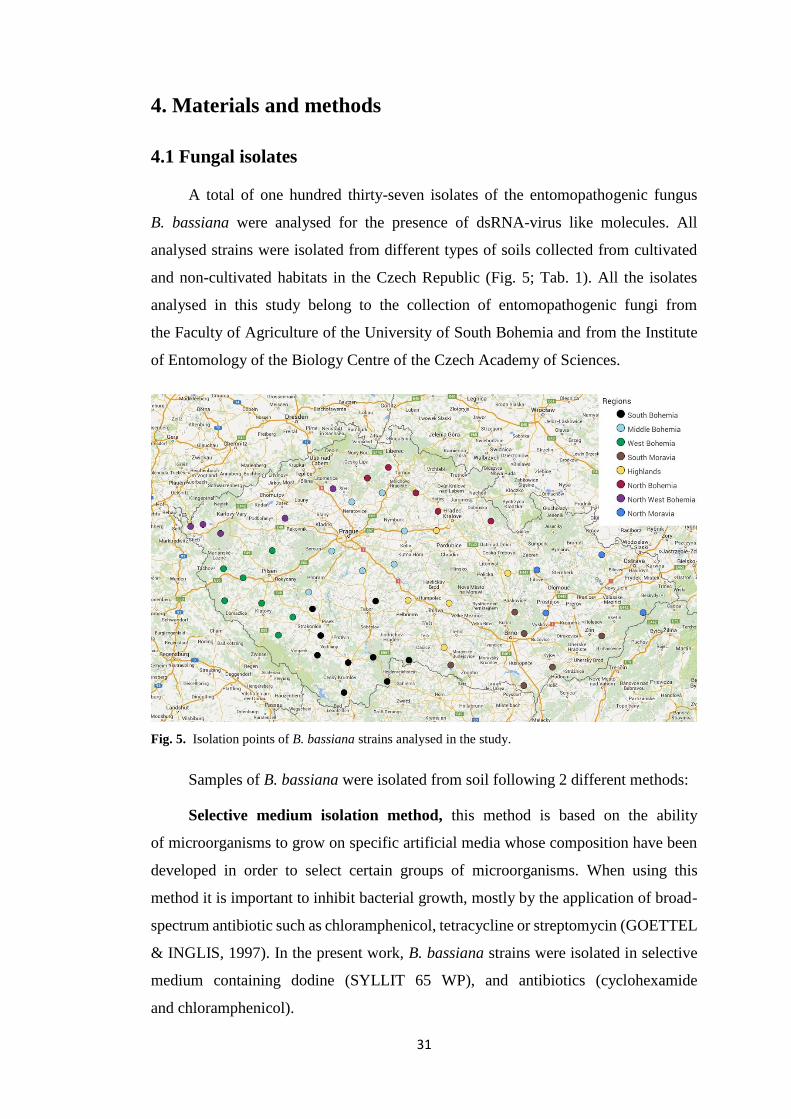
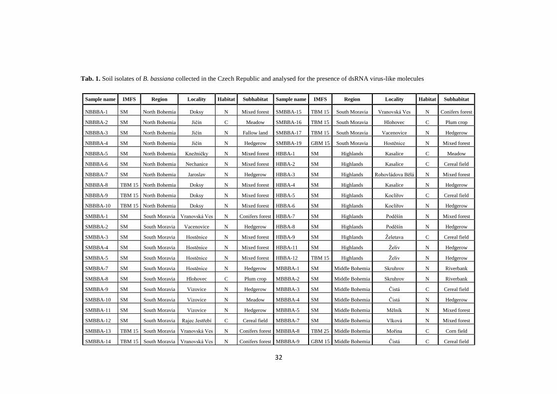
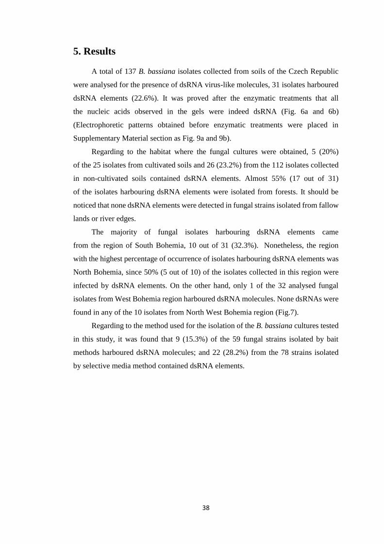
In the present work, a collection of 137 isolates of B. bassiana obtained
at different locations and from different habitats in the Czech Republic was analysed.
These isolates were analysed for the presence of dsRNA elements indicative of viral
infections. The results revealed a high prevalence of viral infections in Czech
B. bassiana isolates, with 22.6% of the isolates containing dsRNA elements with viral
characteristics. Obtained dsRNA electropherotypes showed that virus diversity
in infected isolates was high and that mixed virus infections occurred among them.
Based on the characteristics of the electrophoretic band patterns, it could be
hypothesized that B. bassiana isolates collected in the Czech Republic could harbour
members of the viral families Totiviridae, Partitiviridae, Chrysoviridae
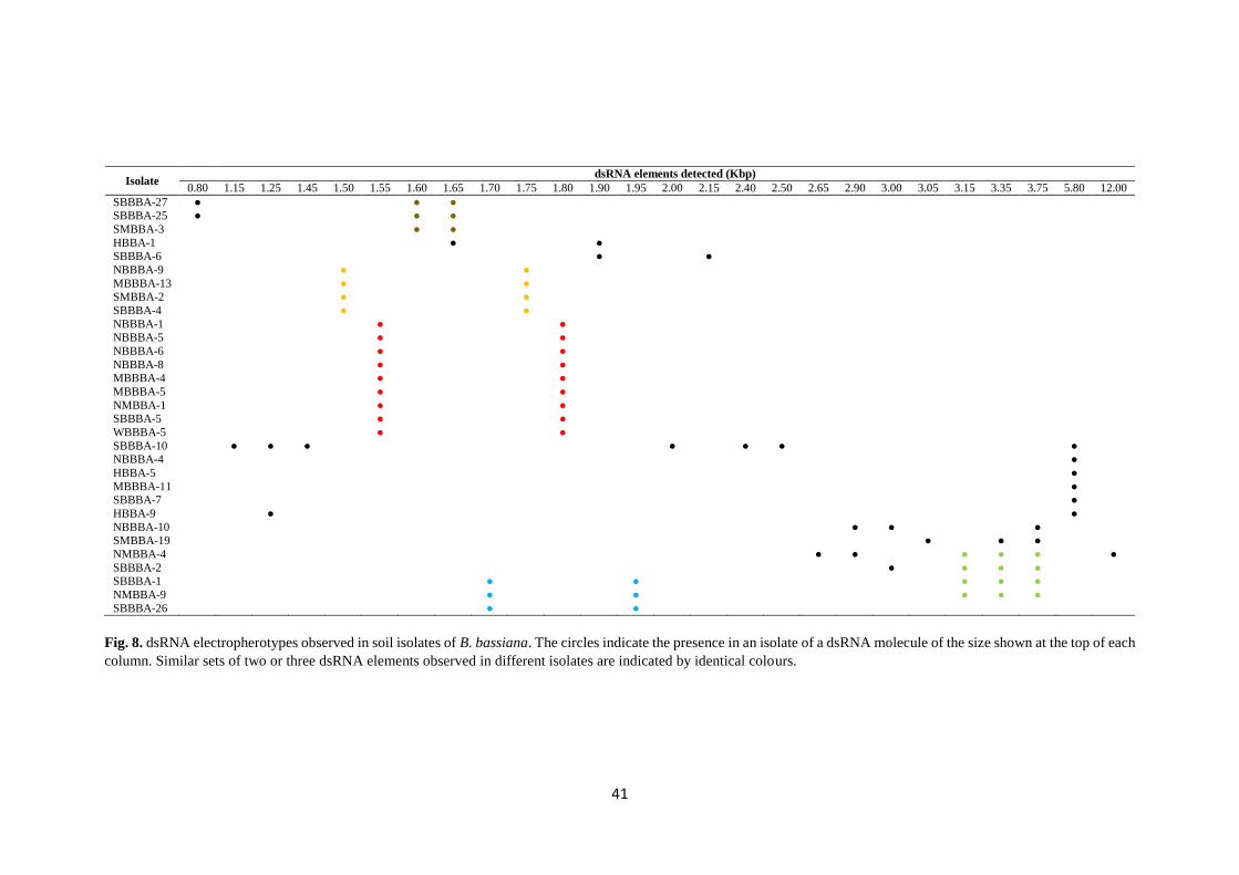
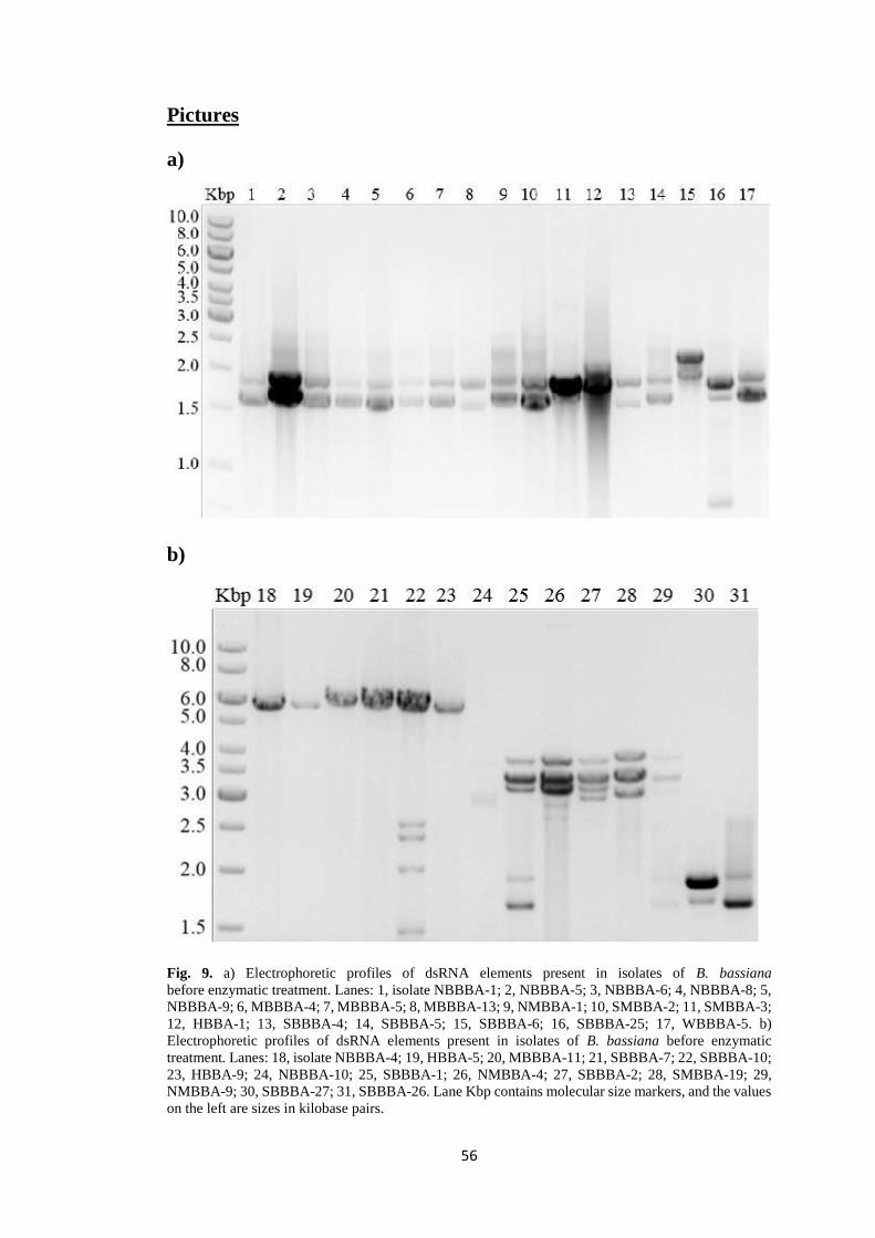
Fig. 8. dsRNA electropherotypes observed in soil isolates of B. bassiana. The circles indicate the presence in an isolate of a dsRNA molecule of the size shown at the top of each
column. Similar sets of two or three dsRNA elements observed in different isolates are indicated by identical colours.
42
6. Discussion
The incidence of dsRNA virus-like molecules found in B. bassiana isolates
collected in different parts of the Czech Republic indicates that mycovirus infections
are common among B. bassiana soil isolates from this country. dsRNA elements were
detected in 22.6% of the 137 analyzed isolates. Higher incidences were found
in previous works, for example, in B. bassiana isolates from Spain and Portugal
incidences of 54.8% were found (40 out of 73 isolates) (HERRERO et al., 2012)
and even higher in isolates from New Zealand 77.8% (7 out of 9 isolates) (YIE et al.,
2014). Nevertheless, other studies achieved lower incidences of dsRNA elements
among B. bassiana isolates, Melzer and Bidochka (1998) found dsRNA presence only
in 2 isolates of the 12 analysed in Canada (16.7 %); similar results were found in Brazil
with 2 out of 13 isolates infected (DALZOTO et al., 2006) and a 20.6% incidence was
found by Castrillo et al. (2004) in isolates of B. bassiana from North America.
Differences between the percentages of infected isolates could be explained
by differences in the number of tested isolates, the habitat, or natural conditions
of countries and regions from which isolates were collected.
Infected isolates were found in B.bassiana isolates coming from all around the
Czech Republic, except from the North West Bohemia region where not infected
isolates were found, or West Bohemia region where only one isolate harbouring
dsRNA was detected. This finding could indicate the direction of propagation of the
virus-like dsRNA molecules in the Czech Republic could have been from east to the
west.
The prevalence of virus-like dsRNA molecules in B. bassiana isolates
from cultivated soils was found slightly lower (20%) than in those from non-cultivated
ones (23.2%), which shows that this feature does not seem to be related
with the incidence of dsRNA elements in B. bassiana. Nevertheless, the majority
(54.8%) of infected isolates coming from the non-cultivated areas were isolated
from forests, which could be explained by the larger number of isolates analysed
from this type of habitat. Herrero et al. (2012) evaluated the prevalence of dsRNA
in B. bassiana strains isolated from soil or plants as endophytes and it was shown that
the prevalence was lower (51.7%) in soil isolates than in the endophytic ones (66.7%).
According to this, the niche from which the fungal host was isolated (soil, plant,
insect…) could influence the incidence of dsRNA elements. Additionally, it was also
43
found a difference in the occurrence of dsRNA elements between B. bassiana strains
isolated with bait methods (15.3%) and strains isolated with selective media (28.2%).
It could indicate that some mycoviruses adversely affect the ability
of entomopathogenic fungi to infect insects.
In this work, 15 different dsRNA profiles were observed among B. bassiana
isolates, slightly less than in similar studies carried out in Spain and Portugal
(19 different profiles observed), but significantly more than in studies by Melzer
and Bidochka (1998), Dalzoto et al. (2006) and Yie et al. (2014), nonetheless, in these
studies lower number of isolates were analysed. There were also detected twenty six
different dsRNA elements in Czech B. bassiana isolates, this is the same number
of different dsRNA elements detected in isolates from Spain and Portugal (HERRERO
et al., 2012). However, they detected more infected isolates, which could explain
the greater variability of dsRNA elements obtained in that study. All the variability
of dsRNA profiles obtained in the present work as well as in similar previous studies,
could be probably influenced by the virus way of transmission among different isolates
from different or the same compatibility groups. Additionally, by hyphal anastomoses,
different rates of transmission of the different viruses could occur.
The estimated sizes of dsRNA elements detected in this work range from 0.8
to 12 kbp. This is the largest range detected among similar published studies
in B. bassiana. Nonetheless, this difference comes mainly from the detected 12 kbp
dsRNA element. This could correspond to the replicative form of a (+) ssRNA member
of family Hypoviridae (monopartite genomes of 9-13 kbp) (ZHANG & NUSS, 2008).
The most common dsRNA profile detected in Beauveria isolates from the Czech
Republic is a set of two dsRNA molecules of 1.55 and 1.80 kbp detected in 9 of the 137
analysed isolates, which roughly corresponds the sizes of 2 dsRNA elements
(1.6 and 1.80 kbp respectively) detected also in 9 B. bassiana isolates from Spain
by Herrero et al., (2012). Similar profiles to this one were found infecting other
8 isolates but with slightly different sizes of the dsRNA elements making up the sets:
1.60 and 1.65 kbp, 1.65 and 1.90 kbp, 1.70 and 1.90; 1.90 and 2.15 kbp. These profiles
could correspond to members of the family Partitiviridae (bipartite genomes
of 1.4-2.4 kbp) (NIBERT et al., 2014). Another dsRNA element of approximately
5.8 kbp was detected in 6 of the analysed isolates, it could correspond to a member
of the Totiviridae family (monopartite genomes 4.6-7 kbp) (GHABRIAL et al., 2015).
This 5.8 dsRNA element could be similar to any of the victorivirus infecting
44
B. bassiana that were recently sequenced and characterized (HERRERO et al., 2012;
YIE et al., 2014). Four additional infected isolates showed a profile of a set of three
dsRNA elements of 3.15, 3.35 and 3.75 kbp respectively. They could correspond to
members of the Chrysoviridae family (genomes composed of 4 segments
of 2.4 to 3.6 kbp) (GHABRIAL et al., 2015). It could happen that the fourth segment
was not able to be distinguished in the electrophoresis gel, since the sizes of the four
segments of chrysovirus genomes are very close. According to this, thanks to
the characteristics of the electrophoretic band patterns, like estimated size and number
of the dsRNA elements, it can be hypothesized that B. bassiana isolates collected in
the Czech Republic could harbor members of the viral families Totiviridae,
Partitiviridae, Chrysoviridae and Hypoviridae. Nevertheless, complete or partial
sequences of all the dsRNAs detected in this study would be necessary for their correct
classification. Additionally, it is important to mention that dsRNA elements similar in
size do not always have the same nucleotide sequence. It means that virus species
diversity among Czech B. bassiana isolates could be higher than that estimated by
dsRNA electropherotypes in this work.
The presence of mixed infections could be also inferred from the dsRNA patterns
observed in this study; in some isolates, one or several dsRNA elements similar in size
occurred, but in other isolates, those elements were accompanied by different dsRNAs.
For example, the dsRNA element of 5.8 kbp which is alone in 4 of the infected isolates,
it is found also accompanied by other 6 different dsRNA elements in SBBBA-10
isolate or by one different element in HBBA-9 isolate (Fig. 8). Similar mixed virus
infections were also detected in B. bassiana isolates from Spain and New Zealand
(HERRERO et al. 2012; YIE et al., 2014) as well as in some isolates
of entomopathogenic species Tolypocladium cylindrosporum and in other fungal
species (HERRERO & ZABALGOGEAZCOA, 2011; GHABRIAL & SUZUKI,
2010).
A clear geographical or habitat distribution of isolates harbouring similar viral
infections was not found, except for some sets of two dsRNA molecules that were
hypothesised as possible members of family Partitiviridae (1.50 and 1.75 kbp,
1.55 and 1.80 kbp, 1.70 and 1.95 kbp) which were detected in isolates coming only
from hedgerows and forests.
In the conditions that all B. bassiana isolates were cultured in the laboratory,
not obvious phenotype associated with virus-infected isolates was observed, but as was
45
commented in the introduction, lack of obvious symptoms is common among virus-
infected fungi. Nonetheless, it can happen that these viruses might induce symptoms
under some unexplored environmental conditions. These effects that dsRNA virus-like
molecules could produce in their Beauveria host were not tested in this study, but it
could be really interesting for the future to assess how these dsRNAs affect B. bassiana
biology in order to improve its use as biological control agent.
46
7. Conclusions
Based on the results of the analysis of the presence of virus like-dsRNA
molecules in a collection of soil isolates of B. bassiana isolated from different
locations and habitats of the Czech Republic, it can be concluded:
1. The incidence of dsRNA virus-like molecules found in the 137 B. bassiana
isolates analysed in this study (22.6%) indicate that mycovirus infections are
common among B. bassiana isolates from all around the Czech Republic.
2. dsRNA electrophoretic profiles of infected isolates indicated high virus
diversity in Czech B. bassiana isolates, nonetheless, this diversity could be
higher, since similar band patterns could correspond to different nucleotide
sequences.
3. A clear geographical or habitat distribution of isolates harbouring similar
viral infections was not found, except for some sets of two dsRNA
molecules, which were hypothesised as possible members of family
Partitiviridae, and occurred only in B. bassiana cultures isolated from forest
of hedgerows.
4. Mixed viral infections occurred in several B. bassiana isolates.
5. Under the conditions of cultivation in the laboratory, B. bassiana isolates
harbouring dsRNA virus-like molecules did not show any obvious
phenotype due to this infection.
47
8. References
ANAGNOSTAKIS, S. L. (1982). ANAGNOSTAKIS BIOLOGICAL CONTROL OF CHESTNUT BLIGHT. SCIENCE.
VOL. 215, P. 466–471.
ARNOLD, A. E. AND LEWIS, L. C. (2005). ECOLOGY AND EVOLUTION OF FUNGAL ENDOPHYTES, AND
THEIR ROLES AGAINST INSECTS. IN: INSECT-FUNGAL ASSOCIATIONS: ECOLOGY AND EVOLUTION, F. E.
VEGA AND M. BLACKWELL (EDS.). OXFORD UNIVERSITY PRESS, NEW YORK, P. 74-96.
ARNOLD, A. E., MEJIA, L. C., KYLLO, D., ROJAS, E. I., MAYNARD, Z., ROBBINS, N. AND HERRE, E.
A. (2003). FUNGAL ENDOPHYTES LIMIT PATHOGEN DAMAGE IN A TROPICAL TREE. PROCEEDINGS OF THE
NATIONAL ACADEMY OF SCIENCES. VOL. 100, P. 15649-15654.
AUGUSTYNIUK-KRAM, A. AND KRAM, K. J. (2012). ENTOMOPATHOGENIC FUNGI AS AN IMPORTANT
NATURAL REGULATOR OF INSECT OUTBREAKS IN FORESTS (REVIEW). IN: FOREST ECOSYSTEMS - MORE
THAN JUST TREES, DR. JUAN A. BLANCO (ED.), ISBN: 978-953-51-0202-1.
AUNG, O. M., SOYTONG, K. AND HYDE, K. D. (2008). DIVERSITY OF ENTOMOPATHOGENIC FUNGI IN
RAINFORESTS OF CHIANG MAI PROVINCE, THAILAND. FUNGAL DIVERSITY. VOL. 30, P. 15-22.
BIDOCHKA, M. J., MENZIES, F. V. AND KAMP, A. M (2002). GENETIC GROUPS OF THE INSECT-
PATHOGENIC FUNGUS BEAUVERIA BASSIANA ARE ASSOCIATED WITH HABITAT AND THERMAL GROWTH
PREFERENCES. ARCHIVES OF MICROBIOLOGY. VOL. 178, P. 531-537.
BISWAS, B., ADHYA, S., WASHART, P., PAUL, B., TROSTEL, A. N., POWELL, B., CARLTON, R. AND
MERRIL, C. R. (2002). BACTERIOPHAGE THERAPY RESCUES MICE BACTEREMIC FROM A CLINICAL
ISOLATE OF VANCOMYCINRESISTANT ENTEROCOCCUS FAECIUM. INFECTION AND IMMUNITY. VOL. 70, P.
204–210.
BOGO, M. R., QUEIROZ, M. V., SILVA, D. M., GIMENÉZ, M. P., AZEVEDO, J. L. AND SCHRANK,
A. (1996). DOUBLE-STRANDED RNA AND ISOMETRIC VIRUS-LIKE PARTICLES IN THE
ENTOMOPATHOGENIC FUNGUS METARHIZIUM ANISOPLIAE. MYCOLOGICAL RESEARCH. VOL. 100, P. 1468-
1472.
BOUCIAS, D. G., FARMERIE, W. G. AND PENDLAND, J. C. (1998). CLONING AND SEQUENCING THE
CDNA OF THE INSECTICIDAL TOXIN. JOURNAL OF INVERTEBRATE PATHOLOGY. VOL. 72, P. 258-261.
BOZARTH, R. F. (1972). MYCOVIRUSES: A NEW DIMENSION IN MICROBIOLOGY. ENVIRONMENTAL
HEALTH PERSPECTIVES. VOL. 2, P. 23 – 39.
BRIDGE, P. D., CLARK, M. S. AND PEARCE, D. A. (2005). A NEW SPECIES OF PAECILOMYCES ISOLATED
FROM THE ANTARCTIC SPRINGTAIL CRYPTOPYGUS ANTARCTICUS. MYCOTAXON. VOL. 92, P. 213-222,
ISSN 0093-4666.
BUCK, K. (1998). MOLECULAR VARIABILITY OF VIRUSES OF FUNGI. IN: MOLECULAR VARIABILITY OF
FUNGAL PATHOGENS, P. D., BRIDGE, Y. COUTEAUDIER AND J. M. CLACKSON (EDS). CAB
INTERNATIONAL. WALLINGFORD, UK, P. 53-72.
CARRUTHERS, R. I. AND SOPER, R. S. (1987). FUNGAL DISEASES. IN: EPIZOOTIOLOGY OF INSECT
DISEASES, J. R. FUXA AND Y. TANADA. JOHN WILEY AND SONS, NEW YORK, P. 357-416.
CASTRILLO, L. A, GRIGGS, M. H. AND VANDENBERG, J. D. (2004). VEGETATIVE COMPATIBILITY
GROUPS IN INDIGENOUS AND MASS-RELEASED STRAINS OF THE ENTOMOPATHOGENIC FUNGUS BEAUVERIA
BASSIANA: LIKELIHOOD OF RECOMBINATION IN THE FIELD. JOURNAL OF INVERTEBRATE PATHOLOGY. VOL.
86, P. 26–37.
CHARNLEY, A. K. (2003). FUNGAL PATHOGENS OF INSECTS: CUTICLE DEGRADING ENZYMES AND
TOXINS. ADVANCES IN BOTANICAL RESEARCH. VOL. 40, P. 241–321.
CHARNLEY, A. K. AND COLLINS, S. A. (2007). ENTOMOPATHOGENIC FUNGI AND THEIR ROLE IN PEST
CONTROL. IN: THE MYCOTA. ENVIRONMENTAL AND MICROBIAL RELATIONSHIP IV, K. ESSER, ET AL.
SPRINGER, 2ND EDITION, P. 159-187.
48
CHIBA, S., KONDO, H., TANI, A., SAISHO, D. SAKAMOTO, W.,KANEMATSU, S. AND SUZUKI, N. (2011).
WIDESPREAD ENDOGENIZATION OF GENOME SEQUENCES OF NON-RETROVIRAL RNA VIRUSES INTO
PLANT GENOMES. PLOS PATHOGENS. VOL. 7(7)
CHOI, G. H., DAWE, A. L., CHURBANOV, A., SMITH, M. L., MILGROOM, M. G. AND NUSS, D. L.
(2012). MOLECULAR CHARACTERIZATION OF VEGETATIVE INCOMPATIBILITY GENES THAT RESTRICT
HYPOVIRUS TRANSMISSION IN THE CHESTNUT BLIGHT FUNGUS CRYPHONECTRIA PARASITICA. GENETICS.
VOL. 190, P. 113–127.
CHUN, S. J. AND LEE, Y. H. (1997). INHERITANCE OF DSRNAS IN THE RICE BLAST FUNGUS,
MAGNAPORTHE GRISEA. FEMS MICROBIOLOGY LETTERS. VOL. 148, P. 159–162.
DALZOTO, P. R., GLIENKE-BLANCO, CH., KAVA-CORDEIRO, V. AND RIBEIRO, J. Z. (2006).
HORIZONTAL TRANSFER AND HYPOVIRULENCE ASSOCIATED WITH DOUBLE-STRANDED RNA IN
BEAUVERIA BASSIANA. MYCOLOGICAL RESEARCH. VOL. 110, P. 1475–1481.
EILENBERG, J., HAJEK, A. AND LOMER, C. (2001). SUGGESTION FOR UNIFYING THE TERMINOLOGY IN
BIOLOGICAL CONTROL. BIOCONTROL. VOL. 46, P. 387-400, ISSN 1573-8248.
EILENBERG, J., SCHMIDT, N. M., MEYLING, N. AND WOLSTED, C. (2007). PRELIMINARY SURVEY FOR
INSECT PATHOGENIC FUNGI IN ARCTIC GREENLAND. IOBC/WPRS BULLETIN. INSECT PATHOGENS AND
INSECT PARASITIC NEMATODES. VOL. 30, P. 117-118, ISSN 1027-3115.
ELMHOLT, S., FRISVAD, J. C. AND THRANE, U. (1991). THE INFLUENCE OF FUNGICIDES ON SOIL
MYCOFLORA WITH SPECIAL ATTENTION TO TESTS OF FUNGICIDE EFFECTS ON SOIL-BORNE PATHOGENS. IN:
PESTICIDE INTERACTIONS IN CROP PRODUCTION: BENEFICIAL AND DELETERIOUS EFFECTS, J. ALTMAN
(ED.). CRC PRESS, BOCA RATON, FL, P. 227–243
FAUQUET, C. M., MAYO, M. A., MANILOFF, J., DESSELBERGER, U. AND BALL, L. A. (EDS.) (2005).
VIRUS TAXONOMY: EIGHTH REPORT OF THE INTERNATIONAL COMMITTEE ON TAXONOMY OF VIRUSES.
ELSEVIER ACADEMIC PRESS. SAN DIEGO. ISBN 978-0-12-249951-7.
FRAVEL, D. R. (1988). ROLE OF ANTIBIOSIS IN THE BIOCONTROL OF PLANT DISEASES. ANNUAL REVIEW
OF PHYTOPATHOLOGY. VOL. 26, P. 75–91.
GABARTY, A., SALEM, H. M., FOUDA, M. A., ABAS, A. A. AND IBRAHIM, A. A (2014): PATHOGENCITY
INDUCED BY THE ENTOMOPATHOGENIC FUNGI BEAUVERIA BASSIANA AND METARHIZIUM ANISOPLIAE IN
AGROTIS IPSILON (HUFN.). JOURNAL OF RADIATION RESEARCH AND APPLIED SCIENCES. VOL. 7, P. 95-100.
GHABRIAL, S. A. (1998). ORIGIN, ADAPTATION AND EVOLUTIONARY PATHWAYS OF FUNGAL VIRUSES.
VIRUS GENES. VOL. 16, P. 119–131.
GHABRIAL, S. A. (2008). TOTIVIRUSES. IN: ENCYCLOPEDIA OF VIROLOGY, 3RD EDITION, B. W. J. MAHY
AND M. H. V.,VAN REGENMORTEL (EDS.). ELSEVIER, P. 163–174.
GHABRIAL, S. A. AND CASTÓN, J. R. (2011). FAMILY CHRYSOVIRIDAE. IN: VIRUS TAXONOMY: NINTH
REPORT OF THE INTERNATIONAL COMMITTEE ON TAXONOMY OF VIRUSES, A. M. Q. KING, M. J. ADAMS,
E. B. CARSTENS, AND E. J. LEFKOWITZ (EDS.). ELSEVIER ACADEMIC PRESS, P. 509-513, ISBN 978-0-12-
384684-6.
GHABRIAL, S. A. AND NIBERT, M. L. (2009). VICTORIVIRUS, A NEW GENUS OF FUNGAL VIRUSES IN THE
FAMILY TOTIVIRIDAE. ARCHIVES OF VIROLOGY. VOL. 154, P. 373–379.
GHABRIAL, S. A. AND SUZUKI, N. (2010). FUNGAL VIRUSES. IN: DESK ENCYCLOPEDIA OF PLANT AND
FUNGAL VIROLOGY, B. W. J MAHY AND M. H. V. VAN REGENMORTEL (EDS). ELSEVIER, P 517-524.
GHABRIAL, S. A., CASTÓN, J. R., JIANG, D., NIBERT, M. L. AND SUZUKI, N. (2015). 50-PLUS YEARS OF
FUNGAL VIRUSES. VIROLOGY [ONLINE]. FEBRUARY 2015, AVAILABLE AT: HTTP://WWW.SCIENCEDIRECT.
F., PARMASTO, E., REEB, V., ROGERS, J. D., ROUX, C., RYVARDEN, L., SAMPAIO, J. P., SCHÜßLER,
A., SUGIYAMA, J., THORN, R. G., TIBELL, L., UNTEREINER, W. A., WALKER, C., WANG, Z., WEIR,
A., WEISS, M., WHITE, M. M., WINKA, K., YAO, Y.-J. AND ZHANG, N. (2007). A HIGHER-LEVEL
PHYLOGENETIC CLASSIFICATION OF THE FUNGI. MYCOLOGICAL RESEARCH, VOL. 111, P. 509-547, ISSN
1469-8102.
HILLMAN, B. I. AND CAI, G. (2013). THE FAMILY NARNAVIRIDAE: SIMPLEST OF RNA VIRUSES.
ADVANCES OF VIRUS RESEARCH. VOL. 86, P. 149–176.
HILLMAN, B. I. AND SUZUKI, N. (2004). VIRUSES OF THE CHESTNUT BLIGHT FUNGUS, CRYPHONECTRIA
PARASITICA. ADVANCES OF VIRUS RESEARCH. VOL. 63, P. 423–472.
HOLLINGS, M. (1962). VIRUSES ASSOCIATED WITH DIEBACK DISEASE OF CULTIVATED MUSHROOMS.
NATURE. VOL. 196, P. 962–965.
50
HOWITT, R. L. J., BEEVER, R. E., PEARSON, M. N. AND FORSTER, R. L. S. (2006). GENOME
CHARACTERIZATION OF A FLEXUOUS ROD-SHAPED MYCOVIRUS, BOTRYTIS VIRUS X, REVEALS HIGH
AMINO ACID IDENTITY TO GENES FROM PLANT POTEX-LIKE VIRUSES. ARCHIVES OF VIROLOGY. VOL. 151,
P. 563–579.
HULL, R. (2014). PLANT VIROLOGY. ELSEVIER ACADEMIC PRESS, 5TH EDITION, P. 1104, ISBN 978-0-12-
384871-0.
HUMBER, R. A. (2012). ENTOMOPHTHOROMYCOTA: A NEW PHYLUM AND RECLASSIFICATION FOR
ENTOMOPHTHOROID FUNGI. MYCOTAXON. VOL. 120, P. 477-492.
IKEDA, K., INOUE, K., KIDA, C., UWAMORI, T., SASAKI, A., KANEMATSU, S. AND PARK, P. (2013).
POTENTIATION OF MYCOVIRUS TRANSMISSION BY ZINC COMPOUNDS VIA ATTENUATION OF HETEROGENIC
INCOMPATIBILITY IN ROSELLINIA NECATRIX. APPLIED AND ENVIRONMENTAL MICROBIOLOGY. VOL. 79, P.
3684–3691.
KANEMATSU, S., ARAKAWA, M., OIKAWA, Y., ONOUE, M., OSAKI, H., NAKAMURA, H., IKEDA, K.,
KUGA-UETAKE, Y., NITTA, H., SASAKI, A., SUZAKI, K., YOSHIDA, K. AND MATSUMOTO, N. (2004).
A REOVIRUS CAUSES HYPOVIRULENCE OF ROSELLINIA NECATRIX. PHYTOPATHOLOGY. VOL. 94, P. 561–
568.
KISHI, K., YASUDA, T. AND TAKESHITA, H. (2001). DNASE I: STRUCTURE, FUNCTION, AND USE IN
MEDICINE AND FORENSIC SCIENCE. LEGAL MEDICINE (TOKYO, JAPAN). VOL. 3, P. 69-83.
KLINGEN, I., EILENBERG, J. AND MEADOW, R. (2002). EFFECTS OF FARMING SYSTEM, FIELD MARGINS
AND BAIT INSECT ON THE OCCURRENCE OF INSECT PATHOGENIC FUNGI IN SOILS. AGRICULTURE,
ECOSYSTEMS & ENVIRONMENT. VOL. 91 (2-3), P. 191-198, ISSN 0167-8809.
KOTTA-LOIZOU, I, SIPKOVA, J AND COUTTS, R. H. (2015). IDENTIFICATION AND SEQUENCE
DETERMINATION OF A NOVEL DOUBLE-STRANDED RNA MYCOVIRUS FROM THE ENTOMOPATHOGENIC
FUNGUS BEAUVERIA BASSIANA. ARCHIVES OF VIROLOGY. VOL. 160, P. 873-875.
LEAL, S. C. M., BERTIOLI, D. J., BALL, B. V. AND BUTT, T. M. (1994). PRESENCE OF DOUBLE‐STRANDED RNAS AND VIRUS‐LIKE PARTICLES IN THE ENTOMOPATHOGENIC FUNGUS METARHIZIUM
ANISOPLIAE. BIOCONTROL SCIENCE AND TECHNOLOGY. VOL. 4, P. 89-94.
LI, Z., LI, C., HUANG, B. AND FAN, M. (2001). DISCOVERY AND DEMONSTRATION OF THE TELEOMORPH
OF BEAUVERIA BASSIANA (BALS.) VUILL., AN IMPORTANT ENTOMOGENOUS FUNGUS. CHINESE SCIENCE
BULLETIN. VOL. 46, P. 751–753.
LINDER-BASSO, D., DYNEK, J. N. AND HILLMAN B. I. (2005). GENOME ANALYSIS OF CHRYPHONECTRIA
HYPOVIRUS 4, THE MOST COMMON HYPOVIRUS SPECIES IN NORTH AMERICA. VIROLOGY. VOL. 337, P.
192–203.
LIU, H. (2012). MICROBIAL CONTROL OF CROP PESTS USING ENTOMOPATHOGENIC FUNGI. IN:
INTEGRATED PEST MANAGEMENT: PRINCIPLES AND PRACTICE. D. P. ABROL AND U. SHANKAR (EDS.). CPI
GROUP, P. 237-280, ISBN 978-1-84593-808-6.
LIU, H., FU, Y., JIANG, D., LI, G., XIE J, CHENG, J., PENG, Y., GHABRIAL, S. A. AND YI, X. (2010).
WIDESPREAD HORIZONTAL GENE TRANSFER FROM DOUBLE-STRANDED RNA VIRUSES TO EUKARYOTIC
NUCLEAR GENOMES. JOURNAL OF VIROLOGY. VOL. 84, P. 11876–87
MACIÁ-VICENTE, J. G., PALMA-GUERRERO, J., GÓMEZ-VIDAL, S. AND LOPEZ-LLORCA, L. V.
(2011). NEW INSIGHTS ON THE MODE OF ACTION OF FUNGAL PATHOGENS OF INVERTEBRATES FOR
IMPROVING THEIR BIOCONTROL PERFORMANCE. IN: BIOLOGICAL CONTROL OF PLANT-PARASITIC
NEMATODES: BUILDING COHERENCE BETWEEN MICROBIAL ECOLOGY AND MOLECULAR MECHANISMS. K.
DAVIES AND Y. SPIEGEL (EDS.), SPRINGER, LONDON, P. 203-227, ISBN 978-1-4020-9647-1
MARQUÉZ, L. M., REDMAN, R. S., RODRIGUEZ, R. J. AND ROOSSINCK M. J. (2007). A VIRUS IN A
FUNGUS IN A PLANT: THREE-WAY SYMBIOSIS REQUIRED FOR THERMAL TOLERANCE. SCIENCE. VOL. 315,
P. 513–515.
51
MAZET, I. AND VEY, A. (1995). HIRSUTELLIN A, A TOXIC PROTEIN PRODUCED IN VITRO BY HIRSUTELLA
THOMPSONII. MICROBIOLOGY. VOL. 141, P. 1343-1348, ISSN 1350-0872.
MCCABE, P. M., PFEIFFER, P. AND VAN ALFEN, N. K. (1999). THE INFLUENCE OF DSRNA VIRUSES ON
THE BIOLOGY OF PLANT PATHOGENIC FUNGI. TRENDS IN MICROBIOLOGY. VOL. 7, P. 377-381.
MCCOY, C. W., SAMSON, R. A. AND BOUCIAS, D. G. (1988). ENTOMOGENOUS FUNGI. IN: HANDBOOK
OF NATURAL PESTICIDES, BOCA, RATON, FLA: MR IC PRESS. VOL. 5, MICROBIAL INSECTICIDES, PART A,
ENTOMOGENOUS PROTOZOA AND FUNGI, C. M. IGNOFFO AND N. B. MANDAVA. ISBN 9780849336607
MCFADDEN, J. J. P., BUCK, K. W. AND RAWLINSON, C. J. (1983). INFREQUENT TRANSMISSION OF
DOUBLE-STRANDED RNA VIRUS PARTICLES BUT ABSENCE OF DNA PROVIRUSES IN SINGLE ASCOSPORE
CULTURES OF GAEUMANNOMYCES GRAMINIS. JOURNAL OF GENERAL VIROLOGY. VOL. 64, P. 927–37.
MELZER, M. J. AND BIDOCHKA, M. J. (1998). DIVERSITY OF DOUBLE-STRANDED RNA VIRUSES WITHIN
POPULATIONS OF ENTOMOPATHOGENIC FUNGI AND POTENTIAL IMPLICATIONS FOR FUNGAL GROWTH AND
VIRULENCE. MYCOLOGIA. VOL. 90, P. 586 –594.
MORRIS, T. J. AND DODDS, J. A. (1979). ISOLATION AND ANALYSIS OF DOUBLE-STRANDED RNA FROM
VIRUS-INFECTED PLANT AND FUNGAL TISSUE. PHYTOPATHOLOGY. VOL. 69, P. 854–858.
MOTA-SANCHEZ, D., BILLS, P.S. AND WHALON, M.E. (2002). ARTHROPOD RESISTANCE TO
PESTICIDES: STATUS AND OVERVIEW. IN: PESTICIDES IN AGRICULTURE AND THE ENVIRONMENT. W. B.
WHEELER (ED.), CRC PRESS, NEW YORK, P. 241-272, ISBN 9780824708092
NAKAYASHIKI, H. AND NGUYEN, Q. B. (2008). RNA INTERFERENCE: ROLES IN FUNGAL BIOLOGY.
CURRENT OPINION IN MICROBIOLOGY. ELSEVIER. VOL. 11, P. 494–502.
NIBERT, M. L., GHABRIAL, S. A., MAISS, E., LESKER, T., VAINIO, E. J., JIANG, D. AND SUZUKI, N.
(2014). TAXONOMIC REORGANIZATION OF FAMILY PARTITIVIRIDAE AND OTHER RECENT PROGRESS IN
PARTITIVIRUS RESEARCH. VIRUS RESEARCH. VOL. 188, P. 128–141.
NUSS, D. L. (2005). HYPOVIRULENCE: MYCOVIRUSES AT THE FUNGAL-PLANT INTERFACE. NATURE
REVIEWS MICROBIOLOGY. VOL. 3, P. 632-642.
NUSS, D. L. (2011). MYCOVIRUSES, RNA SILENCING, AND VIRAL RNA RECOMBINATION. ADVANCES IN
VIRUS RESEARCH. VOL. 80, P. 25–48.
NUSS, D. L. AND HILLMAN, B. I. (2011). FAMILY HYPOVIRIDAE. IN: VIRUS TAXONOMY: NINTH REPORT
OF THE INTERNATIONAL COMMITTEE ON TAXONOMY OF VIRUSES, A. M. Q. KING, M. J. ADAMS, E. B.
CARSTENS, AND E. J. LEFKOWITZ (EDS.). ELSEVIER, P. 1029-1033, ISBN 978-0-12-384684-6.
OWNLEY B. H. AND WINDHAM, M. T. (2007). BIOLOGICAL CONTROL OF PLANT PATHOGENS. IN: PLANT
PATHOLOGY: CONCEPTS AND LABORATORY EXERCISES. R. N. TRIGIANO , M. T. WINDHAM AND A. S.