307/ ACTA CHIRURGIAE ORTHOPAEDICAE ET TRAUMATOLOGIAE ČECHOSL., 79, 2012, p. 307–316 LECTURE FOR CONTINUING EDUCATION PRÁCE DOŠKOLOVACÍHO CHARAKTERU Treatment of Proximal Humeral Fractures – a Review of Current Concepts Enlightened by Basic Principles Léčba zlomenin proximálního humeru – přehled současných přístupů s ohledem na základní principy D. MAIER, M. JÄGER, P. C. STROHM, N. P. SÜDKAMP Universitätsklinikum Freiburg, Dept. Orthopädie und Traumatologie, Klinik für Traumatologie Freiburg im Breisgau, Germany SUMMARY Fractures of the proximal humerus commonly affect elderly patients. The vast majority of proximal humeral fractures result from low-energy trauma in presence of osteoporosis. Incidence of proximal humeral fractures dramatically increa- sed over the last decades. Recent epidemiological studies expect a rather stagnant incidence. Diversity of fracture types attenuates reliability of available classification systems. Even though, predictive morphologic criteria have been detected enabling a prognostic assessment. A short or absent metaphyseal head extension and disruption of the medial periosteal hinge reliably predict ischemia of the humeral head fragment. Still, humeral head necrosis may be prevented in early reduc- tion and fixation. The range of treatment options consists of non-operative therapy, minimal-invasive osteosynthesis, open reduction and plate fixation, intramedullary nailing and primary arthroplasty. Most proximal humeral fractures in the elder- ly are stable injuries and can be successfully treated by non-operative means. Operative treatment of displaced, unstable fractures should resort to the least invasive procedure providing adequate reduction and fixation stability. To date, open reduction and locking plate osteosynthesis represents the standard operative procedure in displaced three- and four-part fractures. However, a number of risk factors may promote fixation failure or impair functional outcome, most important low local bone mineral density, residual varus displacement of the humeral head, insufficient restoration of medial calcar sup- port, humeral head ischemia and insufficient fracture reduction. Innovation of fixation techniques (e. g. angular stable loc- king systems and bone augmentation) will further expand indications for operative fracture treatment. Outcome of hemi- arthroplasty is closely related to anatomical tuberosity healing and restoration of rotator cuff function. Reverse shoulder arthroplasty may provide satisfactory shoulder function in geriatric patients, rotator cuff dysfunction or failure of first-line treatment. Choice of treatment should be individualized and base on careful evaluation of patient-specific, fracture-speci- fic and surgeon-specific aspects. INTRODUCTION Proximal humeral fractures account for about 5% of all extremity fractures and 70% of patients are at least 70 years of age (11). The prevalence of female gender ran- ges between 73 and 85% (4,11). A 60-year-old women with a life expectancy of 81 years has an estimated resi- dual lifetime risk of 8% of fracturing her proximal hume- rus (30). The fracture is caused by low-energy trauma in 87% of cases (typically a fall from stand) suggesting presence of osteoporosis (11). Proximal humeral frac- tures follow femoral neck and distal radius fractures as the third most common osteoporotic fracture type (8). In Finland, the incidence of low-energy proximal hume- ral fractures among women aged 80 years or older was 88 per 100.000 persons in 1970, sharply increased to 304 in 1995, but then stabilized at a level of 298 in 2007 (22).
Transcript
307/ ACTA CHIRURGIAE ORTHOPAEDICAEET TRAUMATOLOGIAE ČECHOSL., 79, 2012, p. 307–316 LECTURE FOR CONTINUING EDUCATION
PRÁCE DOŠKOLOVACÍHO CHARAKTERU
Treatment of Proximal Humeral Fractures –a Review of Current Concepts Enlightened by Basic Principles
Léčba zlomenin proximálního humeru – přehled současných přístupů s ohledem na základní principy
D. MAIER, M. JÄGER, P. C. STROHM, N. P. SÜDKAMP
Universitätsklinikum Freiburg, Dept. Orthopädie und Traumatologie, Klinik für Traumatologie Freiburg im Breisgau, Germany
SUMMARY
Fractures of the proximal humerus commonly affect elderly patients. The vast majority of proximal humeral fracturesresult from low-energy trauma in presence of osteoporosis. Incidence of proximal humeral fractures dramatically increa-sed over the last decades. Recent epidemiological studies expect a rather stagnant incidence. Diversity of fracture typesattenuates reliability of available classification systems. Even though, predictive morphologic criteria have been detectedenabling a prognostic assessment. A short or absent metaphyseal head extension and disruption of the medial periostealhinge reliably predict ischemia of the humeral head fragment. Still, humeral head necrosis may be prevented in early reduc-tion and fixation. The range of treatment options consists of non-operative therapy, minimal-invasive osteosynthesis, openreduction and plate fixation, intramedullary nailing and primary arthroplasty. Most proximal humeral fractures in the elder-ly are stable injuries and can be successfully treated by non-operative means. Operative treatment of displaced, unstablefractures should resort to the least invasive procedure providing adequate reduction and fixation stability. To date, openreduction and locking plate osteosynthesis represents the standard operative procedure in displaced three- and four-partfractures. However, a number of risk factors may promote fixation failure or impair functional outcome, most important lowlocal bone mineral density, residual varus displacement of the humeral head, insufficient restoration of medial calcar sup-port, humeral head ischemia and insufficient fracture reduction. Innovation of fixation techniques (e. g. angular stable loc-king systems and bone augmentation) will further expand indications for operative fracture treatment. Outcome of hemi-arthroplasty is closely related to anatomical tuberosity healing and restoration of rotator cuff function. Reverse shoulderarthroplasty may provide satisfactory shoulder function in geriatric patients, rotator cuff dysfunction or failure of first-linetreatment. Choice of treatment should be individualized and base on careful evaluation of patient-specific, fracture-speci-fic and surgeon-specific aspects.
INTRODUCTION
Proximal humeral fractures account for about 5% of allextremity fractures and 70% of patients are at least 70years of age (11). The prevalence of female gender ran-ges between 73 and 85% (4,11). A 60-year-old womenwith a life expectancy of 81 years has an estimated resi-dual lifetime risk of 8% of fracturing her proximal hume-rus (30). The fracture is caused by low-energy traumain 87% of cases (typically a fall from stand) suggesting
presence of osteoporosis (11). Proximal humeral frac-tures follow femoral neck and distal radius fractures asthe third most common osteoporotic fracture type (8).In Finland, the incidence of low-energy proximal hume-ral fractures among women aged 80 years or older was88 per 100.000 persons in 1970, sharply increased to304 in 1995, but then stabilized at a level of 298 in 2007(22).
Treatment options include non-operative therapy,minimal-invasive osteosynthesis, open reduction andplate fixation, intramedullary nailing and primary arth-roplasty. Not least due to advanced fixation techniques,the rate of surgically treated patients continues to incre-ase. Neer (37) estimated that approximately 20% of pa -tients would benefit from surgery in his 1970 essay. Incontrast, 53% of French patients were surgically treatedin 2001 (34, 37).
This article elucidates presents current concepts intreatment of proximal humeral fractures on the basis ofestablished principles. Common classification systemsare introduced with regard to their clinical feasibilityand relevance. A special focus is put on osteoporosisand its implications for treatment. The survey intendsto assist therapeutic decision-making, which shouldbase on careful case-related analysis and evaluation ofpatient-specific, fracture-specific and surgeon-specificaspects.
CLASSIFICATIONS
As long ago as 1934, Codman (9) proposed a stillroutinely used classification of proximal humeral frac-tures. He distinguished 12 fracture morphologies basedon the configuration of the four main segments, nomi-nally the shaft, the head, the greater and the lessertubercle. Based on Codman’s segment classification,Neer (36) defined 6 subgroups with therapeutic rele-vance. He recommended surgery when two main frag-ments were displaced > 45° or > 1 cm. The AO classi-fication from 1990 differentiates between three maingroups of fractures (extraarticular, partially articularand intraarticular) dividing into a total of 27 subtypes.Resch et al. (44) could demonstrate, that the fractureline in four-part fractures does not run through but 5-10 mm lateral of the bicipital groove. Thus, the sul-cus is regularly attached to the lesser tuberosity frag-ment. They described two basic morphologic fracturetypes, the varus and the valgus fracture. The varus frac-ture may present as an impaction or distraction type.The valgus fracture may be impacted in straight lateralor posterolateral direction. Valgus fractures rarely occurwithout impaction. Hertel et al. (19) introduced a bina-ry (Codman-LEGO) description system comprising 12basic fracture types and examined morphologic risk fac-tors of humeral head ischemia. Good predictors werea short meta physeal head extension (< 8 mm) and dis-ruption of the medial hinge with displacement of> 2 mm. However, Bastian and Hertel (2) pointed out,that initial ischemia may not reliably predict subsequ-ent humeral head necrosis indicating the potential ofreperfusion in early open reduction and osteosynthesis.Majed et al. (33) found only low to moderate inter -observer reliability among available fracture classifi-cation systems despite application of CT-based 3Dreconstruction models. The Codman-Hertel classifica-tion achieved the highest interobserver score (κ= 0.44)followed by the Neer (κ= 0.33), Resch (κ= 0.15) andAO classification system (κ= 0.11).
When is a fracture unstable?Integrity of the medial calcar, the so-called medial
hinge of the humerus, considerably improves biome-chanical stability of proximal humeral fractures (26).The periosteum at the medial cortex starts to tear ata head-to-shaft displacement of about 3 mm. Completerupture of the medial periosteal hinge occurs at displa-cements of about 35 mm. Therefore, varus-distractionfractures with destruction of the medial hinge representhighly unstable fracture types.
When is a fracture displaced?To date, Neer’s criteria for fractures benefiting from
surgical treatment (displacement > 45° and > 1 cm) arecontinuously revised. With respect to the greater tube-rosity, already displacements of > 2-5 mm may repre-sent an indication for surgery (42). Operative treatmentshould be considered in head-to-shaft displacement of> 50% of the diaphyseal diameter and in varus or val-gus deformity of the humeral head with deviation of > 20-30° from the physiological 130° head-to-shaft inclination (10, 35).
TREATMENT MODALITIES
Non-operative treatmentIn view of the high prevalence of proximal humeral
fractures, there is a lack of prospective, randomized-con-trolled trials on non-operative treatment, which can beexplained by the difficulty of establishing comparabili-ty of patient cohorts, fracture types and implementationof treatment. Fjalestad et al. (13) found no significantdifference of functional outcome after 12 months of fol-low-up between open reduction and locking plate osteo -synthesis and non-operative treatment of displacedthree- and four-part proximal humeral fractures in pa -tients aged 60 years or older. The mean absolute Con-stant-Score was 52.3 and 52.2 points, respectively. Theage- and gender-adjusted value was 74.4% in bothgroups. The mean ASES-Score measuring subjectiveoutcome was 14.8 and 15.5 points, respectively. Thisstudy also included fractures with severe displacement,which was defined as malposition of at least 45° angu-lar deviation and displacement of the greater or lessertuberosity of at least 1 cm (Neer’s criteria). Displace-ment between the head and metaphyseal main fragmentsmust not have exceeded 50% of the diaphyseal diame-ter. A prospective study enrolling 160 patients, most ofall with one-, two- and three-part fractures, observeda mean difference of 8.2 points between the injured andcontralateral shoulder in Constant-Scores and of 10.2points in DASH-Scores after 12 months of non-operati-ve therapy (17). Foruria et al. (14) examined the relation -ship of fracture patterns and functional outcomes. Infractures with posteromedial impaction, a poorer outco-me was noted as the articular surface displaced inferi-orly increasing its distance from the acromion. Worseoutcome was noted as a fractured greater tuberosity dis-placed medially overlapping with the posterior articularsurface. Also, patients with lateral impaction fractureshad inferior outcomes.
absolute Constant-Score was 66.8 points correspondingto an age- and gender-related value of 87% after an ave-rage of 17 months of follow-up. Implant-related com-plications (plate impingement, screw perforation into theglenohumeral joint, loosening of screws) occurred in17%. The rate of avascular necrosis was 5.5%. No casesof non-union were observed (47).
Intramedullary locking nail osteosynthesisIdeal indications are displaced two-part fractures.
Further indications are three-part and four-part fractu-res with metaphyseal comminution or diaphyseal invol-vement but no or minor displacement of the tuberositi-es. Open reduction and plate fixation is usually preferredin three- and four-part fractures with marked tuberositydisplacement. Zhu et al. (58) compared intramedullarylocking nail (PHN, Synthes, Switzerland) and lockingplate (LPHP and PHILOS, Synthes, Switzerland) osteo -synthesis for treatment of two-part fractures. After threeyears of follow-up, there were neither significant diffe-rences with regard to the ASES-Score (90.0 versus 94.0points) nor the Constant-Score (93.3 versus 94.5 points).The complication rate was significantly lower in thelocking nail group (4%) compared to the locking plategroup (31%). Screw penetration into the articular sur -face was the most frequent complication in the lockingplate group and required revision surgery in 19% withinthree postoperative months. A multicenter matched-pairs analysis enrolled 152 patients with displaced two-, three- and four-part fractures treated either with ante-rograde angular and sliding stable proximal interlockingnails or locking plates (16). After 1 year, functionalresults were similar with a mean age- and gender-rela-ted Constant-Score of 81% and 75%, respectively. Also,total rates of complications were not statistically diffe-rent (22% and 28%). In the nail group, rotator cuff lesi-ons occurred in 3% being associated with the necessityof arthroscopic treatment and implant removal. Interes-tingly, the rate of secondary fracture displacement was18% in the plate group compared to only 1% in the nailgroup.
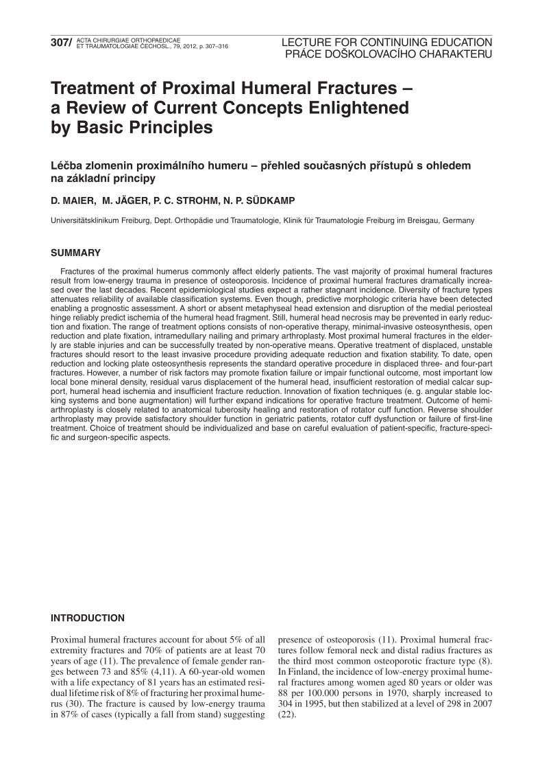
Angular stable locked intramedullary nails enhancebiomechanical stability of fracture fixation which maybe particularly beneficial in osteoporosis (20). Fig. 1demonstrates treatment of a displaced, osteoporotic two-part fracture of the humeral surgical neck in a 79-year-old woman with an intramedullary locking nail (Multi-Loc Proximal Humeral Nail, Synthes, Switzerland). Theimplant provides multiple proximal and distal angularstable locking options.
Open reduction and locking plate osteosynthesis
Typical indications comprise two-, three- and four-part fractures with displacement of the tuberosity or headfragments (52). Reconstruction of compound fracturesshowing tuberosity or humeral head fragmentation usu-ally requires open reduction. Open procedures offersuperior control of reduction and more flexible fixationoptions compared to minimal-invasive procedures. Until
A prospective, observational study reported a predic-ted risk of delayed union or non-union of 7.0% (17).Occurrence of avascular necrosis of the head fragmentis highly variable. Humeral head necrosis may developin 6.5-15.4% (14, 32). However, reliable predictorscould not yet be determined. Interestingly, the inciden-ce of EMG-verified posttraumatic axillary nerve lesionswas 30% after a mean of 13.4 weeks and decreased to15% after 1 year of non-operative treatment (13). Ingeneral, closed reduction without fixation may not effec-tuate substantial improvement of fracture alignment orfunctional outcome. In consideration of potential risksfor soft tissue and neurovascular complications, physi-cians should critically evaluate indications for closedfracture manipulations (17).
The author’s standard therapeutic regimen for non-operative treatment includes shoulder immobilization inneutral or internal rotation for one week. Pendulum exer-cises are started from week two. Passive and active-assis-ted physiotherapy is performed during the fourth to sixthweek and respects a limit of 90° abduction and flexion.From week 7, active range of motion is continuouslyincreased according to patient’s tolerance. Regular x-raycontrols are mandatory and should be performed aftereach progress of the physiotherapeutic regimen. Frequ-ently, non-operative management of proximal humeralfractures tends to be conducted too restrictive. Thereby,it is well known, that prolonged periods of immobiliza-tion (> 2 weeks) delay functional recovery (29). Super-vised physiotherapy should definitely start within 2weeks after trauma (24, 29).
Minimal-invasive osteosynthesisResch and coworkers (5, 45) introduced techniques
of percutaneous reduction and fracture fixation with can-nulated screws and K-wires. From a biomechanical pointof view, the Humerusblock, using two crossed 2.2 mmK-wires, represents a dynamic type of fixation whichenables neutralisation of stress forces occurring at thefracture site. Elasticity of the construct contributes tolower the incidence of secondary fragment or implantdisplacement particulary in elderly patients with osteo-penic or osteoporotic bone structure. Patients with three-part fractures achieved a mean Constant-Score of 61.2points corresponding to an intraindividual Constant-Score of 84.9% (compared to the non-injured arm). Infour-part fractures, the mean Constant-Scores were 49.5points and 68.5%, respectively.
Compared to isolated, percutaneous K-wire osteo-synthesis without locked cortical fixation, the Hume-rusblock achieved considerably lower rates of seconda-ry fragment or implant displacement (9.8%) (47,48). Therate of humeral head necrosis was 7.8%. Other intra-medullary implants without locking mechanism (e.g.Helix wire, Rush pin, Hackethal, Prévot, Ender or Zif-ko nails) do not seem to provide sufficient biomechani-cal stability resulting in high rates of non-union andsecondary fragment or implant displacements (56).
Current results of minimal-invasive (polyaxial) loc-king plate fixation are promising (47, 48). The mean
now, the anatomical, deltopectoral approach representsthe benchmark procedure (37). Extended deltoid-split-ting approaches may improve posterior exposure butalso impose the anterior terminal branch of the axillarynerve at risk (46). A comparative, retrospective studyobserved no statistically significant difference in clini-cal, radiographic, and electrophysiological outcomesbetween the deltopectoral and deltoid-splitting approach(57). It appears crucial to recognize and understand themorphology of the fracture to strategize its reduction andfixation. First, any varus or valgus deformity of the headfragment in relation to the shaft is corrected, which ena-bles anatomical reduction of the tuberosities. The resul-ting head-tuberosity construct may be effectively stabi-
lised by suture fixation. Next, residual ad latus and rota-tional deformities are addressed. Preliminary head-to-shaft K-wire fixation may support definite plate fixa tion.Cloverleaf, buttress and semitubular plates have beenlargely replaced by site-specific (preformed) lockingplates, which provide more secure fixation particularlyin osteoporotic bone (49).
A prospective multicenter, observational study repor-ted a mean Constant-Score of 70.6 points 1 year aftermono-axial locking plate fixation (LPHP and PHILOS,Synthes, Switzerland) corresponding to an intraindivi-dual Constant-Score of 85.1% (53). The mean DASH-Score was 15.2 points. Complications were encounteredin 34% of patients within a 12 months period of follow-up. Notably, 40% of complications were caused by anincorrect surgical technique being already present at theend of the operative procedure. The most common com-plication (14% of patients) was intraoperative screw per-foration of the humeral head. Almost one fifth of pa -tients had to undergo unplanned revision surgery within12 postoperative months. Another multicenter studyreported almost identical results after 1 year of follow-up (7). Secondary screw perforation and avascular nec-rosis occurred in 8%. In a systematic review of mid- andlong-term results, the average Constant-Score was 73.6points after a mean follow-up of 29.2 months (51). Four-part fractures scored significantly lower than two-partfractures (67.7 versus 77.4 points). The overall compli-cation rate was 48.8% including a revision rate of 13.8%.The most common complications were varus malunion(16.3%), avascular necrosis (10.8%), intraarticularscrew perforation (7.5%), subacromial impingement(4.8%) and infection (3.5%). Again, the most commoncause for revision surgery was screw perforation. Re -cently, efforts were made to determine factors predic-ting functional outcome and failure rates in open re -duction and locking plate osteosynthesis (table 1).Hardeman et al. (18) noted superior results in youngerpatients. More displaced fractures, AO type C fractures,varus fracture configuration and reduced head vascula-rity (according to the Hertel criteria) resulted in worse
Fig. 1. Displaced, osteoporotic two-part fracture in a 79-year-old woman treated with intramedullary angular stable lockingnail osteosynthesis.Upper left and right: Osteoporotic two-part fracture of the sur-gical neck with medial disengagement of the shaft due to ten-sion forces exerted by the pectoralis major muscle. Note themetaphyseal comminution zone.Lower left and right: Postoperative radiographs of intrame-dullary osteosynthesis with the MultiLoc Proximal HumeralNail (Synthes, Switzerland). The correct entry point restoreshead-to-shaft alignment. The nail end must not surmount thecartilage layer to prevent rotator cuff dysfunction. Angularstable locking bolts were inserted proximally and distally toenhance stability in osteoporotic bone.
outcome↓ Hardeman et al. 2012local bone mineral density↓ failure rate↑ Krappinger et al. 2011initial fracture Suedkamp et al 2009,displacement outcome↓ Hardeman et al. 2012and complexity↑initial varus deformity failure rate↑ Krappinger et al. 2011deformity↑ outcome↓ Gardner et al. 2007,
Hardeman et al. 2012quality of reduction↑ failure rate↓ Krappinger et al. 2011
outcome↑ Hardeman et al. 2012restoration of medial failure rate↓ Gardner et al. 2007,support↑ Krappinger et al. 2011
loss of reduction↓ Gardner et al. 2007head fragment outcome↓ Bastian, Hertel 2008,vascularity↓ Hardeman et al. 2012
Tab. 1. Selection of important factors influencing outcome in openreduction and locking plate osteosynthesis
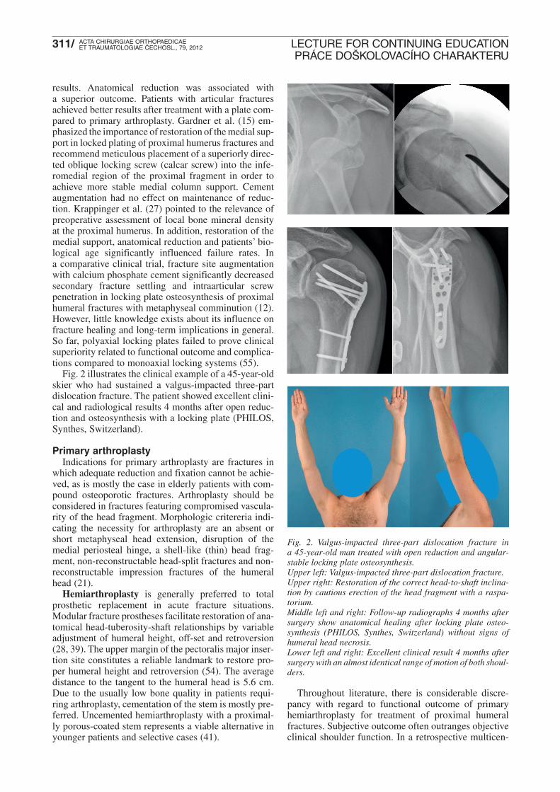
results. Anatomical reduction was associated witha superior outcome. Patients with articular fracturesachieved better results after treatment with a plate com-pared to primary arthroplasty. Gardner et al. (15) em -phasized the importance of restoration of the medial sup-port in locked plating of proximal humerus fractures andrecommend meticulous placement of a superiorly direc-ted oblique locking screw (calcar screw) into the infe-romedial region of the proximal fragment in order toachieve more stable medial column support. Cementaugmentation had no effect on maintenance of reduc -tion. Krappinger et al. (27) pointed to the relevance ofpreoperative assessment of local bone mineral densityat the proximal humerus. In addition, restoration of themedial support, anatomical reduction and patients’ bio-logical age significantly influenced failure rates. Ina comparative clinical trial, fracture site augmentationwith calcium phosphate cement significantly decreasedsecondary fracture settling and intraarticular screwpenetration in locking plate osteosynthesis of proximalhumeral fractures with metaphyseal comminution (12).However, little knowledge exists about its influence onfracture healing and long-term implications in general.So far, polyaxial locking plates failed to prove clinicalsuperiority related to functional outcome and complica-tions compared to monoaxial locking systems (55).
Fig. 2 illustrates the clinical example of a 45-year-oldskier who had sustained a valgus-impacted three-partdislocation fracture. The patient showed excellent clini-cal and radiological results 4 months after open reduc-tion and osteosynthesis with a locking plate (PHILOS,Synthes, Switzerland).
Primary arthroplastyIndications for primary arthroplasty are fractures in
which adequate reduction and fixation cannot be achie-ved, as is mostly the case in elderly patients with com-pound osteoporotic fractures. Arthroplasty should beconsidered in fractures featuring compromised vascula-rity of the head fragment. Morphologic critereria indi-cating the necessity for arthroplasty are an absent orshort metaphyseal head extension, disruption of themedial periosteal hinge, a shell-like (thin) head frag-ment, non-reconstructable head-split fractures and non-reconstructable impression fractures of the humeralhead (21).
Hemiarthroplasty is generally preferred to totalprosthetic replacement in acute fracture situations.Modular fracture prostheses facilitate restoration of ana-tomical head-tuberosity-shaft relationships by variableadjustment of humeral height, off-set and retroversion(28, 39). The upper margin of the pectoralis major inser-tion site constitutes a reliable landmark to restore pro-per humeral height and retroversion (54). The averagedistance to the tangent to the humeral head is 5.6 cm.Due to the usually low bone quality in patients requi-ring arthroplasty, cementation of the stem is mostly pre-ferred. Uncemented hemiarthroplasty with a proximal-ly porous-coated stem represents a viable alternative inyounger patients and selective cases (41).
Throughout literature, there is considerable discre-pancy with regard to functional outcome of primaryhemiarthroplasty for treatment of proximal humeralfractures. Subjective outcome often outranges objectiveclinical shoulder function. In a retrospective multicen-
Fig. 2. Valgus-impacted three-part dislocation fracture ina 45-year-old man treated with open reduction and angular-stable locking plate osteosynthesis.Upper left: Valgus-impacted three-part dislocation fracture. Upper right: Restoration of the correct head-to-shaft inclina-tion by cautious erection of the head fragment with a raspa-torium.Middle left and right: Follow-up radiographs 4 months aftersurgery show anatomical healing after locking plate osteo-synthesis (PHILOS, Synthes, Switzerland) without signs ofhumeral head necrosis.Lower left and right: Excellent clinical result 4 months aftersurgery with an almost identical range of motion of both shoul-ders.
ter study with a minimum follow-up of 1 year 79% ofpatients were asymptomatic or reported only minor pain(25). In contrast, only 41,9% were able to flex and only34,7% of patients to abduct their shoulders above 90°.Anatomical tuberosity healing significantly improvedsubjective and objective shoulder function. In recentyears, functional outcome steadily improved withadvancement of both design of prostheses and operati-ve technique. Bastian and Hertel (3) found a medianConstant-Score of 70 points (39-84) and a median Sub-jective Shoulder Value of 90 points (40-100) at a meanfollow-up of 5 years after hemiprosthetic replacement.In a long-term survey with a mean follow-up of 10.3years not more than 47.4% of patients were satisfied withtheir outcome (1). The results of a randomized-control-led study demonstrated a significant advantage in qua-lity of life in favour of hemiarthroplasty when compa-red to non-operative treatment in elderly patients withdisplaced four-part fractures (40). The main advantageappeared to be less pain while there were no differencesin range of motion. A retrospective cohort study com-pared open reduction and locking plate osteosynthesiswith hemiprosthesis in three- and four-part-fractures(50). After an average follow-up of 3 years, the cohorttreated with locking plate achieved a significantly hig-her mean Constant-Score compared to the hemiprost-hesis cohort (74.6 versus 63.8 points) despite a higheroverall complication rate.
Complications specific for hemiarthroplasty may bedivided into three chronologic categories (43). Intra -operative complications include component malpositio-ning (≤ 39%), iatrogenic fracture and axillary nerve inju-ry (≤ 5%). Early postoperative complications consist oftuberosity pull-off (≤ 23%), glenohumeral stiffness(≤ 5%), instability (≤ 15%) and infection (≤ 6%). Tube-rosity absorption (≤ 7%) or non-union (≤ 17%), rotatorcuff dysfunction (≤ 23%), glenoid erosion or arthrosis(≤ 35%), loosening (≤ 3%), periprosthetic fracture(≤ 2%) and formation of heterotopic ossifications(≤ 30%) represent examples of late complications. Reat-tachment of the tuberosities with cable wire and addi -tional bone grafting produced better radiographic (hea-ling, displacement, absorption) and functional resultsthan isolated suture fixation (28).
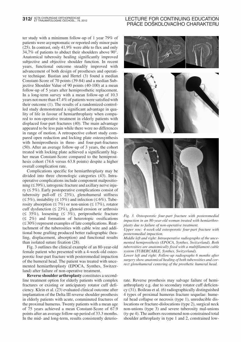
Fig. 3 outlines the clinical example of an 80-year-oldfemale patient who presented with a 4-week-old osteo-porotic four-part fracture with posteromedial impactionof the humeral head. The patient was treated with unce-mented hemiarthroplasty (EPOCA, Synthes, Switzer-land) after failure of non-operative treatment.
Reverse shoulder arthroplasty constitutes a second-line treatment option for elderly patients with complexfractures or existing or anticipatory rotator cuff defi -ciency. Klein et al. (23) evaluated clinical outcome afterimplantation of the Delta III reverse shoulder prosthesisin elderly patients with acute, comminuted fractures ofthe proximal humerus. Twenty patients with a mean ageof 75 years achieved a mean Constant-Score of 67.9points after an average follow-up period of 33.3 months.In the mid- and long-term, results consistently deterio-
rate. Reverse prosthesis may salvage failure of hemi-arthroplasty e.g. due to secondary rotator cuff deficien-cy (31). Boileau et al. (6) radiographically distinguished4 types of proximal humerus fracture sequelae: hume-ral head collapse or necrosis (type 1), unreducible dis-locations or fracture-dislocations (type 2), surgical necknon-unions (type 3) and severe tuberosity mal-unions(ty-pe 4). The authors recommend non-constrained totalshoulder arthroplasty in type 1 and 2, constrained low-
Fig. 3. Osteoporotic four-part fracture with posteromedialimpaction in an 80-year-old woman treated with hemiarthro-plasty due to failure of non-operative treatment.Upper row: 4-week-old osteoporotic four-part fracture withposteromedial impaction. Middle left and right: Intraoperative radiographs of the unce-mented hemiprosthesis (EPOCA, Synthes, Switzerland). Bothtuberosities are anatomically fixed with a multifilament cablesystem (TUBERCABLE, Synthes, Switzerland).Lower left and right: Follow-up radiographs 6 months aftersurgery show anatomical healing of both tuberosities and cor-rect glenohumeral centering of the prosthetic humeral head.
profile fracture prosthesis with additional bone graftingin type 3 and reverse arthroplasty in type 4 sequelae. Todate, scapular notching still represents the most com-mon complication (44-96%) following reverse shoulderarthroplasty leading to polyethylene wear, synovitis (PEdisease), glenoidal bone up to loosening of the gle-nosphere (38). Further specific complications consist ofscapular stress fractures and degeneration of the del toidmuscle.
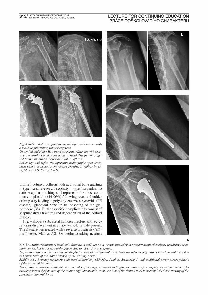
Fig. 4 shows a subcapital humerus fracture with seve-re varus displacement in an 85-year-old female patient.The fracture was treated with a reverse prosthesis (Affi-nis Inverse, Mathys AG, Switzerland) taking account
Fig. 4. Subcapital varus fracture in an 85-year-old woman witha massive preexisting rotator cuff tear.Upper left and right: Two-part (subcapital) fracture with seve-re varus displacement of the humeral head. The patient suffe-red from a massive preexisting rotator cuff tear.Lower left and right: Postoperative radiographs after treat-ment with a cemented-stem reverse prosthesis (Affinis Inver-se, Mathys AG, Switzerland).
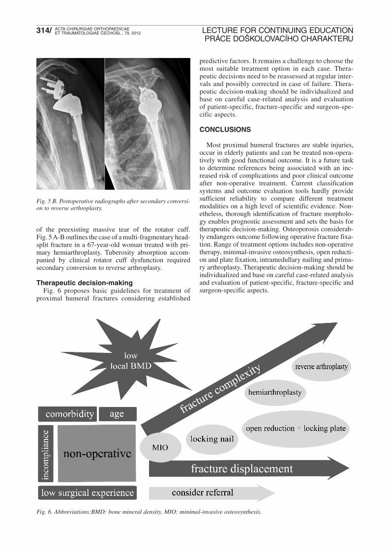
Fig. 5 A. Multi-fragmentary head-split fracture in a 67-year-old woman treated with primary hemiarthroplasty requiring secon-dary conversion to reverse arthroplasty due to tuberosity absorption.Upper row: Non-reconstructable head-split fracture of the humeral head. Note the inferior migration of the humeral head dueto neuropraxia of the motor branch of the axillary nerve.Middle row: Primary treatment with hemiarthroplasty (EPOCA, Synthes, Switzerland) and additional screw osteosynthesis of the coracoid fracture.Lower row: Follow-up examination 18 months after surgery showed radiographic tuberosity absorption associated with a cli-nically relevant dysfunction of the rotator cuff. Meanwhile, reinnervation of the deltoid muscle accomplished recentering of theprosthetic humeral head.
Fig. 5 B. Postoperative radiographs after secondary conversi-on to reverse arthroplasty.
of the preexisting massive tear of the rotator cuff.Fig. 5A-B outlines the case of a multi-fragmentary head-split fracture in a 67-year-old woman treated with pri-mary hemiarthroplasty. Tuberosity absorption accom-panied by clinical rotator cuff dysfunction requiredsecondary conversion to reverse arthroplasty.
Therapeutic decision-makingFig. 6 proposes basic guidelines for treatment of
proximal humeral fractures considering established
predictive factors. It remains a challenge to choose themost suitable treatment option in each case. Thera -peutic decisions need to be reassessed at regular inter-vals and possibly corrected in case of failure. Thera-peutic decision-making should be individualized andbase on careful case-related analysis and evaluation of patient-specific, fracture-specific and surgeon-spe-cific aspects.
CONCLUSIONS
Most proximal humeral fractures are stable injuries,occur in elderly patients and can be treated non-opera-tively with good functional outcome. It is a future taskto determine references being associated with an inc -reased risk of complications and poor clinical outcomeafter non-operative treatment. Current classificationsystems and outcome evaluation tools hardly providesufficient reliability to compare different treatmentmodalities on a high level of scientific evidence. Non-etheless, thorough identification of fracture morpholo-gy enables prognostic assessment and sets the basis fortherapeutic decision-making. Osteoporosis considerab-ly endangers outcome following operative fracture fixa-tion. Range of treatment options includes non-operativetherapy, minimal-invasive osteosynthesis, open reducti-on and plate fixation, intramedullary nailing and prima-ry arthroplasty. Therapeutic decision-making should beindividualized and base on careful case-related analysisand evaluation of patient-specific, fracture-specific andsurgeon-specific aspects.
Fig. 6. Abbreviations:BMD: bone mineral density, MIO: minimal-invasive osteosynthesis.
1. ANTUNA, S. A., SPERLING, J. W., COFIELD, R. H.: Shoulderhemiarthroplasty for acute fractures of the proximal humerus:A minimum five-year follow-up. J. Shoulder Elbow Surg., 17:202–209, 2008.
2. BASTIAN, J. D., HERTEL, R.: Initial post-fracture humeral headischemia does not predict development of necrosis. J. ShoulderElbow Surg., 17: 2–8, 2008.
3. BASTIAN, J. D., HERTEL, R.: Osteosynthesis and hemiarthro-plasty of fractures of the proximal humerus: Outcomes in a con-secutive case series. J. Shoulder Elbow Surg., 18: 216–219, 2009.
4. BELL, J. E., LEUNG, B. C., SPRATT, K. F., KOVAL, K. J., WEIN-STEIN, J. D., GOODMAN, D. C., TOSTESON, A. N.: Trends andvariation in incidence, surgical treatment, and repeat surgery ofproximal humeral fractures in the elderly. J. Bone Jt Surg., 93-A:121–131, 2011.
5. BOGNER, R., HUBNER, C., MATIS, N., AUFFARTH, A., LEDE-RER, S., RESCH, H.: Minimally-invasive treatment of three- andfour-part fractures of the proximal humerus in elderly patients. J.Bone Jt Surg., 90-B: 1602–1607, 2008.
6. BOILEAU, P., CHUINARD, C., LE HUEC, J. C., WALCH, G.,TROJANI, C.: Proximal humerus fracture sequelae: Impact ofa new radiographic classification on arthroplasty. Clin. Orthop.Relat. Res., 442: 121–130, 2006.
7. BRUNNER, F., SOMMER, C., BAHRS, C., HEUWINKEL, R.,HAFNER, C., RILLMANN, P., KOHUT, G., EKELUND, A.,MULLER, M., AUDIGE, L., BABST, R.: Open reduction andinternal fixation of proximal humerus fractures using a proximalhumeral locked plate: A prospective multicenter analysis. J. Ort-hop. Trauma, 23: 163–172, 2009.
8. CALVO, E., MORCILLO, D., FORURIA, A. M., REDONDO-SANTAMARIA, E., OSORIO-PICORNE, F., CAEIRO, J. R.:Nondisplaced proximal humeral fractures: High incidence amongoutpatient-treated osteoporotic fractures and severe impact onupper extremity function and patient subjective health perception.J. Shoulder Elbow Surg., 20: 795–801, 2011.
9. CODMAN, E. A. The shoulder; rupture of the supraspinatus ten-don and other lesions in or about the subacromial bursa. Boston,Mass.: T. Todd company; 1934: 51, 529 p.
10. COURT-BROWN, C. M., CATTERMOLE, H., McQUEEN, M.M.: Impacted valgus fractures (b1.1) of the proximal humerus. Theresults of non-operative treatment. J. Bone Jt Surg., 84-B: 504–508,2002.
11. COURT-BROWN, C. M., GARG, A., McQUEEN, M. M.: The epi-demiology of proximal humeral fractures. Acta Orthop. Scand.,72: 365–371, 2001.
12. EGOL, K. A., SUGI, M. T., ONG, C. C., MONTERO, N., DAVI-DOVITCH, R., ZUCKERMAN, J. D.: Fracture site augmentationwith calcium phosphate cement reduces screw penetration afteropen reduction-internal fixation of proximal humeral fractures. J.Shoulder Elbow Surg., [Epub 2011, Dec. 21], 21: 741–748, 2012.
13. FJALESTAD, T., HOLE, M. O., HOVDEN, I. A., BLUCHER, J.,STROMSOE, K.: Surgical treatment with an angular stable platefor complex displaced proximal humeral fractures in elderly pa -tients: A randomized controlled trial. J. Orthop. Trauma, 26:98–106, 2012.
14. FORURIA, A. M., DE GRACIA, M. M., LARSON, D. R., MUNU-ERA, L., SANCHEZ-SOTELO, J.: The pattern of the fracture anddisplacement of the fragments predict the outcome in proximalhumeral fractures. J. Bone Jt Surg., 93-B: 378–386, 2011.
15. GARDNER, M. J., WEIL, Y., BARKER, J. U., KELLY, B. T.,HELFET, D. L., LORICH, D. G.: The importance of medial sup-port in locked plating of proximal humerus fractures. J. Orthop.Trauma, 21: 185–191, 2007.
16. GRADL, G., DIETZE, A., KAAB, M., HOPFENMULLER, W.,MITTLMEIER, T.: Is locking nailing of humeral head fracturessuperior to locking plate fixation? Clin Orthop Relat Res. 467:2986–2993, 2009.
17. HANSON, B., NEIDENBACH, P., DE BOER, P., STENGEL, D.:Functional outcomes after nonoperative management of fracturesof the proximal humerus. J. Shoulder Elbow Surg., 18: 612–621,2009.
18. HARDEMAN, F., BOLLARS, P., DONNELLY, M., BELLE-MANS, J., NIJS, S.: Predictive factors for functional outcome andfailure in angular stable osteosynthesis of the proximal humerus.Injury, 43: 153–158, 2012.
19. HERTEL, R., HEMPFING, A., STIEHLER, M., LEUNIG, M.: Pre-dictors of humeral head ischemia after intracapsular fracture of theproximal humerus. J. Shoulder Elbow Surg., 13: 427–433, 2004.
20. HORN, J., GUEORGUIEV, B., BRIANZA, S., STEEN, H.,SCHWIE GER, K.: Biomechanical evaluation of two-part surgicalneck fractures of the humerus fixed by an angular stable lockedintramedullary nail. J. Orthop. Trauma, 25: 406–413, 2011.
21. JAEGER, M., MAIER, D., IZADPANAH, K., STROHM, P., SUD-KAMP, N. P.: [Limitations of reconstruction – prostheses]. Unfall-chirurg, 114: 1068–1074, 2011.
22. KANNUS, P., PALVANEN, M., NIEMI, S., SIEVANEN, H.,PARKKARI, J.: Rate of proximal humeral fractures in older fin-nish women between 1970 and 2007. Bone, 44: 656–659, 2009.
23. KLEIN, M., JUSCHKA, M., HINKENJANN, B., SCHERGER,B., OSTERMANN, P. A.: Treatment of comminuted fractures ofthe proximal humerus in elderly patients with the delta iii reverseshoulder prosthesis. J. Orthop. Trauma, 22: 698–704, 2008.
24. KOVAL, K. J., GALLAGHER, M. A., MARSICANO, J. G., CUO-MO, F., McSHINAWY, A., ZUCKERMAN, J. D.: Functional out-come after minimally displaced fractures of the proximal part ofthe humerus. J. Bone Jt Surg.,79-A: 203–207, 1997.
25. KRALINGER, F., SCHWAIGER, R., WAMBACHER, M., FAR-RELL, E., MENTH-CHIARI, W., LAJTAI, G., HUBNER, C.,RESCH, H.: Outcome after primary hemiarthroplasty for fractureof the head of the humerus. A retrospective multicentre study of167 patients. J. Bone Jt Surg., 86-B: 217–219, 2004.
26. KRALINGER, F., UNGER, S., WAMBACHER, M., SMEKAL,V., SCHMOELZ, W.: The medial periosteal hinge, a key structu-re in fractures of the proximal humerus: A biomechanical cadaverstudy of its mechanical properties. J. Bone Jt Surg., 91-B: 973–976,2009.
27. KRAPPINGER, D., BIZZOTTO, N., RIEDMANN, S., KAM-MERLANDER, C., HENGG, C., KRALINGER, F. S.: Predictingfailure after surgical fixation of proximal humerus fractures. Inju-ry, 42: 1283–1288, 2011.
28. KRAUSE, F. G., HUEBSCHLE, L., HERTEL, R.: Reattachmentof the tuberosities with cable wires and bone graft in hemiarthro-plasties done for proximal humeral fractures with cable wire andbone graft: 58 patients with a 22-month minimum follow-up. J.Orthop. Trauma, 21: 682–686, 2007.
29. KRISTIANSEN, B., ANGERMANN, P., LARSEN, T. K.: Func-tional results following fractures of the proximal humerus. A con-trolled clinical study comparing two periods of immobilization.Arch Orthop Trauma Surg. 108: 339–341, 1989.
31. LEVY, J., FRANKLE, M., MIGHELL, M., PUPELLO, D.: Theuse of the reverse shoulder prosthesis for the treatment of failedhemiarthroplasty for proximal humeral fracture. J. Bone Jt Surg.,89-A: 292–300, 2007.
32. LILL, H., BEWER, A., KORNER, J., VERHEYDEN, P., HEPP,P., KRAUTHEIM, I., JOSTEN, C.: [Conservative treatment of dis-located proximal humeral fractures]. Zentralbl. Chir., 126:205–210, 2001.
33. MAJED, A., MACLEOD, I., BULL, A. M., ZYTO, K., RESCH,H., HERTEL, R., REILLY, P., EMERY, R. J.: Proximal humeralfracture classification systems revisited. J. Shoulder Elbow Surg.,20: 1125–1132, 2011.
34. MARAVIC, M., LE BIHAN, C., LANDAIS, P., FARDELLONE,P.: Incidence and cost of osteoporotic fractures in france during2001. A methodological approach by the national hospital data-base. Osteoporos. Int., 16: 1475–1480, 2005.
35. MURRAY, I. R., AMIN, A. K., WHITE, T. O., ROBINSON, C.M.: Proximal humeral fractures: Current concepts in classifica-tion, treatment and outcomes. J. Bone Jt Surg., 93-B: 1–11, 2011.
36. NEER, C. S., 2nd: Displaced proximal humeral fractures. I. Clas-sification and evaluation. J. Bone Jt Surg., 52-A: 1077–1089,1970.
37. NEER, C. S., 2nd: Displaced proximal humeral fractures. II. Tre-atment of three-part and four-part displacement. J. Bone Jt Surg.,52-A: 1090–1103, 1970.
38. NICHOLSON, G. P., STRAUSS, E. J., SHERMAN, S. L.: Sca-pular notching: Recognition and strategies to minimize clinicalimpact. Clin. Orthop. Relat. Res., 469: 2521–2530, 2011.
39. NIJS, S., REUTHER, F., BROOS, P.: Primary fracture arthroplastyof the proximal humerus using a new and freely adjustable modu-lar prosthesis combined with compression osteosynthesis of thetuberosities. Oper. Orthop. Traumatol., 23: 21–28, 2011.
40. OLERUD, P., AHRENGART, L., PONZER, S., SAVING, J.,TIDERMARK, J.: Hemiarthroplasty versus nonoperative treat-ment of displaced 4-part proximal humeral fractures in elderly pati-ents: A randomized controlled trial. J. Shoulder Elbow Surg., 20:1025–1033, 2011.
41. PIJLS, B. G., WERNER, P. H., EGGEN, P. J.: Primary uncemen-ted hemiarthroplasty for severe fractures of the proximal hume-rus. J. Orthop. Trauma, 25: 279–285, 2011.
42. PLATZER, P., THALHAMMER, G., OBERLEITNER, G.,KUTSCHA-LISSBERG, F., WIELAND, T., VECSEI, V., GAEB-LER, C.: Displaced fractures of the greater tuberosity: A compa-rison of operative and nonoperative treatment. J. Trauma, 65:843–848, 2008.
43. PLAUSINIS, D., KWON, Y. W., ZUCKERMAN, J. D.: Compli-cations of humeral head replacement for proximal humeral frac-tures. Instr. Course. Lect., 54: 371–380, 2005.
44. RESCH, H., BECK, E., BAYLEY, I.: Reconstruction of the val-gus-impacted humeral head fracture. J. Shoulder Elbow Surg., 4:73–80, 1995.
45. RESCH, H., POVACZ, P., FROHLICH, R., WAMBACHER, M.:Percutaneous fixation of three- and four-part fractures of the pro-ximal humerus. J. Bone Jt Surg., 79-B: 295–300, 1997.
46. ROBINSON, C. M., KHAN, L., AKHTAR, A., WHITTAKER, R.:The extended deltoid-splitting approach to the proximal humerus.J. Orthop. Trauma, 21: 657–662, 2007.
47. RODERER, G., ERHARDT, J., GRAF, M., KINZL, L., GEB-HARD, F.: Clinical results for minimally invasive locked platingof proximal humerus fractures. J. Orthop. Trauma, 24: 400–406,2010.
48. RUCHHOLTZ, S., HAUK, C., LEWAN, U., FRANZ, D., KUH-NE, C., ZETTL, R.: Minimally invasive polyaxial locking plate
fixation of proximal humeral fractures: A prospective study. J.Trauma, 71: 1737–1744, 2011.
49. SEIDE, K., TRIEBE, J., FASCHINGBAUER, M., SCHULZ, A. P.,PUSCHEL, K., MEHRTENS, G., JURGENS, C.: Locked vs. Unloc-ked plate osteosynthesis of the proximal humerus – a biomechani-cal study. Clin. Biomech. (Bristol, Avon). 22: 176–182, 2007.
50. SOLBERG, B. D., MOON, C. N., FRANCO, D. P., PAIEMENT,G. D.: Surgical treatment of three and four-part proximal humeralfractures. J. Bone Jt Surg., 91-A: 1689–1697, 2009.
51. SPROUL, R. C., IYENGAR, J. J., DEVCIC, Z., FEELEY, B. T.:A systematic review of locking plate fixation of proximal hume-rus fractures. Injury, 42: 408–413, 2011.
52. STROHM, P. C., HELWIG, P., KONRAD, G., SUDKAMP, N. P.:Locking plates in proximal humerus fractures. Acta Chir. orthopTraum. čech., 74: 410–415, 2007.
53. SUDKAMP, N., BAYER, J., HEPP, P., VOIGT, C., OESTERN,H., KAAB, M., LUO, C., PLECKO, M., WENDT, K., KOSTLER,W., KONRAD, G.: Open reduction and internal fixation of proxi-mal humeral fractures with use of the locking proximal humerusplate. Results of a prospective, multicenter, observational study. J.Bone Jt Surg., 91-A: 1320–1328, 2009.
54. TORRENS, C., CORRALES, M., MELENDO, E., SOLANO, A.,RODRIGUEZ-BAEZA, A., CACERES, E.: The pectoralis majortendon as a reference for restoring humeral length and retroversi-on with hemiarthroplasty for fracture. J. Shoulder Elbow Surg.,17: 947–950, 2008.
55. VOIGT, C., GEISLER, A., HEPP, P., SCHULZ, A. P., LILL, H.:Are polyaxially locked screws advantageous in the plate osteo-synthesis of proximal humeral fractures in the elderly? A pro-spective randomized clinical observational study. J. Orthop. Trau-ma, 25: 596–602, 2011.
56. WACHTL, S. W., MARTI, C. B., HOOGEWOUD, H. M., JAKOB,R. P., GAUTIER, E.: Treatment of proximal humerus fracture usingmultiple intramedullary flexible nails. Arch. Orthop. Trauma Surg.,120: 171–175, 2000.
57. WU, C. H., MA, C. H., YEH, J. J., YEN, C. Y., YU, S. W., TU, Y.K.: Locked plating for proximal humeral fractures: Differencesbetween the deltopectoral and deltoid-splitting approaches. J.Trauma, 71: 1364–1370, 2011.
58. ZHU, Y., LU, Y., SHEN, J., ZHANG, J., JIANG, C.: Locking intra-medullary nails and locking plates in the treatment of two-part pro-ximal humeral surgical neck fractures: A prospective randomizedtrial with a minimum of three years of follow-up. J. Bone Jt Surg.,93-A: 159–168, 2011.
Corresponding author:Univ.-Prof. Dr. N.P. SüdkampUniversitätsklinikum FreiburgDept. Orthopädie und Traumatologie, Klinik für TraumatologieHugstetter Starsse 55, 79106 Freiburg im Breisgau Germany


![· 2800 m. ) " Aconitine : JAPAN 18, 17 Aconitine mesa-conitine, japaconitine 1977 ÇRobert H. Dreisbach r Handbook of poisoning : Diagnosis & Treatment] : ; :](https://static.dokumenty.site/doc/80x56/6098c0603823582aac35026c/2800-m-aconitine-japan-18-17-aconitine-mesa-conitine-japaconitine.jpg)


