Page 1
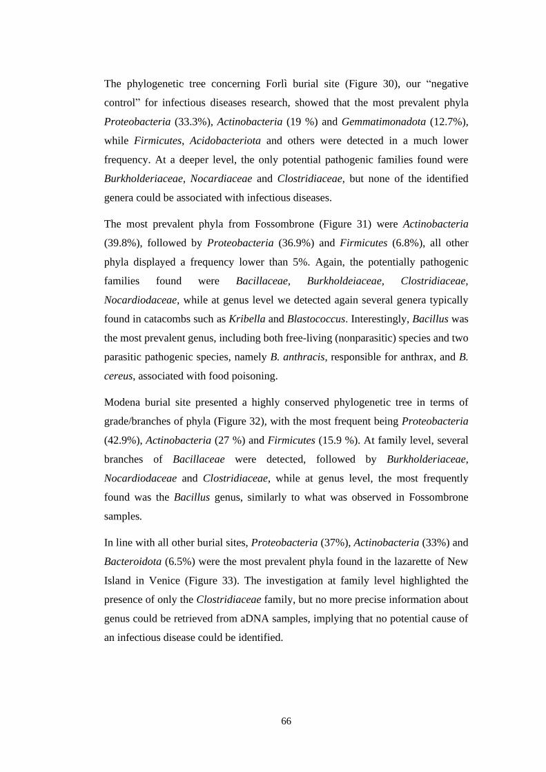
UNIVERSITY OF VERONA
UNIVERSITY OF VERONA
DEPARTMENT OF NEUROSCIENCES, BIOMEDICINE AND MOVEMENT SCIENCES
GRADUATE SCHOOL OF
APPLIED LIFE AND HEALTH SCIENCES
DOCTORAL PROGRAM IN
LIFE AND HEALTH SCIENCES
31° CYCLE
S.S.D. MED/07
TITLE OF THE DOCTORAL THESIS
PALEOGENOMICS AND PALEOMICROBIOLOGY APPROACHES APPLIED TO THE UNDERSTANDING
OF TRUE AND APOCRYPHAL PESTS IN DIFFERENT BIOARCHAEOLOGICAL CONTEXTS: THE
STUDY OF ANCIENT INFECTIOUS DISEASES
Coordinator: Prof. Giovanni Malerba
Tutor: Prof. Giuseppe Cornaglia
Co-Tutor: Dott.ssa Elisabetta Cilli
Co-Tutor: Prof. Nicola Vitulo
Doctoral Student: Dott.ssa Alda Bazaj
Page 2
i
This work is licensed under a Creative Commons Attribution-NonCommercial-
NoDerivs 3.0 Unported License, Italy. To read a copy of the licence, visit the web page:
http://creativecommons.org/licenses/by-nc-nd/3.0/ Attribution — You must give appropriate credit, provide a link to the license, and indicate if changes were
made. You may do so in any reasonable manner, but not in any way that suggests the licensor endorses you
or your use. NonCommercial — You may not use the material for commercial purposes.
NoDerivatives — If you remix, transform, or build upon the material, you may not distribute the modified material.
Page 3
ii
Abstract Ph.D. Course in Applied Life and Health Sciences, 31th cycle
Doctor of Philosophy
Paleogenomics and paleomicrobiology approaches applied to the
understanding of true and apocryphal pests in different bioarchaeological
contexts: the study of Ancient infectious diseases
By Alda BAZAJ
Plague is a bacterial disease caused by Yersinia pestis, which primarily affects wild
rodents. It is spread from one rodent to another by fleas. To date, Plague is probably
the infectious pathology responsible of the largest amount of deaths among all
human history. Unfortunately, it is still persistent in some area of the world, as on
November 4, 2014 the Ministry of Health of Madagascar reported an outbreak of
plague (https://www.epicentro.iss.it/peste/aggiornamenti) to the World Health
Organization. The first case was identified on August 31, a male from
Soamahatamana village in the district of Tsiroanomandidy, who died on September
3. As of November 16, a total of 119 cases of plague were confirmed, including 40
deaths.
Next-generation sequencing (NGS) and metagenomics has recently revolutionized
genomic research, and its combination with high-throughput target enrichment
method can be proficiently applied to the study of ancient DNA (aDNA), thus
providing a powerful tool for understanding the evolution of pandemic infectious
diseases like the plague.
Page 4
iii
Al professor Giuseppe Cornaglia
Grazie per la fiducia che hai avuto in me e per avermi insegnato a
non arrendermi mai…
(1958-2020)
Page 5
iv
INDEX
1. INTRODUCTION 1
1.1. Ancient DNA 1
1.1.1. aDNA specificities 2
1.1.2. aDNA degradation 2
1.1.2.1. Cytosine Deamination 4
1.1.2.2. aDNA depurination 5
1.1.2.3. Fragment length 6
1.1.3. Contamination 6
1.2. Paleomicrobiology 8
1.3. The plague 11
1.4. Yersinia pestis 14
2. OBJECTIVES 17
3. METHODS 19
3.1. Setting up an “ad hoc” Paleomicrobiology laboratory 19
3.2. Biological specimens’ collection and manipulation 22
3.3. DNA extraction 27
3.4. Polymerase Chain Reaction (PCR):
using “specific primers” to detect Y. pestis 30
3.5. Next Generation Sequencing (NGS) 34
3.6. Metagenomics 36
3.7. 16S rRNA Metagenomics and aDNA 37
3.8. Preparation of NGS Libraries from aDNA 40
3.9. Microbial taxonomic profiling 43
3.10. Phylogenetic trees 43
4. RESULTS 45
4.1. aDNA extraction and purity 45
4.2. PCR and Sanger Sequencing 46
Page 6
v
4.3. Metagenomics 49
4.3.1. Paleomicrobiome analysis on V3 region 51
4.3.2. Paleomicrobiome analysis on V5 region 61
5. CONCLUSIONS AND FUTURE PERSPECTIVES 67
6. PUBLICATION LIST 70
BIBLIOGRAPHY 72
SUPPLEMENTARY INFORMATION 81
Page 7
vi
To my husband Valerio, who fostered my love for learning, and my child Daniele.
A.B.
Page 8
1
Chapter 1
INTRODUCTION
1.1 Ancient DNA
Ancient DNA (aDNA) provides direct insights onto the past that modern DNA or
paleontological studies alone cannot provide. It has been proven to address
questions regarding history relationships, population dynamics and diversity
through time. Although aDNA can be a very powerful tool, it should be handled
with care [Fulton et al., 2012]. The first studies about aDNA began in 1984, when
the DNA of an extinct Quagga, a relative of a zebra, were recovered [Higuchi et al.,
1984]. History started changing from that moment, as technical and biological
advances permitted to evolve Paleogenomics through time.
The state-of-the-art genomic technologies, through the combination of high-
throughput sequencing and the most recent bioinformatics tools, has allowed the
study of whole genome sequences of large population datasets, the identification of
ancient pathogens and their evolution [Allentoft et al., 2015, Fu et al., 2016, Olalde
et al., 2018]. Next-generation sequencing (NGS) has completely revolutionized
aDNA research, when is well combined with high-throughput target enrichment
methods. On the other side, aDNA present specific limitations that require careful
consideration during data analysis.
Page 9
2
1.1.1 aDNA specificities
Ancient DNA can be defined as any genomic sequence retrieved from dead
organisms. The DNA of each living organism is frequently damaged through time,
but such damages are repaired by mechanisms that preserve the integrity of the
genetic material. While after death the damage of DNA still continues, the repairing
mechanisms can no longer maintain the DNA intact. Therefore, most of the aDNA
sequences found nowadays are in different stages of degradation.
1.1.2 aDNA degradation
DNA degradation is influenced mostly by atmospheric conditions such as
temperature or humidity, and other variables linked with the burial environment,
namely salt concentration, soil pH or the chemical composition of the ground.
The degradation process reduces the amount of endogenous aDNA present in the
samples, usually accounting for less than 1% of the total sequenced reads [Fu et al.,
2013]. On the other hand, in some exceptional cases, the retrieved endogenous
DNA was found to exceed 70% [Meyer et al., 2012, Raghavan et al., 2014,
Rasmussen et al., 2010, Gamba et al., 2014, Keller et al., 2012, Carpenter et al.,
2013, Lazaridis et al., 2014, Olalde et al., 2014].
Nevertheless, environmental factors are still the biggest threat to DNA integrity.
Upon exposure to climatic and environmental agents, DNA is subjected to chemical
reactions such as deamination, depurination or hydrolysis that damage DNA
structure (Figure 1), ultimately leading to its degradation [Höss et al., 1996]. In
addition, oxidation can induce DNA lesions that impair Polymerase Chain Reaction
Page 10
3
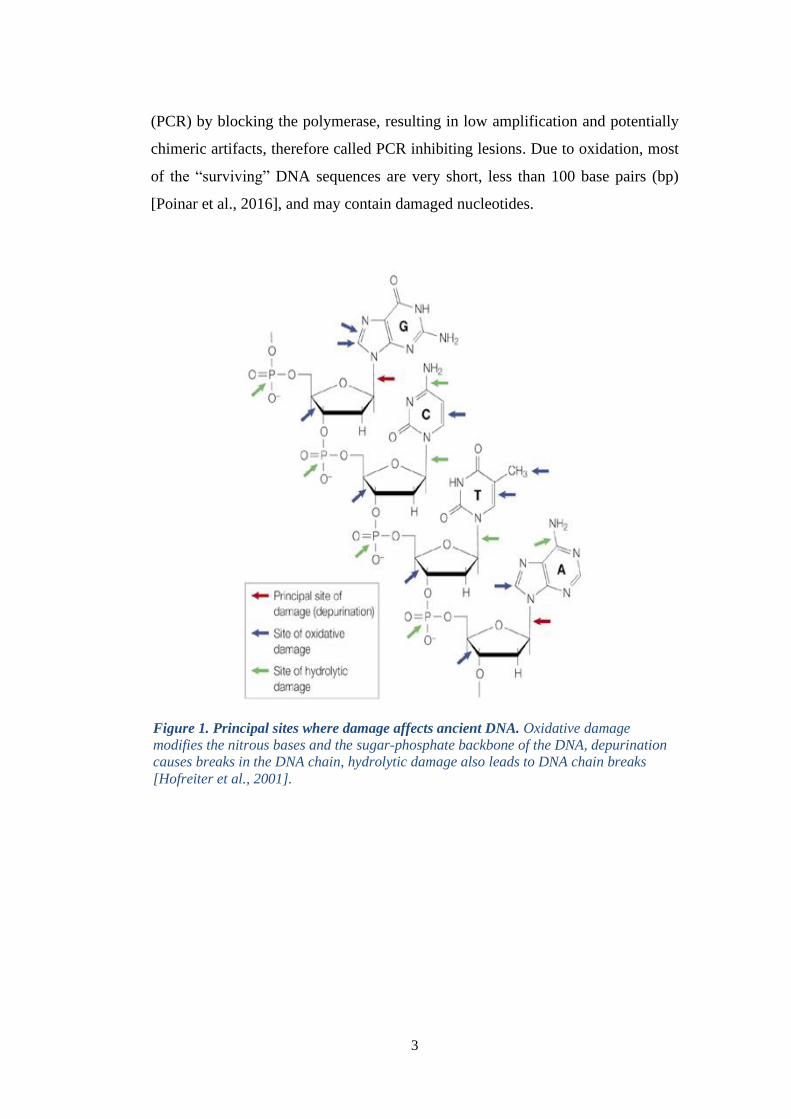
(PCR) by blocking the polymerase, resulting in low amplification and potentially
chimeric artifacts, therefore called PCR inhibiting lesions. Due to oxidation, most
of the “surviving” DNA sequences are very short, less than 100 base pairs (bp)
[Poinar et al., 2016], and may contain damaged nucleotides.
Figure 1. Principal sites where damage affects ancient DNA. Oxidative damage
modifies the nitrous bases and the sugar-phosphate backbone of the DNA, depurination
causes breaks in the DNA chain, hydrolytic damage also leads to DNA chain breaks
[Hofreiter et al., 2001].
Page 11
4
1.1.2.1 Cytosine Deamination
The most characteristic degradation of DNA molecules due to post-mortem damage
is the hydrolytic deamination of Cytosine (C) to Uracil (U) [Gilbert et al., 2007],
which codes as Thymine (T). During DNA replication, C deamination causes the
misincorporation of an Adenine (A) instead of the original Guanine (G), which is
followed by C to T substitutions in the 5’ ends of the sequences [Hofreiter et al.,
2001]. The C to T replacement at the 5’ ends of the DNA fragments, results in a
higher frequency of G to A substitutions at the 3’ ends of the complementary strands
[Briggs et al., 2007, Rasmussen et al., 2014]. It is unclear whether any other
miscoding lesions may be relevantly frequent in aDNA molecules or their
distribution along the sequence.
Cytosines deamination particularly affects aDNA reads ends, where the percentage
of deaminated C can exceed 40% [Briggs et al., 2007]. Specific techniques have
been implemented to minimize errors in sequencing and mapping procedures due
to aDNA damage. Indeed, single stranded library building protocol can be an
efficient method to analyse poor quality samples, characterized by a low rate of
endogenous DNA and highly degraded strands [Gansauge et al., 2013]. On the other
side, the presence of cytosine deamination can also be used to discriminate between
reads obtained by real aDNA or modern contaminant [Rohland et al., 2009].
Page 12
5
1.1.2.2 aDNA Depurination
DNA fragmentation is mainly caused by depurination, namely the disruption of an
N-glucosyl bond between a purine and the sugar of the DNA chain, resulting in a
chain with an abasic site. The chain is then fragmented through 𝛽 elimination,
leaving 3′-aldehydic and 5′-phosphate ends (Figure 2) [Briggs et l., 2007].
Depurination, considered the most critical chemical damage to aDNA structure,
ultimately causes the underrepresentation of purines (G and A) usually at the 5’
fragment ends [Briggs et al., 2007, Orlando et al., 2011 and Meyer et al., 2012].
A recent study demonstrated that depurination occurs at both ends of the aDNA
fragments [Meyer et al., 2012] and suggested an explanation for the observation in
previous publications of depurination only at the 5’ ends. Such reason was
hypothesized to reside in the pre-processing step required by most library building
protocols, namely the blunt-ends repair, an enzymatic process that extends recessed
and degrades overhanging 3′ ends of DNA fragments [Briggs et al., 2007]. Thus,
only with the development of single-stranded DNA libraries, this process could be
observed also at the 3’ ends of the aDNA fragments [Meyer et al., 2012].
Figure 2. Depurination. A chemical reaction in which an N-glycosyl bond is broken
resulting in an abasic site. The abasic site is later removed, fragmenting the DNA
through 𝛽 elimination [Dabney et al., 2013].
Page 13
6
1.1.2.3 Fragment length
One of first published studies describing the aDNA peculiarities showed that even
in well-preserved specimens, only very short fragments (50-150 bp) of aDNA could
be retrieved. The entity of aDNA degradation is inversely correlated with the
amplification efficiency and the length of the amplification product [Pääbo et al.,
1989]. The same consideration was reported by the one of the first studies on aDNA
involving high-throughput sequencing, describing the difficulties of both aDNA
extraction and library preparation [Green et al., 2010].
1.1.3 Contamination
Research on endogenous aDNA of an extracted sample can result particularly
challenging due to the small portion of survived copies of endogenous aDNA in an
extract, compared with modern DNA present in the environment. The PCR
amplifies not only those small portions of aDNA, but also DNA from different
sources, which may contaminate the sample during different manipulation steps of
aDNA specimen. Indeed, the sample itself can be contaminated by adhering
microorganisms residing in the environment, as bones, ribs and teeth are extremely
porous. The highest risk of contamination resides in the collection of specimens,
particularly in human and microbials studies, yet another source of contamination
is represented by sample handling procedures in the laboratory (DNA extraction
and PCR). The air filtering system of the building may be contaminated by the
presence of insects or other biological entities, although the major concern is
represented by the contamination with exogenous DNA. Laboratory personnel and
reagents may introduce exogenous human or animal DNA, as living organisms are
constantly shedding DNA-bearing tissues in the form of skin cells, hair, saliva, and
other secretions. Archaeological skeletal remains, for example (Figure 3), may be
Page 14
7
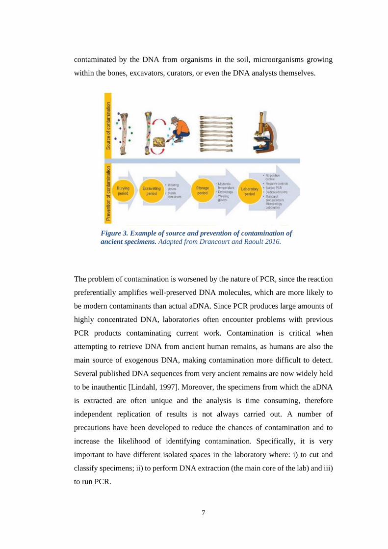
contaminated by the DNA from organisms in the soil, microorganisms growing
within the bones, excavators, curators, or even the DNA analysts themselves.
The problem of contamination is worsened by the nature of PCR, since the reaction
preferentially amplifies well-preserved DNA molecules, which are more likely to
be modern contaminants than actual aDNA. Since PCR produces large amounts of
highly concentrated DNA, laboratories often encounter problems with previous
PCR products contaminating current work. Contamination is critical when
attempting to retrieve DNA from ancient human remains, as humans are also the
main source of exogenous DNA, making contamination more difficult to detect.
Several published DNA sequences from very ancient remains are now widely held
to be inauthentic [Lindahl, 1997]. Moreover, the specimens from which the aDNA
is extracted are often unique and the analysis is time consuming, therefore
independent replication of results is not always carried out. A number of
precautions have been developed to reduce the chances of contamination and to
increase the likelihood of identifying contamination. Specifically, it is very
important to have different isolated spaces in the laboratory where: i) to cut and
classify specimens; ii) to perform DNA extraction (the main core of the lab) and iii)
to run PCR.
Figure 3. Example of source and prevention of contamination of
ancient specimens. Adapted from Drancourt and Raoult 2016.
Page 15
8
1.2 Paleomicrobiology
Paleomicrobiology is defined as the study of microorganisms in ancient remains
that were naturally present in healthy organisms as well as those that were
responsible for infectious diseases [Rivera-Perez et al., 2016]. This discipline
includes branches of medical microbiology, history, anthropology, and
archaeozoology, and is aimed at evaluating the evolution of ancient pathogens
(recently including also ancient microbiota) through their identification and the
analysis of functional data such as antimicrobial resistance [Drancourt et al., 2016
chap. 5 Paleomicrobiology of Humans]. Molecular analysis of ancient pathogens
can also provide useful information to reconstruct past epidemic trends and help
refining even the most recent models of emerging infections, thus giving an
important contribution to the development of adequate preventive measures. In
addition, the combination of microbial and human metagenomics data has a huge
potential to extend the paleomicrobiology field to a wider community of scientists
and scholars [Bazaj et al., 2015].
Before the development of genomic techniques, the identification of infectious
pathogens in ancient remains was restricted to the visual identification of bone
injuries and its correlation with ancient written proofs [Kousoulis et al., 2012].
There are many pathogen casualties, though, for which it is impossible to visually
identify the etiological cause of the reported mortality, for example Syphilis (caused
by Treponema pallidum) can be confused with skeletal lesions [Rothschild et al.,
2005], or Brucellosis (caused by Brucella melitensis) can be misclassified as
Tuberculosis (caused by Mycobacterium tuberculosis) [Mutolo et al., 2012, Kay et
al., 2014]. Another paradigmatic example of this approach is that of Medieval Black
Death, which could be attributed to a viral pandemic with an aerosol transmission
pattern, based on the descriptions from historical records [Bossak et al., 2007]. For
such cases, the etiological cause can only be univocally identified through genetic
markers.
Page 16
9
The first genetic studies that targeted ancient pathogens were performed with PCR
techniques [Kolman et al., 1999, Gernaey et al., 2001, Drancourt et al., 2003, Zink
et al., 2003 and Nguyen-Hieu et al., 2010]. Such studies required a prior
pathological diagnosis, as PCR relies on specific primers to amplify each possible
pathogen [Willerslev et al., 2007], however as bacteria and virus are ubiquitous, the
specificity of these tests is usually underrated and false positive results are frequent
[Päabo et al., 2004, Gilbert et al., 2005, Gilbert et al., 2006].
The publication of the draft genome of Y. pestis from British individuals of the 14th
century [Bos et al., 2011] is a milestone as it was the first draft genome of a
pathogen obtained from ancient human remains. Other strains of Y. pestis were then
characterized, from the Bronze Age [Rasmussen et al., 2015, Spyrou et al., 2018]
to the 19th century pandemics [Stenseth et al., 2008] in Europe and from China [Cui
et al., 2013]. The next step for paleomicrobiology is the retrieval of aDNA in
unexplored environments, the development of protein-based approaches and their
integration to unravel genetic adaptation through time.
The discoveries in the field have been always related with the ultimate technical
improvements, indeed Second Generation Sequencing (SGS) provided a huge
amount of data to analyse, whereas the advent of third generation sequencing
methods might supply the basis for de-novo assembly of aDNA remains, the
ultimate goal that was left unreached so far. Another alternative and promising
approach is represented by metagenomics performed on aDNA, which could
potentially lead to the recovery of complete extinct environments.
Page 17
10
Figure 4. Overview and Timeline of Historically Notable Disease Outbreaks in Human
History. Adapted from Andam et al., 2016.
Page 18
11
1.3 The plague
Plague is an infectious disease of bacterial origin still present in many parts of the
world, including some regions of industrialized countries. It is caused by Yersinia
pestis bacterium, which is typically hosted by the parasitic fleas of rodents, rats,
squirrels and in some cases also pets such as cats. Normally, Y. pestis has low
mortality rates in these species which can therefore be considered as long-term
infectious reserves, indeed it is still present in 22 rodent reservoirs. The origin of
the plague is very ancient, and due to its destructive force, it has been collectively
named “the black death”, a disease that has accompanied humanity over the
centuries and was often present in the great literary works and art (Figure 5).
Three main Y. pestis epidemics have affected Europe in historical times: The First
Pandemic started with the Plague of Justinian (541-543 AD) and continued until
Figure 5. The Triumph of Death (P. Bruegel, 1562). Oil panel painting showing an
allegory of the Last Judgment influenced by the medieval plague scenes.
Page 19
12
~750 AD [Russell et al., 1968]. The Second Pandemic, named the Black Death, was
probably the most famous and was responsible for killing up to 40% of the
European population [Rasmussen et al., 2015] during the 14th century (1346-1352
AD). The second wave of the Black Death was named the Great Plague (1665-1666
AD), which caused the death of almost one third of the European population and
literally infected all the countries from the Mediterranean to Scandinavia and Russia
within five years. In Europe it remained endemic, bouncing back into cycles of 10-
12 years at least for the next three centuries until the 18th century [Zietz et al., 2004].
Finally, the Third Pandemic started in China in 1860s, with the outbreak of a serious
epidemy in 1894, before spreading all over the world as series of pestilences
between the 18th and 19th centuries [Cohn et al., 2008; Stenseth et al., 2008] until
the end of the 20th century [Bos et al., 2011; Cui et al., 2013; Drancourt et al., 1998;
Wagner et al., 2014].
Y. pestis strains responsible for all three major epidemics were identified, namely
the strains from the Justinian outbreak [Wagner et al., 2014], the 14th century Black
Death strains [Bos et al., 2011, Schuenemann et al., 2014] and 18th century
pandemic [Bos et al., 2016]. Based on literature records, earlier Y. pestis outbreaks
may have occurred in Europe before the Justinian Plague, such as the Plague of
Athens (427-430 BC) and Antonine Plague (165-180 AD). The lack of DNA
evidence, though, does not allow either the confirmation of such events or the
identification of the pathogen linked with the historical records [Drancourt et al.,
2002; Drancourt and Raoult 2002]. The earliest evidence of Y. pestis DNA presence
in human remains was detected in Late Neolithic and Bronze Age individuals from
the steppe and eastern Europe (5000-3500 BP) [Rasmussen et al., 2015]. The
analysis of these strains revealed that most recent common ancestor of all European
Y. pestis strains lived up to ~6000 years BC, indicating that the Black Death causal
strain was present in Europe at least since the Bronze Age.
Although as of 2018 [WHO 2018] it is still endemic in only 17 countries (Figure
6), the plague has a remarkable evocative power and immediately brings back
images of horror and devastation.
Page 20
13
Figure 6. Reported Plague Cases by Country (2013-2018 WHO)
Page 21
14
1.4 Yersinia pestis
The genus Yersinia, member of the family Enterobacteriaceae, consists of 11
species, 3 of them are human pathogens (Y. pestis, Y. pseudotuberculosis and Y.
enterocolitica).
Yersinia pestis was discovered in 1894 and named Pasteurella pestis by Alexandre
Yersin, a French/Swiss physician and bacteriologist from the Pasteur Institute,
during an endemic of plague in Hong Kong, and was renamed Yersinia pestis in
1944 after his discoverer. Yersin noted that rats were affected by plague even before
epidemics in humans and that plague was regarded by many locals as a disease of
rats; indeed, villagers in China and India asserted that when large numbers of rats
were found dead, plague outbreaks soon followed [Yersin and Treill 1894].
Y. pestis is a Gram-negative, rod-shaped, non-motile, non-lactose fermenting, non-
spore forming, facultative anaerobic coccobacillus presenting cell wall, lipid
composition and antigens typical of enterobacteria. The plague coccobacillus is a
mandatory parasite growing at temperatures from 4°C to 40°C, optimally growing
between 28°C and 30°C, with a remarkably stable and vigorous virulence, ability
to multiply in the tissues of its host and cause death. Its lipopolysaccharide is
characterized rough, there is no true capsule, however there is a carbohydrate-
protein envelope, named capsular antigen or fraction 1 (F1), which forms during
growth above 33°C and confers antiphagocytic properties [Perry and Fetherston
1997].
Human plague has three clinical forms: pneumonic, bubonic and septicemic.
Bubonic plague is the most-known form in popular lore, it constitutes about three-
fourths of plague cases. It is also the least dangerous form of plague, accounting
today for virtually no deaths and in the past killing only half of its victims (at a time
when contracting the other forms of plague brought almost certainly to death).
Page 22
15
Typically, bubonic plague appears two-to-six days after Yersinia infection with
symptoms like shivering, vomiting, headache, giddiness, light-susceptibility, back
pain, limbs pain and sleeplessness with apathy or delirium in complicated cases.
The most characteristic sign, however, is the appearance of one or more tender,
swollen lymph nodes, or buboes, usually distributed in the groin area and armpits.
The temperature rises rapidly above 40 °C and frequently falls slightly on the
second or third day, with marked fatigue. Bubonic plague is not directly infectious
from person to person; the bacillus is carried from rodent to person or from person
to person by infected fleas (Xenopsylla cheopis). Once ingested by the flea, it
multiplies until the insect’s digestive tract is blocked. When the flea bites another
rodent or a human, bacilli are released into the new host and migrate through the
lymphatic system to lymph nodes, where they produce proteins that impair the
normal inflammatory response by preventing the intervention of infection-fighting
macrophages. After weakening the host’s immune response system, the bacilli
quickly colonize the lymph nodes, producing a painful swelling and, eventually,
destroying the tissue. Finally, they cause general septicaemia or blood poisoning by
entering the blood stream either directly, or from the lymph nodes, where they can
be found in abundance together with spleen, bone marrow and liver.
At odds with bubonic plague, pneumonic plague can directly be transmitted through
human-to-human contact [Treille and Yersin 1894], as the bacillus can be passed to
other people in droplets expelled by coughing or sneezing, therefore it is highly
infectious. Pneumonic plague can also develop as a complication of bubonic plague
and displays the same symptoms as severe pneumonia (fever, weakness, and
shortness of breath) followed by pulmonary edema and eventually death in 3-4 days
if not treated properly. Additional symptoms include insomnia, stupor, staggering
gait, speech disorder, and loss of memory. Extensive control measures against rats
and their fleas have eliminated plague from Europe, but it is still occurring in other
regions of the world.
Yersinia pestis is one of the most studied pathogenic microorganisms in
paleomicrobiology, having the largest number of published strains recovered from
ancient remains, as well as the most complete timeline due to its famous outbreaks
Page 23
16
[Bos et al., 2011, Wagner et al., 2014]. For such reasons Y. pestis can be considered
as the paradigmatic pathogen in the research field of infectious diseases in past
populations. Y. pseudotuberculosis is the much less pathogenic ancestor (Figure 7)
of Y. pestis [Achtman et al., 1999].
Figure 7. Global Phylogeny for Y. pestis [Harbeck et al., 2013]
Page 24
17
Chapter 2
OBJECTIVES
The main objective of this thesis was to investigate ancient specimens from three
different bioarchaeological contexts presumably related to Yersinia pestis and to
discriminate between true and apocryphal plagues using a paleomicrobiological
approach, through the application of state-of-the-art techniques such as NGS and
Metagenomics.
This approach is quite expensive and technically demanding, but it can be
proficiently applied to investigate the structure of microbial communities not only
within the paleomicrobiological context, but it can also be applied to different fields
like archaeology, anthropology and forensic medicine [Bazaj et al., 2015].
Unfortunately, no suitable aDNA laboratory where to properly perform the
experiments was present at the University of Verona. Therefore, during the two
years of my PhD program, I was involved in the design and realization of an aDNA
laboratory at the Microbiology Section of the Department of Diagnostics and Public
Health at the University of Verona.
In collaboration with the aDNA Laboratory at the Cultural Heritage Department of
the University of Bologna, Ravenna-Campus, we had the possibility to work with
teeth (most of which presenting decay) samples (Figures 12 and 13) collected from
three different burial sites of presumed plague victims in Italy. Specifically, 3
samples related to the 1576 plague outbreak were collected from the Cemetery of
Lazzaretto New Island (Venice); 9 samples belonged to the “ Necropoli Tardoantica
a Forum Semproni” in Fossombrone, which exhibited a peculiar burial method
Page 25
18
typical of pandemic events like the plague; finally 13 samples were collected from
“Necropoli Romana” in Modena - NOVISAD Park.
As previously stated, the broad goal of this project was to shed a light on infectious
diseases of the past to predict the evolution of the potential pandemic events in the
future. Such goal could only be reached upon achieving the following milestones:
• Building a dedicate aDNA laboratory in the Microbiology section
• Creation of a clean room to possibly isolate samples from modern DNA
• Setup of an “operator guideline” to avoid any kind of contamination
• Handling and proper manipulation of ancient specimens
• Optimization of the aDNA extraction protocols
• Setup of PCR reaction to maximize amplification of short fragments typical
of aDNA (150-200 bp)
• NGS sequencing and Metagenomic analysis to identify the microorganism
responsible for the presumed plague victims of the three different burial
sites.
Page 26
19
Chapter 3
METHODS
3.1 Setting up an “ad hoc” Paleomicrobiology
laboratory
A critical requirement for this project was the presence of a suitable lab where to
handle samples properly and to perform the analyses, because of the fragility of
aDNA and of the high risk of external modern DNA contamination. Since
paleomicrobiology specimens are non-reproducible and often of historical interest,
it is fundamental to follow carefully each step of the analysis pipeline. The aDNA
facility must be isolated from any location where PCR is routinely performed. After
numbering, classifying, crashing, drilling, pulverizing (Figure 8) and sampling the
specimens, the most important step that was the DNA extraction from the bones,
paying the utmost attention to avoid contamination from external DNA.
Figure 8. Drilling and pulverization of a rib specimen
Page 27
20
For this reason, we introduced a negative control sample consisting of
Hydroxyapatite (HA), which is a naturally occurring mineral form of calcium
apatite (Ca5(PO4)3(OH)). Hydroxyapatite is the hydroxyl endmember of the
complex apatite group, where the OH− ion can be replaced by fluoride, chloride or
carbonate, producing fluorapatite or chlorapatite. Pure HA appears as a white
powder constituted by hexagonal crystals, while its modified form, known as bone
mineral, constitutes up to 50% volume and 70% weight of human bones. Moreover,
carbonated calcium-deficient HA is the main mineral of which dental enamel and
dentin are composed, making pure HA the best compound to use as a negative
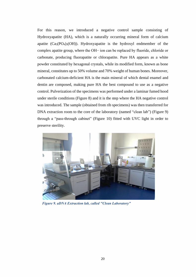
control. Pulverization of the specimens was performed under a laminar fumed hood
under sterile conditions (Figure 8) and it is the step where the HA negative control
was introduced. The sample (obtained from rib specimens) was then transferred for
DNA extraction room to the core of the laboratory (named “clean lab”) (Figure 9)
through a “pass-through cabinet” (Figure 10) fitted with UVC light in order to
preserve sterility.
Figure 9. aDNA Extraction lab, called "Clean Laboratory”
Page 28
21
In order to assess the suitability of the newly built “ad hoc” paleomicrobiology lab,
we replicated the experiments performed in the established aDNA Laboratory at the
Cultural Heritage Department of the University of Bologna, Ravenna-Campus. All
the amplifications and sequencing reactions were replicated at least twice in each
laboratory in order to authenticate the results and carefully check the mutations
found. All the steps of the analysis were conducted under strict guidelines for
contamination control and detection and reproducibility of data [Cooper & Poinar,
2000; Gilbert et al., 2005; Llamas et al., 2017].
Finally, we were able to reproduce their analysis in our laboratory and to evaluate
the authenticity of the results obtained in all the samples included in the project
[Cilli et al., 2020 article in press], meaning that our laboratory fulfilled all the
requirements for proper aDNA manipulation.
The analysis of aDNA is time-consuming and very expensive, but it can provide a
powerful tool for investigating evolutionary processes that cannot be approached
using only modern data.
Figure 10. Picture of the “pass-through cabinet” to preserve the
sterility of the specimen during manipulation steps
Page 29
22
3.2 Biological specimens’ collection and
manipulation
The study of skeletal remains recovered in a catastrophic death assemblage can
provide information about the health status of the population in different
archaeological contexts, which may be useful to answer some questions about the
plague, a still present disease in several parts of the world. This study, in
collaboration with the aDNA Laboratory at the Cultural Heritage Department of the
University of Bologna, Ravenna-Campus, was based on teeth samples (most of
which with no signs of damage) collected from three different burial site of
presumed plague victims in Italy. Three samples related to the Plague of 1576
(Table 1) were collected from the Cemetery of Lazzaretto New Island (Venice).
Figure 11. New Island Lazzaretto Venezia
Page 30
23
The analysis of the composition of the samples allowed to reconstruct the diet of
16th century venetian population. Moreover, the ratio between trace elements
indicated that the subjects belonged to different social classes, suggesting that the
flagellum of Y. pestis didn’t make any social distinction [M. Borrini, F. Bartoli et
al., 2010].
Nine samples came from the “Necropoli Tardoantica a Forum Semproni” in
Fossombrone (Figure 12), which exhibited a peculiar burial method.
Indeed, no individual burial was found at this site, instead all the corpses were
buried together in a hasty, superficial and careless manner (see Figure 13), typical
of pandemic events like the plague. In addition, based on historical events
contemporary to the burial, a lethal epidemic event presumably occurred. Such
event was characterized by a fairly large incubation period (up to 12 days for
plague) and a few days period between the clinical manifestation and the death of
the individual [M. Luni, O. Mei and P. Gobbi 2013].
Figure 12. Teeth samples collected from Fossombrone
Page 31
24
Thirteen samples were collected from “Necropoli Romana” in Modena- NOVISAD
Park (Figure 14). This burial site was found by chance in the city center of Modena
during the construction of an underground parking area. The big cemetery was
found to be constituted by several sepulchral nucleons of different ages, indeed the
most ancient burial sites dated around the 2nd century BC, while the necropolis of
the presumably plague victims dated
back to the 17th century. The burial
architecture was typical of the Christian
Romans, the only population that took
care of the sepulture of the individuals
who died from the plague instead of
incinerating them, the other treatment
to which infected individuals were
subjected. The graveyard consisted of
69 tombs arranged in several parallel
rows each housing several bodies, often
laid hurriedly one on top of the other,
both wrapped in bandages and without
Figure 13. Detail view of burial site of plague victims
Bodies are tossed from carts into hastily dug pits and covered with a layer of
dirt thin enough that animals might dig up body parts [Ranson Riggs 2010].
Figure 14. Teeth samples collected from
Modena
Page 32
25
clothing under a pile of quicklime. Some signs of burns on the bones were also
found suggesting sterilization practices even before burial, probably to avoid the
diffusion of the epidemic. The remains found are often incomplete and there are no
signs of deposition according to religious canons, despite the land being religious
property [Labate et al., 2010].
Bones and dental pulp represent the best material for research on microbial
pathogens, because their aDNA content better preserved. Before the analysis, each
sample was subjected to a decontamination procedure consisting of 45 minutes of
UVC irradiation on each side [Haensch et al., 2010]. Each lab operator had to wear
a full body suit and a new overall dedicated only to aDNA laboratory before
entering, and a second sterile overall on top of the first one every time the operator
moved to a different room.
Handling of the samples followed the guidelines for contamination precautions
described in the literature [Cooper & Pionar, 2000, Pääbo et al., 2004], consisting
of changing gloves every time in between samples, sterilization of the bench with
UVC for at least 45 minutes after sample manipulation, transfer of the samples
using a pass-through cabinet (Figure 10) between the processing room and the DNA
extraction room. Furthermore, all the equipment, benches, reagents, pipettes, scale,
plasticware, masks, helmets, were incubated for 45 min under UVC light (250 nm)
before and after usage and cleaned with bleach, which causes oxidative damage to
DNA, producing chlorinated base products [Fulton et al., 2012].
All three rooms constituting the aDNA lab were irradiated with UVC light from 21
to 6 to ensure a contamination-free environment. At the end of the sample
processing, pulverized bones were aliquoted (0.1-0.2 g) and stored at 4 °C until use.
Unfortunately, since most of the specimens were collected by external
archaeologists, we could not guarantee that the protocols for avoiding
contamination were followed during the collection of the samples.
Page 33
26
Table 1. Dataset of all samples received
ID SAMPLE_ID ITEM_ID DATA NOTE NOTE2 STANDARD DATA ESTRAZIONE
Lazzaretto Isola Nuova-VeneziaLN 1 18082150 LN08 T37 21/08/2018 T: 0,047| P1C: 0,052| P2R:0,099 CAPO S. CIS 1 SETT IV SI 23/08/2018
LN 2 18082151 LN08 T38 21/08/2018 T:0,051| P1C:0,080| P2C:0,077| P3R: 0,093 SET IV N C/O ID 15 SI 23/08/2018
LN 3 18082152 LN08 T48 21/08/2018 T:0,019| P1C:0,076 |P2R:0,093 CAMPO SANTO SETIV N CIS1 SI 23/08/2018
LN_CN
Modena NOVISAD
NP 1 18070509 NP_TB_245_US 3399 24/07/2018 PTR: 71 mg M1; M2 DESTRO E SINISTRO SI 24/07/2018
NP 2 180705010 NP_TB_245_US 3400 24/07/2018 P: 36 mg M2; M3 SINISTRI SI 24/07/2018
NP 3 180705011 NP_TB_246_US 3334 24/07/2018 P: 77 mg | R: 124 mg 2 MOLARI SI 24/07/2018
NP 4 180705012 NP_TB_246_US 3335 24/07/2018 P: 72 mg C. 1172 SI 24/07/2018
NP 5 180705013 NP_TB_247 _US 3338 24/07/2018 PC: 30 mg C. 1173 campioni aDNA SI 24/07/2018
NP 6 180705014 NP_TB_248_US 3362 24/07/2018 R: 40 mg C. 1166 campioni aDNA SI 24/07/2018
NP 7 180705015 NP_TB_255_US 3403 24/07/2018 PR: 48 mg C. 1124 campione aDNA SI 24/07/2018
NP 8 180705016 NP_TB_256_US 3407 24/07/2018 R: 78 mg M1;M2 DESTRI SI 24/07/2018
NP 9 180705017 NP_TB_257_US 3410 24/07/2018 D: 242 mg DENTE bambino (?) SI 24/07/2018
NP 10 180705018 NP_TB_259_US 3418 24/07/2018 DR: 10 mg M1;M2; M3 DESTRI SI 24/07/2018
NP 11 180705019 NP_TB_278_US 3481_IND 1 24/07/2018 PR: 37 mg 2 MOLARI SI 24/07/2018
NP 12 180705020 NP_TB_278_US 3481_IND 2 24/07/2018 P: 77 mg 1 MOLARE SI 24/07/2018
NP 13 18070521 NP_TB_280_US 3484 24/07/2018 P: 46 mg 2 PREMOLARI SI 24/07/2018
NP_CN
FossombroneFS1A; FS1B 18070501 FS_TB 3 23/07/2018 MR:_159mg | PR: 94MG_| 1 MOLARE | 1 PREMOLARE SI 24/07/2018
FS 2 18070502 FS_TB 31 23/07/2018 R: 90 mg| 1 MOLARE SI 24/07/2018
FS 3 18070503 FS_TB 34 23/07/2018 R: 189 mg| 1 MOLARE | 1 PREMOLARE SI 24/07/2018
FS 4 18070504 FS_TB 35B 23/07/2018 R 142 mg 2 PREMOLARI SI 24/07/2018
FS 5 18070505 FS_TB 114 23/07/2018 P: 103 mg 1 MOLARE | 1 PREMOLARE SI 24/07/2018
FS 6 18070506 FS_TB121 23/07/2018 P: 127 mg 1 MOLARE | 1 PREMOLARE SI 24/07/2018
FS 7 18070507 FS_TB 204_A 23/07/2018 P: 77 mg 1 MOLARE | 1 PREMOLARE SI 24/07/2018
FS 8 18070508 FS_TB 204_B 23/07/2018 P: 116 mg 2 PREMOLARI SI 24/07/2018
FS 9 18070509 FS_TB 212 23/07/2018 P: 59 mg 1 MOLARE | 1 PREMOLARE SI 24/07/2018
FS_CN
Page 34
27
3.3 DNA extraction
The most limiting factor in aDNA retrieval and analysis is the percentage of
endogenous DNA found.
The content of endogenous aDNA is less than the 1% of the total sequenced reads,
even though the rate of endogenous aDNA of different samples from the same
individual can differ by orders of magnitude [Green et al. 2010]. In addition, the
pathogens to be identified may require different extraction protocols depending on
their life cycle and the specific tissue they target. For instance, aDNA extraction of
Y. pestis or Mycobacterium leprae is often achieved from bone tissue, teeth
[Schuenemann et al., 2013, Bos et al., 2011] or dental cementum, which exhibits a
rate of endogenous DNA similar to that of the petrous bone [Hansen et al., 2017].
The petrous bone is the densest bone in the human skeleton, exhibiting the highest
rate of aDNA [Gamba et al., 2014, Pinhasi et al., 2015], although in the study of
pathogens from ancient remains its targeting is usually not an option [Margaryan et
al., 2018]. On the other hand, the presence of Mycobacterium tuberculosis is usually
assessed by extracting the DNA from ribs due to the pulmonary tuberculosis
[Bowman et al., 2012]. Pathogens that do not infect bones, such as Vibrio cholera,
can be particularly challenging to identify, as the DNA can be extracted only from
soft tissues, which is restricted to rare mummified or dissected specimens [Devault
et al., 2014].
DNA extraction protocols are typically divided in two main steps: the first is the
solubilization of DNA-bearing tissues and the consequential release of the DNA
molecules, the second consists in the purification of such DNA [Heintzman et al.,
2015]. The release of the genetic material is achieved through compounds like
proteinase K which hydrolyses collagen and Ethylenediaminetetraacetic acid
(EDTA) which demineralizes hydroxyapatite [Rohland & Hofreiter, 2007] by
chelating Ca2+ ions. DNA is then purified and separated from other organic and
inorganic molecules by silica-based methods [Rohland & Hofreiter 2007, Dabney
Page 35
28
et al., 2013, Allentoft et al., 2015], or alternatively by the less commonly used
phenol-chloroform methods [Barnett et al., 2012].
Silica-based methods can be divided between in-solution based and column-based.
In the in-solution based method the calcified tissue is digested, the DNA is
electrostatically captured by a silica pellet [Rohland & Hofreiter 2007] which is
finally washed to allow the release and recovery of the DNA. This method was
recently improved by replacing the in-solution silica with silica columns [Dabney
et al., 2013], which increased the recovery of extra-short endogenous aDNA
fragments (<80 bp) [Gamba et al., 2016] representing the larger fraction of aDNA
reads [Orlando et al., 2015]. Aliquots of 0.1-0.2 g were tested with a modified
version of 3 aDNA extraction protocols [Rohland & Hofreiter 2007, Dabney et al.,
2013, Allentoft et al., 2015]. The adapted protocol (described in detail in the
supplementary data) was based on the “The MinElute PCR Purification Kit”
commercial kit (Qiagen), which provides spin columns, buffers, and collection
tubes for silica-membrane-based purification of PCR products (70 - 4000 bp in
size), yielding large amounts of highly concentrated DNA in very small volumes
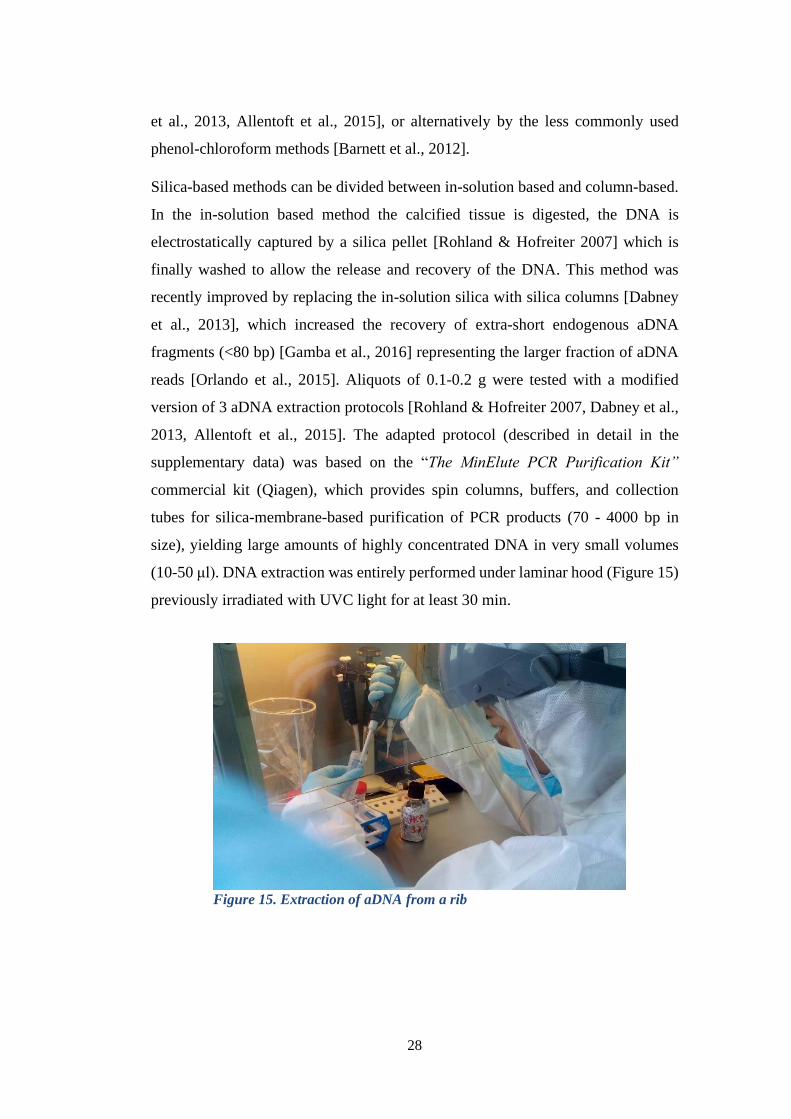
(10-50 μl). DNA extraction was entirely performed under laminar hood (Figure 15)
previously irradiated with UVC light for at least 30 min.
Figure 15. Extraction of aDNA from a rib
specimen
Page 36
29
Quantification of 1 µL extracted aDNA sample was performed on a NanoDrop™
2000 UV-Vis spectrophotometer (Thermo Scientific) according to the manufacturer
instructions. The instrument is specifically intended for small volumes and
measures the absorbance at different wavelengths to evaluate sample concentration
(via Lambert-Beer’s law) and to estimate the degree of contamination.
Indeed, the absorbance ratio at 260 and 280 nm (A260/A280) determines the presence
of proteins in the sample; the optimal value for the study of mDNA is between 2.1
and 1.8 but in the case of aDNA such ratio ranges between 1.4 and 1.8, higher values
determine the presence of protein contaminants. On the other hand, the absorbance
ratio at 260 and 230 nm (A260/A230) indicates the presence of carbohydrates and
phenols, values below 1.8 indicate the presence of contamination.
As the quantification method is absorbance-based, no distinction can be made
between modern, human and microbial aDNA, moreover, the high level of
fragmentation of aDNA makes its precise quantification particularly complex.
Nevertheless, previous studies established this analysis of the fragments as the
reference method for aDNA quantification [Brzobohatá et al., 2017].
Page 37
30
3.4 Polymerase Chain Reaction (PCR): using
“specific primers” to detect the presence of
Y. pestis
A turning point for forensic science and DNA typing laboratories is represented by
the Polymerase Chain Reaction (PCR), a technique described for the first time in
1985 by Kary Mullis, for which he was awarded with Nobel Prize in 1993. PCR
has revolutionized molecular biology, as it allowed to generate hundreds of millions
of copies of a specific sequence of DNA in only few hours. Specifically, due to the
low number of surviving aDNA molecules, no aDNA analysis would be possible
without PCR and therefore it would be impossible to reconstruct and have a clear
view of the pandemic events in the past.
PCR is an enzymatic process in which a specific region of DNA is replicated over
and over again to reproduce many copies of a particular sequence [Bulter J. M.
2012]. The amplification is done with two synthetic oligodeoxynucleotide primers,
each about 25 bases long, a thermostable DNA polymerase, and the four
deoxyribonucleotide triphosphates. PCR is an ideal tool to amplify a small number
of intact aDNA sequences present in a vast excess of damaged molecules [Pääbo et
al., 1989]. During enzymatic amplification, most damaged molecules will either not
be replicated at all, e.g. due to interior intramolecular cross-links, or will be at a
replicative disadvantage with respect to intact molecules, because lesions such as
baseless sites, slow down the DNA polymerase. Moreover, the strong inverse
correlation between amplification efficiency and the size of the amplification
product observed for aDNA, but not for modern DNA, can be employed as another
criteria for discriminating authentic aDNA from contaminating exogenous DNA.
Another critical issue to assess is the specificity of the primers used to amplify the
sequence of interest, as some doubts about the specificity of pla gene for Y. pestis
were raised [Janse et al., 2013].
Page 38
31
Here, three PCR analyses using SimpliAmp™ Thermal Cycler, were performed:
the first, based on a paper by Hänsch and colleagues [Hänsch et al., 2015], to detect
the plasminogen activator/coagulase (pla) gene, located on the pPCP1 plasmid, that
was assumed to be specific for detecting Y. pestis, but was present also in
Citrobacter koseri and Escherichia coli. The second pair of presumably specific
primer used for detection of Y. pestis [Raoult et al., 2000], were used to perform a
“suicide PCR”, that is a PCR where the couple of primers is used only for the first
cycle. In this PCR reaction there was not a positive control, so the amplicons
obtained were sequenced to confirm the presence of Y. pestis. The third PCR
reaction was performed using specific primers designed at the aDNA Laboratory of
Ravenna (Table 2). As previously stated, the samples were independently analysed
by two aDNA labs, using reagents manufactured by the same company, in detail:
AmpliTaq Gold™ DNA Polymerase with Buffer II and MgCl2 (Thermofisher)
consisting in: 5 U/µl Ampli Taq™ Gold Hot Start; 10x Gold Buffer; 25 mM MgCl2;
10 mM dNTP; 50 mg/ml Bovine Serum Albumin (BSA) as an enhancer; for each
set of primer: 10 µM Primer Fw 10 µM Primer Rv and DNAse/RNAse free H2O,
with the following cycling conditions:
Pla_1 Fw and Pla_1 Rv [Hänsch et al., 2015]
Activation: 15 min at 95 °C
Amplification (50 cycles):
• Denaturation: 30 s at 94 °C
• Annealing: 30 s at 60 °C
• Elongation: 1 min at 72 °C
Final extension: 10 min at 72 °C
Cooling and storage: 8 °C until analysis
YP12D and YP11R [Didier and Raoult, 2000]
Activation: 5 min at 95 °C
Amplification (50 cycles):
• Denaturation: 45 s at 95 °C
• Annealing: 45 s at 55 °C
• Elongation: 1 min at 72 °C
Page 39
32
Final extension: 10 min at 72 °C
Cooling and storage: 8 °C until analysis
pst_Fw and pst_Rv [Ravenna Campus]
Activation: 5 min at 95 °C
Amplification (50 cycles):
• Denaturation: 45 s at 95 °C
• Annealing: 45 s at 60 °C
• Elongation: 1 min at 72 °C
Final extension: 10 min at 72 °C
Cooling and storage: 8 °C until analysis
Name Seq 5’→3’
Annealing temperatu
re (°C)
Amplicon size (bp) Ref.
Notes
pla_1 Fw
GACTGGGTTCGGGCACATG 60
70 (33)
Hänsch et al.,
2015 qPCR (pla)
pla_1 Rv CGGATGTCTTCTCACGGA 60
70 (33)
Hänsch et al.,
2015 qPCR (pla)
YP12D CAGCAGGATATCAGGAAACA 55
148 (106)
Raoult et al., 2000
pla gene
YP11R GCAAGTCCAATATATGGCATAG 55
148 (106)
Raoult et al., 2000
pla gene
pst_Fw CTGTGGGAGCAGTTCTGGAT 60
73 (33) Ravenna Campus
pst_Rv TTGAGAACCCGTACAGCACT 60
73 (33) Ravenna Campus
Table 2. Primers used for PCR analyses
The amplicons were separated on a 1.5% agarose gel to evaluate the presence of
real aDNA, providing optimal resolution for small DNA fragments (0.2 – 1 kbp).
Page 40
33
Gel electrophoresis is a method for separation and analysis of biological
macromolecules (DNA, RNA and proteins), based on their size and charge. DNA
is a negatively charged molecule due to the presence of phosphate groups, therefore,
when immersed in an electric field, it will move towards the anode. The mobility
of the substance on a gel depends on its charge and mass, implying that a larger
strand of DNA (potentially mDNA, or contaminant) will travel a shorter distance
compared to a smaller fragment, which is what usually aDNA consists of. For a
correct identification of the amplicons we used Gene Ruler 50 bp DNA Ladder
(Thermofisher) which ensures high mass and size resolution for double-stranded
DNA in the 50 to 1000 bp range. Each amplicon was visually quantified according
to the DNA standard, finally the 70, 73 and 148 bp fragments were sent for
sequencing using traditional Sanger method [Sanger et al., 1975].
The obtained sequences were used as query strings for BLAST (Basic Local
Alignment Statistical Tool), a program that compares nucleotide or protein
sequences to sequence databases to find regions of local similarity between
sequences and calculates the statistical significance of each match. BLAST matches
can be used to infer functional and evolutionary relationships between sequences
as well as to help identifying members of gene families.
Page 41
34
3.5 Next Generation Sequencing (NGS)
Next-generation DNA sequencing (NGS) involves rapid, high-throughput
collection of short DNA sequences ranging from 25 to 250 bp. Recently, pathogen
bacteria genome sequencing has been used as an epidemiological tool to trace
contemporary outbreaks, like the cholera outbreak in Haiti in 2010 [Eppinger et al.,
2014] and the Ebola epidemy in west Africa in 2014 [Carroll et al., 2015, Gire et
al., 2014]. The majority of pathogen genome sequencing efforts have been focused
on contemporary DNA, due to the easier handling and conservation and the higher
availability of specimens with respect to aDNA. Indeed, the application of NGS to
aDNA remains a big challenge due to the high degree of degradation caused by
endonucleases and environmental factors that may result in modified bases or strand
breaks [Briggs A. W, Heyn P. 2012]. Thus, obtaining high sequencing depth and
accuracy from such samples is often difficult, as the combination of the presence of
uracil (caused by cytosine deamination) and the low copy of number of aDNA can
potentially lead to miscoding errors.
Despite all these drawbacks, NGS represented a promising technique for
unravelling aDNA due to the similarity between the length of the fragments
required for NGS and that available from aDNA samples, usually too short for
traditional sequencing techniques. Indeed, recent studies showed successful
applications of NGS to aDNA belonging to the Neanderthal mitochondrial genome
[Green et al., 2008] and the extinct woolly mammoth [Miller et al., 2008, Poinar et
al., 2006].
Page 42
35
It is still unclear whether NGS techniques may be applied to traditional forensic
DNA as proficiently as to aDNA analysis [Blow et al., 2008]. Currently, NGS
techniques cannot accurately identify repetitive sequences and thus, unless future
improvements are made, they cannot reliably deal with the short tandem repeat
regions (STR) which forensic DNA analysis is based on [Hert et al., 2008].
Moreover, the amount of data produced by NGS approaches, consisting of millions
of short reads, makes bioinformatics support crucial for forensic DNA laboratories,
which might also require to switch the analysis of genetic markers from STRs to
single nucleotide polymorphisms (SNPs) to fully exploit the potentiality of NGS.
Page 43
36
3.6 Metagenomics
Metagenomics is a branch of genomics that simultaneously studies a complex
community of microorganisms present in a sample, avoiding the growth on
selective media. Indeed, growth on culture media it allows to identify only 1-3% of
the microorganisms actually present in natural samples, losing 97-99% of the
information [Gordon, 2012; Hugenholtz et al., 1998], because of their particular
growth conditions, such as specific nutrients and anaerobic conditions.
The drawbacks of the classical microbiology techniques to identify microorganisms
can be overcome by metagenomics approaches, consisting of extracting genomic
DNA from the samples. Followed by sequencing of the 16S rRNA. In such way it
is possible not only to identify simultaneously every single microorganism
belonging to the community, but also to analyze their interaction with each other,
with the environment (microbial ecology) or with the hosting organism.
As microbial communities are involved in a variety of complex biological
processes, metagenomics can be fruitfully applied to numerous fields in order to
unravel their specific function within each context.
A great contribution was provided by Carl Woese, who in 1967 separated the
Archea and the Bacteria dominia, using molecular phylogeny techniques applied to
ribosomal RNA 16S [Woese et al., 1990, 1978 and 1977]. The molecular analysis
of the gene sequence that encodes the minor ribosomal subunit (16S) is still today
considered the most relevant sequence for the classification of Bacteria and Archea.
Page 44
37
3.7 16S rRNA Metagenomics and aDNA
16S rRNA stands for 16S Ribosomal
Ribonucleic Acid (rRNA) (Figure 16),
where S (Svedberg) is a unit of
measurement for the sedimentation rate.
The 16S rRNA encodes the small subunit
of prokaryotic ribosomal RNA and
contains nine hypervariable regions (V1-
V9) separated by ten highly conserved
regions (Figure 17).
The hypervariable regions characterize the diversity between species and allow
bacterial taxonomy studies. Several studies showed that each of the 9 hypervariable
regions provide specific information for bacterial classification [Petrosino et al.,
2009].
Figure 17. Full length of 16S rRNA gene. Adapted from
https://help.ezbiocloud.net/wp/wp-content/uploads/2017/05/16s_var_pcr.png
Figure 16. 16S rRNA subunit of prokaryotic
ribosome
Page 45
38
The 16S rRNA gene is the most used gene in microbial metataxonomic analysis
because it is conserved across members of the paraphyletic prokaryotic domains
Bacteria and Archaea [Ziesemer et al., 2015], therefore allowing the design of
“universal” primers for microbial PCR amplification, yet also sufficiently variable
to allow the classification at an approximate species level [Roh et al., 2010]. The
full-length 16S gene is usually amplified using 27F and 1492R primers, although
multiple primers on both strands are required for accurate DNA sequencing.
Archaea-specific primer sequences typically lack in specificity. The V3 region
(primers F333 5’- TCCTACGGGAGGCAGCAG-3’ and U592R 5’-
ACCGCGGCKGCTGGC-3’), was used for first time for the preparation of 16S
libraries from thermophile communities [Baker et al., 2004]. Such libraries
suggested that the V3 region was an excellent candidate for aDNA amplification
for two main reasons: i) it is the shortest 16S rRNA region (~100 bp shorter than
V4 region), ii) it exhibits high sequence heterogeneity, resulting in a good
taxonomic variability [Ziesemer et al., 2015].
Region V4 (universal primers 515F 5’-GTGYCAGCMGCCGCGGTAA-3’ and
806R28 5’-GGACTACNVGGGTWTCTAAT-3’) was considered not a suitable
choice for aDNA studies, as the highly fragmented aDNA molecules, rarely
exceeding 200 bp in length, were shorter than the entire V4 sequence (~292 bp
including primers).
To guarantee the exclusive analysis of aDNA in our samples we focused also on
the V5 region. Indeed, the V5 hypervariable region (primers F784 5’-
AGGATTAGATACCCTGGTA-3’ and R934 5’-
TGTGCGGGCCCCCGTCAATT-3’) performs well on a number of metrics: it has
very good predicted taxonomic coverage and is relatively short (144–148 bp) with
little amplicon length variation. The sequence encompassing V3 and V5 regions
(primers F333 and R934) was used for the construction of the libraries of bacterial
16S rRNA gene [Weil et al., 2017].
In addition, since the V3-V5 amplicon would be much longer than the expected
sequences for aDNA, the two regions were amplified individually, as the combined
analysis of such regions was found to be a suitable discriminant for microbial
Page 46
39
research. We analysed eight different samples from the three burial sites
presumably associated with the presence of Y. pestis (see figures in supplementary
information), and two additional samples, from a different bio-archaeological
context (Forlì) in which bisome burials were found, were considered as negative
control because they cannot be attributed to pestilential periods (see Material and
Methods). PCR reactions were performed with the same reagents as previously
described for conventional PCR, with the following thermal cycling profiles:
Amplification for V3 region of 16S gene with F333/U529R primer [Baker
et al., 2004]
Activation: 10 min at 95 °C
Amplification (25-35 cycles):
• Denaturation: 45 s at 94 °C
• Annealing: 1 min at 56 °C
• Elongation: 1 min at 72 °C
Final extension: 10 min at 72 °C
Cooling and storage: 4 °C until analysis
Amplification for V5 region of 16S gene with F784/R934 primer
Activation: 10 min at 94 °C
Amplification (35cycles):
• Denaturation: 45 s at 94 °C
• Annealing: 1 min at 55 °C
• Elongation: 1 min at 72 °C
Final extension: 10 min at 72 °C
Cooling and storage: 4 °C until analysis
Page 47
40
3.8 Preparation of NGS Libraries from aDNA
Library preparation consists of all the chemical reactions and procedures that
modify DNA fragments to meet the experimental requirements for NGS
sequencing. In detail, the DNA fragments to be sequenced are end-repaired and
ligated with universal sequencing-adaptors.
Adaptor-ligated libraries are convenient for aDNA studies because they can help
overcoming the main inconvenience of NGS which is the short reads length.
Moreover, adaptor primers outside the aDNA sequence allow the recovery of
information from molecules too short for traditional PCR. Finally, universal
adaptors provide higher amplification of the entire library before any downstream
experiments. Even though NGS presents advantages for aDNA studies, post-
mortem aDNA damage, consisting of strand-breaks and base modification (see
aDNA degradation), still represent a challenge to face.
There are no standard protocols for the preparation and NGS sequencing of aDNA
libraries, they are usually tailored on the specific requirements of each project, but
they follow the same pipeline. The first step usually consists of DNA fragmentation,
but it can be considered unnecessary for aDNA due to its intrinsic fragmentation.
Then, the ends of aDNA fragments have to be repaired and ligated with short
sequences (adapters) which will be eventually recognized by NGS platforms
(Figure 18). The reparation reaction basically consists in the degradation of the
overhanging 3’ ends and the filling of 5’ overhanging ends. Unfortunately, fragment
ends are frequently affected by one of the most common miscoding lesions in
aDNA molecules, namely cytosine deamination to uracil (resulting in C to T
transition). The elimination of deamination products on one hand reduces the
number of mismatches and the genotyping errors, but on the other hand prevents
the recognition of specific patterns used to validate the presence of aDNA.
The following step is common for both treated and non-treated reads and
represented by the adapter ligation. This process can be accomplished either using
Page 48
41
two different adapters (blunt-end ligation) to the read ends, or a single Y-shaped
adapter with a T-overhang to both ends of DNA. However, the Y-shaped adapter
requires an additional pre-processing step consisting of the addition of A-overhangs
through the so-called A-tailed ligation [Willmann et al., 2018], and may lead to the
misincorporation of T in the read ends [Seguin-Orlando 2013]. It is also
recommended to include a negative (blank) library control during library
preparation to assess the quality of the library.
Figure 18. Library preparation method for double strand libraries. The DNA is
fragmented and the reads ends are repaired by adding A bases, then the adapters are
ligated to the repaired ends. Finally reads are amplified with PCR technique (Ilumina).
Page 49
42
Finally, the DNA sequences obtained are amplified through a limited number of
PCR cycles, in order to preserve the variability of the library, moreover, the choice
of an adequate PCR polymerase is also crucial to avoid GC and read length biases
[Dabney and Meyer 2012]. To differentiate several libraries sequenced in the same
run, barcodes are attached to the adapters during the amplification process [Craig
et al., 2008, Knapp et al., 2012].
The amplicon obtained from the two PCR reactions were sent to the Paleogenetic
laboratory at University of Firenze for the library preparation and DNA sequencing
[Modi et al., 2017].
Page 50
43
3.9 Microbial taxonomic profiling
Bioinformatic analyses and microbial taxonomic profiling were conducted using
MALT (MEGAN [Huson et al., 2007] ALignment Tool) software, by aligning the
sequences obtained from teeth samples against SILVA database (https://www.arb-
silva.de/).
3.10 Phylogenetic trees
Phylogenetic trees are one of the most common representations of the biological
diversity, displaying the relations of biological samples or species using branches,
nodes and taxa. The branching pattern of a tree is define its topology, while the
group of the descendants of a node defines a clade. Clades composed by all the
descendants of a common ancestor are called monophyletic, otherwise they are
named paraphyletic.
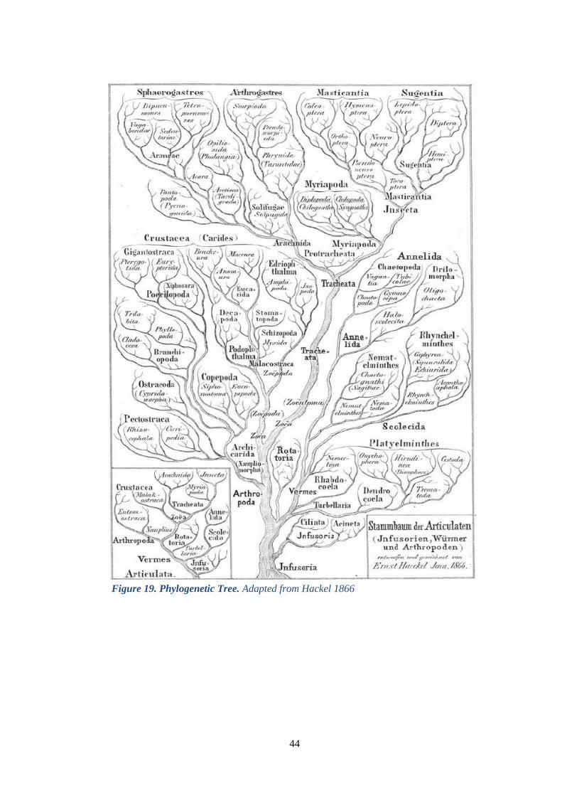
Phylogenetic trees can be built based on genetic or morphological diversity. One of
the most used criteria to build a genetic phylogenetic tree is called Maximum
Likelihood (ML), which is a statistical method for estimating unknown parameters
in a probability model. In terms of phylogeny, the likelihood represents the
probability of an observed sequence on a particular tree assuming a specific
evolutionary model (see figure 19). If the ancestor of all the taxa present in the
phylogeny is unknown, the phylogenetic tree is considered unrooted, otherwise it
is called rooted and the branches length can be interpreted as time estimates.
Moreover, if an outgroup is added, it is possible to identify the ancestral node of the
phylogeny and root it.
Page 51
44
Figure 19. Phylogenetic Tree. Adapted from Hackel 1866
Page 52
45
Chapter 4
RESULTS
4.1 aDNA extraction and purity
Three different bioarchaeological context from Italy (Venice, Fossombrone,
Modena) were taken under analyses for research of Y. pestis, the bacterium causing
the plague. The specimens were treated with extreme caution, as detailed in the
methods section, to prevent any cross-contamination with modern DNA. Extraction
and purification of aDNA was performed with MinElute PCR purification Kit
(Qiagen GmbH, Hilden, Germany), for each extraction reaction, a negative control
based on hydroxyapatite powder was introduced to detect possible DNA
contamination from exogenous sources during both pulverization and extraction
procedures.
Quantification results showed an overestimation of the amount of DNA present in
all samples, with a measured concentration range from 12.7 to 43.4 ng/µL, with an
average A260/A230 ratio ranging from 0.79 to 3.03 and an average A260/A280 ratio
range between 1.38 and 1.55. Overall, our results suggest that the samples do not
exhibit a high degree of purity, most probably due to the internal degradation of
aDNA and the exposition to environmental factors. Nevertheless, such values can
still be considered satisfactory, as they are comparable with previously reported
data on aDNA samples [Brzobohatá et al., 2017].
Page 53
46
4.2 PCR and Sanger Sequencing
All the aDNA samples were subjected to conventional PCR reactions using three
sets of supposedly “specific primers” to assess primer specificity (Figure Sx). The
first set was used detect the plasminogen activator/coagulase (pla) gene (pla_1_FW
5’-GACTGGGTTCGGGCACATG-3’, pla_1_RV 5’-
CGGATGTCTTCTCACGGA-3’) located on the pPCP1 plasmid, that was assumed
to be specific for detecting Y. pestis [Hänsch et al., 2015], but was found in
Citrobacter koseri and Escherichia coli. The second set of primers was employed
for a “suicide PCR” (YP12D 5’-CAGCAGGATATCAGGAAACA-3’, YP11R 5’-
GCAAGTCCAATATATGGCATAG-3’), where the primers were used only for the
first cycle [Raoult et al., 2000].
The third couple of primers (pst_FW 5’-CTGTGGGAGCAGTTCTGGAT, pst_RV
5’- TTGAGAACCCGTACAGCACT-3’) was specifically designed at the aDNA
Laboratory of Ravenna for this research. The amplification products were purified
with the MinElute PCR purification Kit (Qiagen GmbH, Hilden, Germany), and
sent for Sanger sequencing on both strands.
The obtained sequences were used as query strings for BLAST (Basic Local
Alignment Statistical Tool) against all non-redundant nucleotide sequences.
Unfortunately, due to shortness of the query sequences, ascribable to the high
degree of fragmentation of aDNA, the detection of Y. pestis with traditional PCR
amplification was not successful. Indeed, most of the sequenced samples consisted
only of short fragments, not informative enough to retrieve reliable information
from the databases available online. Indeed, the most frequently identified species
from BLAST analysis were environmental bacteria such as Streptomyces
bingchenggensis (strain BCW-1) and Rhodococcus hoagii (strain 103S, also called
Rhodococcus equi).
Page 54
47
S. bingchenggensis (strain BCW-1) belongs to the Streptomyces genus, consisting
of soil and water Gram positive filamentous bacteria, well known for their ability
to produce complex secondary metabolites including many antibiotics. It was
isolated in Harbin, China (China General Microbiology Culture Collection Center
CGMCC1734) and has one of the largest sequenced bacterial genomes at almost 12
Mb.
Figure 20. Streptomyces bingchenggensis (strain BCW-1) taxonomy adapted from
https://www.uniprot.org/taxonomy/749414
Page 55
48
R. hoagii (strain 103S, or R. equi) belongs to the Rhodococci genus, consisting of
aerobic, Gram positive actinomycetes characterized by a high G/C content and by
an environmental-dependent morphological differentiation (e.g., cocci or
filaments). Moreover, Rhodococci display long-term survival in soil, an exceptional
tolerance for high levels of heavy metals and a metabolic propensity towards
hydrophobic pollutants even in the presence of more readily assimilable carbon
sources, which make Rhodococci particularly suitable for bioremediation
applications [Kämpfer et al., 2014]. R. equi is an important pathogen causing
pneumonia in foals, wild boars, domestic pigs and immunocompromised humans
such as HIV-AIDS patients or transplant recipients, whose infections symptoms
resemble clinical and pathological signs of pulmonary tuberculosis.
Figure 21. Rhodococcus hoagii (strain 103S) taxonomy adapted from
https://www.uniprot.org/taxonomy/685727
Page 56
49
4.3 Metagenomics
As conventional PCR allowed the identification of only environmental bacteria, a
large number of questions still remained unanswered, such as which
bacteria/pathogens were present during particular historical periods and if the
diversity of commensal microorganisms was affected by modern diet, lifestyle and
especially environmental and climatic changes over time. We therefore analyzed
taxonomic profiles generated by amplicon sequencing in temporally and
geographically diverse archaeological teeth specimens.
We analyzed aDNA samples from 8 different subjects belonging to 3 burial sites: 2
from Modena (NP), 3 from Venice (LN), 3 from Fossombrone (FS). Two samples
collected in a burial site in Forlì (FODI) not dated back to a plague outbreak were
added to the analysis and considered as the “negative control” for Y. pestis and the
“positive control” for environmental/soil bacteria. The authenticity of aDNA was
confirmed by the presence of typical molecular signatures of post-mortem DNA
damage, such as fragmentation patterns consistent with depurination and mis-
incorporation patterns compatible with cytosine deamination within overhangs.
The relative phylum abundance analysis of both V3 and V5 regions highlighted a
considerable presence of Proteobacteria (Figures 22 and 23), a major phylum of
Gram-negative bacteria including a wide variety of pathogenic genera, such as
Escherichia, Salmonella, Vibrio, Helicobacter, Legionellales, Yersinia and many
others. Due to its large variety of genera, the Proteobacteria phylum was named
after Proteus, a Greek god of the sea capable of assuming many different shapes,
while its classification, informally called the "purple bacteria and their relatives",
was established in 1987 [Woese, 1987]. Most of the other identified phyla, such as
Acidobacteria, Actinobacteria, Chloroflexi and others, include soil bacteria
commonly found in buried specimens.
Page 57
50
Figure 22. Relative phylum abundance according to V3 region Figure 23. Relative phylum abundance according to V5 region
Page 58
51
4.3.1 Paleomicrobiome analysis on V3 region
In 16S metagenomics approaches, OTU (Operational Taxonomic Unit) are cluster
of similar sequence variants of the 16S rDNA marker gene sequence. Each of these
cluster represents a taxonomic unit of a bacteria species or genus, depending on the
sequence similarity threshold. Typically, OTU clusters are defined by a 97%
identity threshold of the 16S gene sequences to distinguish bacteria at the genus
level. Here, after deep sequencing of 16S rRNA gene V3 we described for each
specimen we the microbiome profile in terms of the different OTUs identified at
the Phylum, Class, Family and Genus taxonomic levels.
The analysis of relative abundances (Figures 22 and 23) revealed that, at phylum
level, the most representative OTUs belong to the Actinobacteria (17.3 %),
Proteobacteria (16.5 %) and Chloroflexi (13.1 %), followed by Acidobacteriota (9.7
%), Bacteroidota (5.8 %), Firmicutes (5 %), Gemmatimonadota (4 %) and
Patescibacteria (3.1 %). Cyanobacteria, Dadabacteria, Deinococcota,
Dependentiae, Desulfobacterota, Elusimicrobiota, Entotheonellaeota,
Euryarchaeota, Fibrobacterota, Verrucomicrobiota, Nitrospira, Dependentiae,
Elusimicrobiota, Spirochaetota and other non-classifiable were found in very lower
frequency (0.1%-2.7%).
A closer inspection of the prevalence at class level (Figure 24) highlighted that the
most abundant classes were Actinobacteria, Alphaproteobacteria, Bacilli,
Acidobacteriae, together with Clostridia and Gammaproteobacteria, particularly
interesting as they include several potential pathogens.
Page 59
52
Indeed, Clostridia is a class of ubiquitous strictly anaerobic to aerotolerant spore-
forming bacilli, usually found in soil as well as in normal intestinal flora of humans
and animals. This class includes both Gram-positive and Gram-negative species,
although the majority of isolates are Gram-positive. Clostridial wound infections
are typically polymicrobic, where the clostridial species, including C. perfringens,
C. novyi, C. septicum, and others [Wells and Wilkins 1996], represent the primary
pathogens.
Gammaproteobacteria is the class of Vibrionales, inhabitants of fresh or saltwater,
which include several pathogenic species such as Vibrio cholerae, the agent
responsible for cholera. Moreover, most bioluminescent bacteria belong to this
family, typically found as symbionts of deep-sea animals [Devault et al., 2014].
Gammaproteobacteria also the class of Legionellales, consisting of the Legionella
and Coxiella families, both of which include notable pathogens. Indeed,
legionellosis (acute pneumonia), is usually caused by Legionella pneumophila,
although potentially any Legionella species may be responsible for such disease.
Less often, legionellosis presents as a non-pneumonic, epidemic, influenza-like
Figure 24. Relative class abundance according to V3 region
Page 60
53
illness called Pontiac fever, while extrapulmonary Legionella infections (e.g.,
pericarditis and endocarditis) are rare. Legionella was first recovered from the
blood of a soldier more than 50 years ago, but its importance as a human pathogen
was not recognized until 1976, when a mysterious epidemic of pneumonia struck
members of the Pennsylvania American Legion [Winn WC Jr. 1996].
Finally, Gammaproteobacteria includes also the class of Yersina pestis (formerly
Pasteurella pestis), the cocco-bacillus responsible for plague outbreaks, which was
isolated and described by Yersin in 1894 [Yersin and Treille. 1894, Collins. 1996].
These results encouraged us to proceed with a deeper examination in order to
classify also family and genus.
Page 61
54
Figure 25. Phylogenetic tree based on V3 region of Forlì (FODI) cemetery at phylum, family and genus levels
Page 62
55
Figure 26. Phylogenetic tree based on V3 region of Fossombrone (FS) burial site at phylum, family and genus levels
Page 63
56
Figure 27. Phylogenetic tree based on V3 region of Modena cemetery (NP) at phylum, family and genus levels
Page 64
57
Figure 28. Phylogenetic tree based on V3 region of New Lazarette in Venice (LN) at phylum, family and genus levels
Page 65
58
We reconstituted the phylogenetic tree for each burial site independently, focusing
on potential pathogenic bacteria that could represent the cause or co-cause of death.
As previously described, the cemetery in Forlì (FODI, Figure 25) was considered
as a negative control for Y. pestis identification but a positive control for aDNA
analyses and soil bacteria investigation. Results showed that the most abundant
phyla present in FODI samples were Proteobacteriota (31.3%), Actinobacteriota
(18.8%), Acidobacteriota (14.1%), Nitrospirota (7.8%), Gemmatimonadota
(7.8%), Chloroflexi (6.3%) and Methylomirabilota (6.3%), all normally found in
soil and water. Unfortunately, due to the degradation and fragmentation of aDNA,
most of the samples family and genus could not be identified (Figure 25).
Nonetheless, the most prevalent family of the Nitrospiriota phylum was that of the
Nitrospiraceae (13.5%), whereas the most frequent families belonging to
Proteobacteria phylum were Gemmatimonadaceae (13.5%) and
Nitrosomonadaceae (10.8%), while other families like Hyphomicrobiaceae,
Xanthobacteriaceae, Steroidobacteriaceae, Dongiaceae displayed a very low
frequency. Finally, also at the genus level, the most prevalent genera identified were
Nitrospira, Dongia, Steroidobacter and Hyphomicrobium, all microorganisms
normally present in soil and water.
The results for the samples belonging to Fossombrone (FS) burial site (Figure 26)
show more homogenic phyla, with the most prevalent being Actinobacteriota
(40.9%), Proteobacteria (34.1%), and Firmicutes (9.1%), followed by
Acidobacteriota, Gemmatimonadota, Chloroflexi and Bacteroidota, all showing a
much lower frequency (3.4% to 4.5%).
Among the Actinobacteriota the Nocardiodaceae, Micrococcaceae,
Pseudonocardaceae families were identified, and in particular the Kribbella [Urzì
et al., 2008] and Blastococcus [Hezbri et al., 2016] genera, whose first discovery
was related to catacombs. Interestingly, the Bacillaceae and Clostridiaceae
families, belonging to the phylum of Firmicutes were also identified.
The Proteobacteria phylum included families of nitrogen-fixing (diazotrophic)
bacteria such as Oxalobacteraceae, Rhizobiaceae, Sphingomonadaceae,
Nitrosomonadaceae, present in various habitats like water, soil, and plant-
Page 66
59
associated environments. Some species/strains belonging to the Oxalobacteraceae
family are mild plant pathogens or are claimed to be opportunistic human pathogens
[Baldani et al., 2014].
One of the most interesting genera found in all samples from Fossombrone was
Brucella, whose infection results in Brucellosis (also known as undulant fever,
Malta fever, or Mediterranean fever [Di Pierdomenico et al., 2011]), a highly
contagious zoonosis caused by ingestion of unpasteurized milk or undercooked
meat from infected animals, or by close contact with their secretions.
Another potential pathogen identified was the Clostridium_sensu_stricto_18 genus,
which was also recently found in the microbiome of HIV-positive patient [Ahmed
et al., 2020]. Unfortunately, no deeper investigation could be performed about the
species of such potential pathogens due to aDNA degradation. We can though
hypothesize that Fossombrone subjects died because of Brucella infection, rather
than from a Y. pestis infection, as it was not identified, moreover we can also
exclude an infection caused by Clostridium, because it was found in only 2 out of
the 3 samples analysed.
As far as Modena (NP) samples are concerned (see figure 27), the Proteobacteria,
Chloroflexi, Firmicutes and Actinobacteria were the four most abundant phyla,
accounting for 32.8 %, 14.8 %, 13.1 % and 11.5% respectively. Again, a deeper
investigation at family and genus level of Proteobacteria, Chloroflexi and
Actinobacteria confirmed the presence of several groups normally found in soil and
water, such as Nocardioides, Nitrospira, Dongia, Flavihumbacterium.
Although both samples belonging to Modena burial site displayed the presence of
Clostridium_sensu_stricto_15 of the Clostridiaceae family, no conclusions could
be drawn regarding potential diseases attributed to this genus as the fundamental
information about the specie was missing. Modena samples also presented
Fretibacterium genus, isolated from subgingival plaque, suggesting that the
samples were overall handled properly.
Finally, the most relevant phyla found in samples belonging to the New Lazarette
(LN) of New Island in Venice (Figure 28) were Proteobacteria (44.3%) and
Page 67
60
Actinobacteriota (28.3%), while all other phyla showed very low frequency.
Concerning family classification, Burkholderiaceae and Clostridiaceae were the
most interesting families, detected in 3 and 2 samples, respectively, while at genus
level the presence of the Nonomurieae suggested that the samples underwent
several climatic and atmospheric variations, as a generic abundance of such genus
was found to be dependent on geographical changes [Sungthong and Nakaew
2015]. Similarly, to Fossombrone, also the catacombs-associated genus Kribbella
was detected in 2 out of 3 samples. Unfortunately, none of the identified genera
could be associated with any possible cause of death.
Page 68
61
4.3.2 Paleomicrobiome analysis on V5 region
The same analysis presented for V3 region was performed on the other informative
region of the 16S-rRNA sequence, that is the V5 region. The analysis of relative
abundances revealed that the most abundant phyla were Proteobacteria (27 %),
Actinobacteria (13.7 %), Acidobacteriota (7.2 %), Bacteroidota (6.7%),
Plancomycetota (6.6%) and Firmicutes (6.3%), while all other phyla, such as
Verrucomicrobiota displayed an abundance lower than 5%.
The prevalence analysis at class level (Figure 29) highlighted the presence of
several Gram-positive and Gram-negative bacteria particularly relevant from a
clinical microbiology standpoint. Indeed, the largest fraction of classes was related
to Gram-negative bacteria, represented by Gammaproteobacteria and
Alphaproteobacteria, while the Gram-positive bacteria were represented by
Clostridia and Bacilli classes. It is worth mentioning, though, that the Bacilli class
includes not only Gram-positive families like Staphylococcaceae,
Streptococcaceae and Listeria, but also for example Escherichia coli, which is a
Gram-negative bacteria.
Figure 29. Relative phylum abundance according to V5 region
Page 69
62
Figure 30. Phylogenetic tree based on V5 region of Forlì (FODI) cemetery at phylum, family and genus levels
Page 70
63
Figure 31. Phylogenetic tree based on V5 region of Fossombrone (FS) burial site at phylum, family and genus levels
Page 71
64
Figure 32. Phylogenetic tree based on V5 region of Modena cemetery (NP) at phylum, family and genus levels
Page 72
65
Figure 33. Phylogenetic tree based on V5 region of New Lazarette in Venice (LN) at phylum, family and genus levels
Page 73
66
The phylogenetic tree concerning Forlì burial site (Figure 30), our “negative
control” for infectious diseases research, showed that the most prevalent phyla
Proteobacteria (33.3%), Actinobacteria (19 %) and Gemmatimonadota (12.7%),
while Firmicutes, Acidobacteriota and others were detected in a much lower
frequency. At a deeper level, the only potential pathogenic families found were
Burkholderiaceae, Nocardiaceae and Clostridiaceae, but none of the identified
genera could be associated with infectious diseases.
The most prevalent phyla from Fossombrone (Figure 31) were Actinobacteria
(39.8%), followed by Proteobacteria (36.9%) and Firmicutes (6.8%), all other
phyla displayed a frequency lower than 5%. Again, the potentially pathogenic
families found were Bacillaceae, Burkholdeiaceae, Clostridiaceae,
Nocardiodaceae, while at genus level we detected again several genera typically
found in catacombs such as Kribella and Blastococcus. Interestingly, Bacillus was
the most prevalent genus, including both free-living (nonparasitic) species and two
parasitic pathogenic species, namely B. anthracis, responsible for anthrax, and B.
cereus, associated with food poisoning.
Modena burial site presented a highly conserved phylogenetic tree in terms of
grade/branches of phyla (Figure 32), with the most frequent being Proteobacteria
(42.9%), Actinobacteria (27 %) and Firmicutes (15.9 %). At family level, several
branches of Bacillaceae were detected, followed by Burkholderiaceae,
Nocardiodaceae and Clostridiaceae, while at genus level, the most frequently
found was the Bacillus genus, similarly to what was observed in Fossombrone
samples.
In line with all other burial sites, Proteobacteria (37%), Actinobacteria (33%) and
Bacteroidota (6.5%) were the most prevalent phyla found in the lazarette of New
Island in Venice (Figure 33). The investigation at family level highlighted the
presence of only the Clostridiaceae family, but no more precise information about
genus could be retrieved from aDNA samples, implying that no potential cause of
an infectious disease could be identified.
Page 74
67
Chapter 5
CONCLUSIONS AND FUTURE
PERSPECTIVES
Ancient DNA research is a complicate and expensive discipline to approach, but
the continue development of sophisticated analytical techniques such as NGS,
Metagenomics and WGS allowed to achieve a considerable success in studying
ancient pathogens. Although aDNA analysis was established as the gold standard
for unravelling the evolution of bacteria, such approach has to deal with critical
problems like the retrieval, detection and characterization of aDNA molecules.
Moreover, the overwhelmingly higher presence of modern DNA and PCR products
in all the steps of the specimen handling can lead to potential contaminations.
However, when all possible precautions are taken, the constitution of an aDNA
repository can also provide important additional information on post-mortem DNA
base modification, particularly on the interpretation and update of ancient genomes
datasets [Der Sarkissian C et al., 2015].
Metagenomic analysis is facing a wide range of challenges, in particular the
comparison of huge amounts of sequencing data against a steadily increasing
number of reference sequences. While the overall composition can be characterized
even without precise alignments, more sophisticated analyses require the
assessment of complete alignments of the metagenomic sequencing reads against a
comprehensive reference database.
Indeed, 16S rRNA amplicon sequencing has been the primary tool for
characterizing ancient microbiome samples since 1998, yet no study has
systematically investigated the effect of aDNA fragmentation on the fidelity of
amplicon-based ancient microbiome reconstructions. Such issue results in an
Page 75
68
unclear interpretation of the differences observed between modern and ancient
microbial communities. Overamplification of archaeal taxa and altered microbial
diversity estimates are predictable artifacts observed in poorly preserved (highly
fragmented) but relatively uncontaminated aDNA samples.
We demonstrated that although amplicon-based 16S rRNA gene sequencing may
be a useful high-throughput screening tool for qualitative characterization of the
preservation and contamination of ancient microbiome samples, it cannot be used
to reliably reconstruct qualitative information about microbial diversity. In addition,
always due to the high fragmentation of DNA, no quantification of the taxonomic
frequency in ancient microbial communities cannot be obtained, resulting in the
impossibility to identify the precise species of microorganisms that could have been
the cause of death.
The most abundant taxon in all burial sites resulting from the investigation of both
V3 and V5 regions of the 16S rRNA was Proteobacteria, more specifically the class
of Gammaproteobacteria, which includes several clinically relevant microbial
genera such as Legionella, Vibrio and Yersinia.
For the samples belonging to Fossombrone, a possible cause of death was found in
Brucellosis [Kay GL et al., 2014; Mutolo MJ et al., 2012], as Brucella was detected
in this burial site, moreover also aDNA related to Bacillus genus was found in both
V3 and V5 analysis, suggesting that probably this cemetery was erroneously
associated with the plague.
Unfortunately, no traces of Y. pestis DNA were found in the teeth samples either in
V3 or in V5 region. We also showed that the putatively specific primers were not
as specific as expected, since the atmospheric and soil condition may have
contributed to the accumulation of mutations in the targeted regions, leading to their
a-specificity. Besides, the evolutionary and epidemic history of this bacterium
could not be easily addressed due to the degradation of endogenous DNA of Y.
pestis, which may have been present at that time but lost over time.
As reported in Y. pestis related literature, a single small genetic mutation
fundamentally influenced the evolution of the deadly pathogen, and thus the course
Page 76
69
of human history, and such single variant may have not survived the passing of
time.
On the other hand, the metagenomic analysis revealed to be successful in
identifying the variability of bacteria found in “a closed box” as the tooth is defined.
Finally, teeth samples were found to be much more informative than any other bone
belonging to the same individual, in terms of microbiome analysis.
Page 77
70
Chapter 6
PUBLICATIONS LIST
Publications related to aDNA research
• Bazaj A, Turrina S, De Leo D, Cornaglia G. Palaeomicrobiology meets
forensic medicine: time as a fourth-dimension for the crime scene. New
Microbes New Infect. 2015;4:5–6. Published 2015 Jan 12.
doi:10.1016/j.nmni.2014.12.006
• Saegeman V, Cohen MC, Alberola J, et al. How is post-mortem
microbiology appraised by pathologists? Results from a practice survey
conducted by ESGFOR. Eur J Clin Microbiol Infect Dis.
2017;36(8):1381–1385. doi:10.1007/s10096-017-2943-6
• Drancourt M, Barbieri R, Cilli E, et al. Did Caravaggio die of
Staphylococcus aureus sepsis?. Lancet Infect Dis. 2018;18(11):1178.
doi:10.1016/S1473-3099(18)30571-1
• Cilli E, Gabanini G, Ciucani M M, et al. 2020 Investigating childbirth
deaths in double burials: Anthropology, paleopathology and ancient
DNA. In press in Journal of Archaeological Science
Page 78
71
Other publications
• Fan X, Wu Y, Xiao M, et al. Diverse Genetic Background of Multidrug-
Resistant Pseudomonas aeruginosa from Mainland China, and Emergence
of an Extensively Drug-Resistant ST292 Clone in Kunming. Sci Rep.
2016;6:26522. Published 2016 May 20. doi:10.1038/srep26522
• Mazzariol A, Bazaj A, Cornaglia G. Multi-drug-resistant Gram-negative
bacteria causing urinary tract infections: a review. J Chemother.
2017;29(sup1):2–9. doi:10.1080/1120009X.2017.1380395
• Bazaj A, Bombiero E, Naso L D, Lo Cascio G, Cornaglia G. Fighting
antibiotic resistance, it’s in your hands: mobile phones are a fertile ground
for microorganisms’ growth. Published 2019 Journal Current trends in
Microbiology
• Brandi J, Di Carlo C, Manfredi M, et al. Investigating the Proteomic Profile
of HT-29 Colon Cancer Cells After Lactobacillus kefiri SGL 13 Exposure
Using the SWATH Method. J Am Soc Mass Spectrom. 2019;30(9):1690–
1699. doi:10.1007/s13361-019-02268-6
• Piccirilli A, Perilli M, Piccirilli V, et al. Molecular characterization of
carbapenem-resistant Klebsiella pneumoniae ST14 and ST512 causing
bloodstream infections in ICU and surgery wards of a tertiary university
hospital of Verona (northern Italy): co-production of KPC-3, OXA-48, and
CTX-M-15 β-lactamases. Diagn Microbiol Infect Dis. 2020;96(3):114968.
doi:10.1016/j.diagmicrobio.2019.114968
• Brandi J, Cheri S, Manfredi M, Di Carlo C, Federici F, Bombiero E, Bazaj
A, Rizzi E, Manna L, Cornaglia G, Marini U, Valenti M T, Marengo E,
Cecconi D. Exploring the Wound Healing, Anti-Inflammatory, Anti-
Pathogenic and Proteomic Effects of Lactic Acid Bacteria on Keratinocytes.
Submitted to Scientific Reports
Page 79
72
Bibliography
Achtman M, Zurth K, Morelli G, Torrea G, Guiyoule A, Carniel E, et al. Yersinia pestis,
the cause of plague, is a recently emerged clone of Yersinia pseudotuberculosis. Proc
Natl Acad Sci USA. 1999;96(24):14043–14048.
Allentoft ME, Sikora M, Sjögren KG, Rasmussen S, Rasmussen M, Stenderup J, et al.
Population genomics of Bronze Age Eurasia. Nature. 2015;522(7555):167-172.
Andam CP, Worby CJ, Chang Q, Campana MG. Microbial Genomics of Ancient Plagues
and Outbreaks. Trends Microbiol. 2016;24(12):978–990.
doi:10.1016/j.tim.2016.08.004
Baker GC, Cowan DA. 16 S rDNA primers and the unbiased assessment of thermophile
diversity. Biochem Soc Trans. 2004;32(Pt 2):218–221. doi:10.1042/bst0320218
Barnett R, Larson G. A phenol-chloroform protocol for extracting DNA from ancient
samples. Methods Mol Biol. 2012;840:13–19. doi:10.1007/978-1-61779-516-9_2
Bazaj A, Turrina S, De Leo D, Cornaglia G. Palaeomicrobiology meets forensic
medicine: time as a fourth-dimension for the crime scene. New Microbes New Infect.
2015;4:5–6. Published 2015 Jan 12. doi:10.1016/j.nmni.2014.12.006
Bennett EA, Massilani D, Lizzo G, Daligault J,Geigl EM, Grange T. Library construction
for ancient genomics: single strand or double strand? BioTechniques. 2014;56:289–
298.
Blow MJ, Zhang T, Woyke T, et al. Identification of ancient remains through genomic
sequencing [published correction appears in Genome Res. 2008 Nov;18(11):1859].
Genome Res. 2008;18(8):1347–1353. doi:10.1101/gr.076091.108
Borrini M, Bartoli F. Analisi paleonutrizionale su alcuni campioni dalla mass grave
dell’Isola del Lazzaretto Nuovo (Venezia) Archivio per l’Antropologia e la Etnologia
Vol. CXL 2010
Bos KI, Herbig A, Sahl J, et al. Eighteenth century Yersinia pestis genomes reveal the
long-term persistence of an historical plague focus. Elife. 2016;5:e12994. Published
2016 Jan 21. doi:10.7554/eLife.12994
Bos KI, Schuenemann VJ, Golding GB, Burbano HA, Waglechner N, Coombes BK,
McPhee JB, DeWitte SN, Meyer M, Schmedes S, Wood J, Earn DJ, Herring DA,
Bauer P, Poinar HN, Krause J. A draft genome of Yersinia pestis from victims of the
Black Death. Nature. 2011 Oct 12;478(7370):506-10. doi: 10.1038/nature10549.
Erratum in: Nature. 2011 Dec 8;480(7376):278.
Bouwman AS, Kennedy SL, Müller R, Stephens RH, Holst M, Caffell AC, et al.
Genotype of a historic strain of Mycobacterium tuberculosis. Proc Natl Acad Sci U S
A. 2012;109(45):18511-18516.
Briggs AW, Heyn P. Preparation of next-generation sequencing libraries from damaged
DNA. Methods Mol Biol. 2012;840:143–154. doi:10.1007/978-1-61779-516-9_18
Page 80
73
Briggs AW, Stenzel U, Johnson PL, et al. Patterns of damage in genomic DNA sequences
from a Neandertal. Proc Natl Acad Sci U S A. 2007;104(37):14616–14621.
doi:10.1073/pnas.0704665104
Carpenter ML, Buenrostro JD, Valdiosera C, Schroeder H, Allentoft ME, Sikora M, et al.
Pulling out the 1%: Whole-Genome Capture for the Targeted Enrichment of Ancient
DNA Sequencing Libraries. Am J Hum Genet. 2013;93(5): 852–864.
Carroll MW, Matthews DA, Hiscox JA, et al. Temporal and spatial analysis of the 2014-
2015 Ebola virus outbreak in West Africa. Nature. 2015;524(7563):97–101.
doi:10.1038/nature14594
Cilli E, Gabanini G, Ciucani M.M, De Fanti S, Serventi P, Bazaj A, Sarno S, Ferri G,
Fregnani A, Cornaglia G, Gruppioni G, Luiselli D, Traversari M. A multifaceted
approach towards investigating childbirth deaths in double burials: Anthropology,
paleopathology and ancient DNA. Submitted to Journal of Archaeological Science.
Cohn SK Jr. Epidemiology of the Black Death and successive waves of plague. Med Hist
Suppl. 2008;(27):74-100
Collins FM. Pasteurella, Yersinia, and Francisella. In: Baron S, editor. Medical
Microbiology. 4th edition. Galveston (TX): University of Texas Medical Branch at
Galveston; 1996. Chapter 29. Available from:
https://www.ncbi.nlm.nih.gov/books/NBK7798/
Cooper A, Poinar HN. Ancient DNA: do it right or not at all. Science.
2000;289(5482):1139. doi:10.1126/science.289.5482.1139b
Cowan DA, Arslanoglu A, Burton SG, et al. Metagenomics, gene discovery and the ideal
biocatalyst. Biochem Soc Trans. 2004;32(Pt 2):298–302. doi:10.1042/bst0320298
Cui Y, Yu C, Yan Y, Li D, Li Y, Jombart T, et al. Historical variations in mutation rate in
an epidemic pathogen, Yersinia pestis. Proc Natl Acad Sci USA. 2013; 110(2):577–
582.
Dabney J, Meyer M, Pääbo S. Ancient DNA damage. Cold Spring Harb Perspect Biol.
2013;5(7):1–7.
Der Sarkissian C, Allentoft ME, Ávila-Arcos MC, et al. Ancient genomics. Philos Trans
R Soc Lond B Biol Sci. 2015;370(1660):20130387. doi:10.1098/rstb.2013.0387.
Devault AM, Golding GB, Waglechner N, Enk JM, Kuch M, Tien JH, et al. Second-
pandemic strain of Vibrio cholerae from the Philadelphia cholera outbreak of 1849. N
Engl J Med. 2014;370(4):334-340.
Di Pierdomenico A, Borgia SM, Richardson D, Baqi M (2011). "Brucellosis in a returned
traveller". CMAJ. 183 (10): E690-2. doi:10.1503/cmaj.091752. PMC 3134761. PMID
21398234
Drancourt M and Raoult D. Paleomicrobiology of Humans. ISBN-13: 978-1555819163.
First publication 2016
Page 81
74
Drancourt M, Aboudharam G, Signoli M, Dutour O, Raoult D. Detection of 400-year-old
Yersinia pestis DNA in human dental pulp: an approach to the diagnosis of ancient
septicemia. Proc Natl Acad Sci USA. 1998;95(21):12637–12640.
Drancourt M, and Raoult D. Cause of Black Death. Lancet Infect Dis. 2002
Aug;2(8):459.
Drancourt M, and Raoult D. Palaeomicrobiology: current issues and perspectives. Nature
Reviews Microbiology. 2005;3(1):23-55.
Eppinger M, Pearson T, Koenig SS, et al. Genomic epidemiology of the Haitian cholera
outbreak: a single introduction followed by rapid, extensive, and continued spread
characterized the onset of the epidemic. mBio. 2014;5(6):e01721. Published 2014
Nov 4. doi:10.1128/mBio.01721-14
Fu Q, Meyer M, Gao X, Stenzel U, Burbano HA, Kelso J, et al. DNA analysis of an early
modern human from Tianyuan Cave, China. PNAS. 2013;110(6): 2223-2227.
Fu Q, Posth C, Hajdinjak M, Petr M, Mallick S, Fernandes D, et al. The genetic history of
Ice Age Europe. Nature. 2016;534(7606):200-205.
Fulton TL, Stiller M. PCR amplification, cloning, and sequencing of ancient DNA.
Methods Mol Biol. 2012;840:111–119. doi:10.1007/978-1-61779-516-9_15
Gamba C, Hanghøj K, Gaunitz C, Alfarhan AH, Alquraishi SA, Khaled AS, et al.
Comparing the performance of three ancient DNA extraction methods for high-
throughput sequencing. Mol Ecol Resour. 2016;16:459–469.
Gamba C, Jones ER, Teasdale MD, McLaughlin RL, Gonzalez-Fortes G, Mattiangeli V,
et al. Genome flux and stasis in a five millennium transect of European prehistory.
Nat Commun. 2014;5:5257.
Gansauge MT, Meyer M. Single-stranded DNA library preparation for the sequencing of
ancient or damaged DNA. Nat Protoc. 2013;8(4):737–748.
doi:10.1038/nprot.2013.038
Gernaey AM, Minnikin DE, Copley MS, Dixon RA, Middleton JC, Roberts CA. Mycolic
acids and ancient DNA confirm an osteological diagnosis of tuberculosis.
Tuberculosis. 2001;81:259–265.
Gilbert MTP, Hansen AJ, Willerslev E, Turner-Walker G, Collins M. Insights into the
processes behind the contamination of degraded human teeth and bone samples with
exogenous sources of DNA. Int J Osteoarchaeol. 2006;16:156–164.
Gilbert MTP, Rudbeck L, Willerslev E, Hansen AJ, Smith C, Penkman KEH, et al.
Biochemical and physical correlates of DNA contamination in archaeological human
bones and teeth excavated at Matera, Italy. J Arch Sci. 2005;32:785–793.
Gire SK, Goba A, Andersen KG, et al. Genomic surveillance elucidates Ebola virus
origin and transmission during the 2014 outbreak. Science. 2014;345(6202):1369–
1372. doi:10.1126/science.1259657
Green RE, Krause J, Briggs AW, Maricic T, Stenzel U, Kircher M, et al. A draft sequence
of the Neandertal genome. Science. 2010;328(5979):710-722.
Page 82
75
Green RE, Malaspinas AS, Krause J, Briggs AW, Johnson PLF, Uhler C, et al. A
complete Neandertal mitochondrial genome sequence determined by high-throughput
sequencing. Cell. 2008; 134(3):416–426.
Hänsch S, Cilli E, Catalano G, et al. The pla gene, encoding plasminogen activator, is not
specific to Yersinia pestis. BMC Res Notes. 2015;8:535. Published 2015 Oct 5.
doi:10.1186/s13104-015-1525-x
Hansen HB, Damgaard PB, Margaryan A, et al. Comparing Ancient DNA Preservation in
Petrous Bone and Tooth Cementum. PLoS One. 2017;12(1):e0170940. Published
2017 Jan 27. doi:10.1371/journal.pone.0170940
Harbeck M, Seifert L, Hänsch S, et al. Yersinia pestis DNA from skeletal remains from
the 6(th) century AD reveals insights into Justinianic Plague. PLoS Pathog.
2013;9(5):e1003349. doi:10.1371/journal.ppat.1003349
Heintzman PD, Zazula GD, Cahill JA, Reyes AV, MacPhee RDE, Shapiro B, et al.
Genomic Data from Extinct North American Camelops Revise Camel Evolutionary
History. Molecular Biology and Evolution. 2015;32(9):2433–2440.
Hert DG, Fredlake CP, Barron AE. Advantages and limitations of next-generation
sequencing technologies: a comparison of electrophoresis and non-electrophoresis
methods. Electrophoresis. 2008;29(23):4618–4626. doi:10.1002/elps.200800456
Hezbri, K; Louati, M; Nouioui, I; Gtari, M; Rohde, M; Spröer, C; Schumann, P; Klenk,
HP; Ghodhbane-Gtari, F; Montero-Calasanz, MD (November 2016). "Blastococcus
capsensis sp. nov., isolated from an archaeological Roman pool and emended
description of the genus Blastococcus, B. aggregatus, B. saxobsidens, B. jejuensis
and B. endophyticus". International Journal of Systematic and Evolutionary
Microbiology. 66 (11): 4864–4872. doi:10.1099/ijsem.0.001443. PMID 27553620.
Higuchi R, Bowman B, Freiberger M, Ryder OA, Wilson AC.DNA sequences from the
quagga, an extinct member of the horse family. Nature. 1984;312(5991):282-284.
Hofreiter M, Serre D, Poinar HN, Kuch M, Pääbo S. Ancient DNA. Nat Rev Genet.
2001;2(5):353–359. doi:10.1038/35072071
Höss M, Jaruga P, Zastawny TH, Dizdaroglu M, Pääbo S. DNA damage and DNA
sequence retrieval from ancient tissues. Nucleic Acids Res. 1996;24(7): 1304–1307.
Höss M, Pääbo S. DNA extraction from Pleistocene bones by a silica-based purification
method. Nucleic Acids Res. 1993; 21:3913–3914.
Huson, D. H., Auch, A. F., Qi, J., & Schuster, S. C. (2007). MEGAN analysis of
metagenomic data. Genome research, 17(3), 377-386.
Janse I, Hamidjaja RA, Reusken C. Yersinia pestis plasminogen activator gene homolog
in rat tissues. Emerg Infect Dis. 2013;19(2):342–344. doi:10.3201/eid1902.120659
Kämpfer P, Dott W, Martin K, Glaeser SP. Rhodococcus defluvii sp. nov., isolated from
wastewater of a bioreactor and formal proposal to reclassify [Corynebacterium
Page 83
76
hoagii] and Rhodococcus equi as Rhodococcus hoagii comb. nov. Int J Syst Evol
Microbiol. 2014;64(Pt 3):755–761. doi:10.1099/ijs.0.053322-0
Kay GL, Sergeanta MJ, Giuffra V, Bandiera P, Milanesed M, Bramanti B, et al. Recovery
of a Medieval Brucella melitensis Genome Using Shotgun Metagenomics. mBio.
2014;5 (4): e01337-14. Published 2014 Jul 15. Doi: 10.1128/mBio.01337-14
Keller A, Graefen A, Ball M, Matzas M, Boisguerin V, Maixner F, et al. New insights
into the Tyrolean Iceman's origin and phenotype as inferred by whole-genome
sequencing. Nat Commun. 2012;3(698).
Kolman CJ, Centurion-Lara A, Lukehart SA, Owsley DA, Tuross N. Identification of
Treponema pallidum subspecies pallidum in a 200-year-old skeletal specimen. J
Infect Dis. 1999;180(6):2060–2063.
Kousoulis AA, Economopoulos KP, Poulakou-Rebelakou E, Androutsos G, Tsiodras S.
The plague of Thebes, a historical epidemic in Sophocles' Oedipus Rex. Emerg Infect
Dis. 2012;18(1):153–157. doi:10.3201/eid1801.AD1801
Labate D, Malnati L, et al., 2017 PARCO NOVI SAD DI MODENA: DALLO SCAVO
AL PARCO ARCHEOLOGICO. Archeologia, antropologia, storia e ambiente di un
insediamento periurbano di età romana e medievale. Ministero dei Beni Culturali e il
Turismo.
Lazaridis I, Patterson N, Mittnik A, Renaud G, Mallick S, Kirsanow K, et al. Ancient
human genomes suggest three ancestral populations for present-day Europeans.
Nature. 2014;513(7518):409-413.
Lindahl T. Instability and decay of the primary structure of DNA. Nature.
1993;362(6422):709-715.
Llamas B, Willerslev E, Orlando L. Human evolution: a tale from ancient genomes.
Philos Trans R Soc Lond B Biol Sci. 2017;372(1713):20150484.
doi:10.1098/rstb.2015.0484
Luni M, Mei O, Gobbi P. Necropoli Tardoantica a Forum Sempronii cum bibl. 2013
Margaryan A, Hansen HB, Rasmussen S, Sikora M, Moiseyev V, Khoklov A, et al.
Ancient pathogen DNA in human teeth and petrous bones. Ecol Evol.
2018;8(6):3534-3542.
Meyer M, Kircher M, Gansauge MT, Li H, Racimo F, Mallick S, et al. A High-Coverage
Genome Sequence from an Archaic Denisovan Individual. Science.
2012;338(6104):222-226.
Miller W, Drautz DI, Ratan A, Pusey B, Qi J, Lesk AM, et al. Sequencing the nuclear
genome of the extinct woolly mammoth. Nature. 2008;456(7220):387-90.
Modi A, Tassi F, Susca RR, et al. Complete mitochondrial sequences from Mesolithic
Sardinia. Sci Rep. 2017;7:42869. Published 2017 Mar 3. doi:10.1038/srep42869
Page 84
77
Mutolo MJ, Jenny LL, Buszek AR, Fenton TW, Foran DR. Osteological and molecular
identification of brucellosis in ancient Butrint, Albania. Am J Phys Anthropol.
2012;147:254–263.
Nguyen-Hieu T, Aboudharam G, Signoli M, Rigeade C, Drancourt M, Raoult D.
Evidence of a louse-born outbreak involving typhus in Douai, 1710–1712 during the
War of Spanish Succession. PLoS One. 2010;5:e15405.
Olalde I, Allentoft ME, Sánchez-Quinto F, Santpere G, Chiang CW, DeGiorgio M, et al.
Derived immune and ancestral pigmentation alleles in a 7,000-year-old Mesolithic
European. Nature. 2014;507(7491):225-8.
Olalde I, Brace S, Allentoft ME, Armit I, Kristiansen K, Booth T, et al. The Beaker
phenomenon and the genomic transformation of northwest Europe. Nature.
2018;555(7695):190-196.
Orlando L, Gilbert MT, Willerslev E. Reconstructing ancient genomes and epigenomes.
Nat Rev Genet. 2015;16(7):395–408. doi:10.1038/nrg3935
Pääbo S, Higuchi RG, Wilson AC. Ancient DNA and the polymerase chain reaction. The
emerging field of molecular archaeology. J Biol Chem. 1989;264(17):9709–9712.
Pääbo S, Poinar H, Serre D, Jaenicke-Després V, Hebler J, Rohland N, et al. Genetic
analyses from ancient DNA. Annu Rev Genet. 2004;38:645–679.
Pääbo S. Ancient DNA: extraction, characterization, molecular cloning, and enzymatic
amplification. Proc Natl Acad Sci USA. 1989;86(6):1939-1943.
Peltzer A, Jäger G, Herbig A, Seitz A, Kniep C, Krause J, et al. EAGER: efficient ancient
genome reconstruction. Genome Biol. 2016;17:60.
Perry RD, Fetherston JD. Yersinia pestis--etiologic agent of plague. Clin Microbiol Rev.
1997 Jan;10(1):35-66.
Poinar HN, Schwarz C, Qi J, Shapiro B, Macphee RD, Buigues B, et al. Metagenomics to
paleogenomics: large-scale sequencing of mammoth DNA. Science.
2006;311(5759):392-394.
Raghavan M, Skoglund P, Graf KE, Metspalu M, Albrechtsen A, Moltke I, et al. Upper
Palaeolithic Siberian genome reveals dual ancestry of Native Americans. Nature.
2014;505(7481):87-91.
Rasmussen M, Anzick SL, Waters MR, Skoglund P, DeGiorgio M, Stafford TW Jr, et al.
The genome of a Late Pleistocene human from a Clovis burial site in western
Montana. Nature. 2014;506(7487):225-229.
Rasmussen M, Li Y, Lindgreen S, Pedersen JS, Albrechtsen, MoltkeI, et al. Ancient
human genome sequence of an extinct Palaeo-Eskimo. Nature. 2010; 463(7282):757-
762.
Rasmussen S, Allentoft ME, Nielsen K, Orlando L, Sikora M, Sjögren KG, et al. Early
divergent strains of Yersinia pestis in Eurasia 5,000 years ago. Cell. 2015;163(3):571-
582.
Page 85
78
Rivera-Perez JI, Santiago-Rodriguez TM, Toranzos GA. Paleomicrobiology: a Snapshot
of Ancient Microbes and Approaches to Forensic Microbiology. Microbiol Spectr.
2016;4(4):10.1128/microbiolspec.EMF-0006-2015. doi:10.1128/microbiolspec.EMF-
0006-2015
Rohland N, Hofreiter M. Ancient DNA extraction from bones and teeth. Nature
Protocols. 2007; 2:1756–1762.
Rothschild BM. History of syphilis. Clin Infect Dis. 2005;40(10):1454–1463.
doi:10.1086/429626
Sanger F, Coulson AR. A rapid method for determining sequences in DNA by primed
synthesis with DNA polymerase. J Mol Biol. 1975;94(3):441-448.
Schuenemann VJ, Avanzi C, Krause-Kyora B5, Seitz A, Herbig A, Inskip S, et al.
Ancient genomes reveal a high diversity of Mycobacterium leprae in medieval
Europe. PLoS Pathog. 2018;14(5):e1006997.
Schuenemann VJ, Bos K, DeWitte S, Schmedes S, Jamieson J, Mittnik A, et al. Targeted
enrichment of ancient pathogens yielding the pPCP1 plasmid of Yersinia pestis from
victims of the Black Death. Proc Natl Acad. Sci USA. 2011;108(38):E746-52.
Schuenemann VJ, Peltzer A, Welte B, van Pelt WP, Molak M, Wang CC, et al. Ancient
Egyptian mummy genomes suggest an increase of Sub-Saharan African ancestry in
post-Roman periods. Nat Commun. 2017;8:15694.
Schuenemann VJ, Singh P, Mendum TA, et al. Genome-wide comparison of medieval
and modern Mycobacterium leprae. Science. 2013;341(6142):179–183.
doi:10.1126/science.1238286
Seguin-Orlando A, Korneliussen TS, Sikora M, et al. Paleogenomics. Genomic structure
in Europeans dating back at least 36,200 years. Science. 2014;346(6213):1113–1118.
doi:10.1126/science.aaa0114
Seguin-Orlando A, Schubert M, Clary J, et al. Ligation bias in illumina next-generation
DNA libraries: implications for sequencing ancient genomes. PLoS One.
2013;8(10):e78575. Published 2013 Oct 29. doi:10.1371/journal.pone.0078575
Spyrou MA, Tukhbatova RI, Feldman M, Drath J, Kacki S, Beltrán de Heredia J, et al.
Historical Y. pestis Genomes Reveal the European Black Death as the Source of
Ancient and Modern Plague Pandemics. Cell Host Microbe. 2016;19(6):874-881.
Spyrou MA, Tukhbatova RI, Wang CC, Valtueña AA, Lankapalli AK, Kondrashin VV,
et al. Analysis of 3800-year-old Yersinia pestis genomes suggests Bronze Age origin
for bubonic plague. Nat Commun. 2018;9(1):2234.
Stenseth NC, Atshabar BB, Begon M, Belmain SR, Bertherat E, Carniel E, Gage KL,
Leirs H, Rahalison L. Plague: past, present, and future. PLoS Med. 2008 Jan
15;5(1):e3. doi: 10.1371/journal.pmed.0050003
Stiller M, Fulton TL. Multiplex PCR amplification of ancient DNA. Methods Mol Biol.
2012;840:133–141. doi:10.1007/978-1-61779-516-9_17
Page 86
79
Sungthong R, Nakaew N. The genus Nonomuraea: A review of a rare actinomycete taxon
for novel metabolites. J Basic Microbiol. 2015;55(5):554–565.
doi:10.1002/jobm.201300691
Treille G-F, and Yersin A. La peste bubonique à Hong-Kong. Ann. Inst. Pastur. 2, 428-
430, 1894
Urzì C; De Leo, F; Schumann, P; et al. (2008). "Kribbella catacumbae sp. nov. and
Kribbella sancticallisti sp. nov., isolated from whitish-grey patinas in the catacombs
of St Callistus in Rome, Italy". International Journal of Systematic and Evolutionary
Microbiology. 58 (Pt 9): 2090–2097. doi:10.1099/ijs.0.65613-0. PMID 18768610.
Wagner DM, Klunk J, Harbeck M, Devault A, Waglechner N, et al. Yersinia pestis and
the plague of Justinian 541-543 AD: a genomic analysis. Lancet Infect Dis. 2014
Apr;14(4):319-26. doi: 10.1016/S1473-3099(13)70323-2. Epub 2014 Jan 28.
Wagner S, Lagane F, Seguin-Orlando A, Schubert M, Leroy T, Guichoux E, et al. High-
Throughput DNA sequencing of ancient wood. Mol Ecol. 2018;27(5):1138-1154.
Weil T, De Filippo C, Albanese D, et al. Legal immigrants: invasion of alien microbial
communities during winter occurring desert dust storms. Microbiome. 2017;5(1):32.
Published 2017 Mar 10. doi:10.1186/s40168-017-0249-7
Wells CL, Wilkins TD. Clostridia: Sporeforming Anaerobic Bacilli. In: Baron S, editor.
Medical Microbiology. 4th edition. Galveston (TX): University of Texas Medical
Branch at Galveston; 1996. Chapter 18. Available from:
https://www.ncbi.nlm.nih.gov/books/NBK8219/
Willerslev E, Cappellini C, Boomsma W, Nielsen R, Hebsgaard MB,1 Brand TB, et al.
Ancient biomolecules from deep ice cores reveal a forested southern Greenland.
Science. 2007;317(5834):111–114.
Willmann C, Mata X, Hanghoej K, et al. Oral health status in historic population:
Macroscopic and metagenomic evidence. PLoS One. 2018;13(5):e0196482.
Published 2018 May 16. doi:10.1371/journal.pone.0196482
Winn WC Jr. Legionella. In: Baron S, editor. Medical Microbiology. 4th edition.
Galveston (TX): University of Texas Medical Branch at Galveston; 1996. Chapter 40.
Available from: https://www.ncbi.nlm.nih.gov/books/NBK7619/
Woese CR, Kandler O, Wheelis ML. Towards a natural system of organisms: proposal
for the domains Archaea, Bacteria, and Eucarya. Proc Natl Acad Sci U S A.
1990;87(12):4576–4579. doi:10.1073/pnas.87.12.4576
Woese CR. Bacterial evolution. Microbiol Rev. 1987;51(2):221–271.
World Health organization (WHO). Plague report 2018 Available from
https://www.who.int/health-topics/plague#tab=tab_1
Zaremba-Niedźwiedzka K, Andersson SG. No ancient DNA damage in Actinobacteria
from the Neanderthal bone. PLoS One. 2013;8(5):e62799. Published 2013 May 3.
doi:10.1371/journal.pone.0062799
Page 87
80
Zhou B, Wen S, Wang L, Jin L, Li H, Zhang H. Ant Caller: an accurate variant caller
incorporating ancient DNA damage. Molecular Genetics and Genomics.
2017;292(6):1419–1430
Ziesemer KA, Mann AE, Sankaranarayanan K, et al. Intrinsic challenges in ancient
microbiome reconstruction using 16S rRNA gene amplification [published correction
appears in Sci Rep. 2016 Jun 02;6:27163]. Sci Rep. 2015;5:16498. Published 2015
Nov 13. doi:10.1038/srep16498
Zietz BP, Dunkelberg H. The history of the plague and the research on the causative
agent Yersinia pestis. Int J Hyg Environ Health. 2004 Feb;207(2):165-78.
Zink AR, Sola C, Reischl U, Grabner W, Rastogi N, Wolf H, Nerlich AG.
Characterization of Mycobacterium tuberculosis complex DNAs from Egyptian
mummies by spoligotyping. J Clin Microbiol. 2003;41(1):359–367.
Page 88
81
Supplementary Information
Ancient DNA extraction (Rohland & Hofreiter, 2007; Dabney et al., 2013;
Allentoft et al., 2015)
MATERIAL
Gloves
Micropipettes
Tips
Serological pipettes
Germicidal wipes
NaClO
NaCl solution 5M in H2O
H2O2
Falcon 15/50 ml
Eppendorf 1.5 / 2 ml
Heater
Stove
Centrifuge 14,000 rpm
MiniElute PCR® purification kit (Qiagen) Buffer TE and BP
Ethanol at 80%
HPLC water
QG buffer Qiagen
TE Buffer (Tris-EDTA pH 8.0)
EDTA (Ethylenediaminetetraacetic acid 0.5 M)
Page 89
82
PROTOCOL
DAY 1
Sterilize reagents and UV materials for at least 60 minutes
1. Calculate the number of samples to be extracted plus a negative
control
2. In a 15 ml falcon, add at maximum 100 mg of bone or tooth
powder and 480µl of the extraction Buffer as it is shown in Table
below
3. Ensure that the tubes are closed with Parafilm
4. Incubate overnight at 37 ° C in continuous agitation
Preparation of the extraction Buffer
EDTA 0.5 M (chelating agent for Ca ions) Proteinase K (enzyme that digests proteins)
Reagents Stock Conc. Final Conc. Q.ty for 1
sample (µl)
EDTA pH8 0,5 M 0,45 M 432
Proteinase K 20 mg/ml 0.25mg/ml 6
H2O 42
Final Volume 480
Page 90
83
DAY 2
Sterilize all reagents and materials under UVC rays for at least 60 minutes
1. Check the personal rates of Buffer PE and PB, if they are sufficient for
extraction;
2. Remove the samples from the stove and centrifuge at 10,000 rpm for 3
min;
3. Transfer the supernatant to a new Eppendorf tube
4. Add 960 µl of Buffer PB and mix for 10 s with a vortex and a short spin of
centrifuge;
5. Transfer 750 µl of liquid into the MinElute® Qiagen Vials and centrifuge
at 14,000 rpm for 2 min. (in this way the liquid passes through the
membrane in the lower part, while instead the DNA will be linked to the
siliceous membrane), then throw the eluate, dab the tube and put it back
under the column.
6. Repeat the operation until the solution is exhausted in each Eppendorf;
7. Add 750 µl of Buffer PE (washing buffer for DNA cleaning), then
centrifuge at 6,000 rpm for 2 min, the liquid passes through the membrane
in the tube, then throw the eluate and put the tube back under the column;
Repeat the operation;
8. Centrifuge at 14,000 rpm for one minute, to dry the filter, and then throw
tube;
9. Place the silica column in a new 1.5 ml Eppendorf, cut the caps and elute
with 20 µl of Buffer TE or water and then incubate the whole at 37 ° C or
room Temperature for 5 minutes;
10. Finally centrifuge at 14,000 rpm for 1 min;
11. elute with another 20 µl of Buffer TE or water and then incubate the whole
at 37 ° C or room T for 5 minutes;
12. Centrifuge at 14,000 rpm for 1 min. Now throw the silica column and keep
the Eppendorf with the eluate in the freezer at - 20 ° C for further use.
Page 91
84
N.B. It is also advisable to make aliquots of the sample in 0.5 ml Eppendorf to
avoid contamination and frequent thawing / freezing.
After the procedure:
• Clean any instrument / material used with sodium hypochlorite
• Rinse / clean with H2Od and possibly germicidal wipes
• Turn on UVC in working spaces and under the hoods
Page 92
85
Electrophoresis results of traditional PCR:
Figure 1. Traditional PCR performed for Modena burial site.
On the left primers from Ravenna campus. On the right “Single suicide PCR” primer [Raoult et al., 2000].
Figure 2. Traditional PCR performed for Modena burial site.
On the left “Single suicide PCR” primer[Raoult et al., 2000] On the right the “specific” pla gene primer [Hänsch et al., 2015].
Page 93
86
Figure 4. Electrophoresis of traditional PCR for Venezia on the left and Fossombrone on the right burial sites.
On the left with Primer from Ravenna campus, and on the right with “specific” pla gene primer [Hänsch et al., 2015].
Figure 3. Electrophoresis of traditional PCR for Fossombrone burial site.
On the left primers from Ravenna campus. On the right “Single suicide PCR” primer [Raoult et al., 2000].
Page 94
87
Figure 5. 16s rRNA V3-V5 PCR amplification of specimens from Venezia Burial site.