78
Morphologie parodontu, jazyka, měkkého a tvrdého patra. Svaly napojené na dolní čelist, čelistní kloub Ivo Klepáček IKKI
| Date post: | 04-Jul-2019 |
| Category: |
Documents |
| Upload: | truongdang |
| View: | 235 times |
| Download: | 0 times |

Morphologie parodontu, jazyka, měkkého a tvrdého
patra. Svaly napojené na dolní čelist, čelistní
kloub
Ivo Klepáček
IKKI

Cavitas oris dutina ústní
zubními oblouky rozdělena na vestibulum oris a cavitas oris propria
ohraničení:
ventrálně: rty – labia štěrbina-rima oris laterálně: tváře – buccae
strop: patro – palatum
spodina: m. mylohyoideus + m. geniohyoideus - jazyk - lingua
dorsálně: isthmus faucium IKKI

Specializovaná sliznice: volná zakončení (bolest, teplota), Paccini (vibrace), Ruffini (tah), chuťové pohárky Vystýlající (tvářová) sliznice: volná zakončení (bolest, teplota) , Paccini (vibrace), Meissner (tlak), Merkel (tlak)
Mucosa Žvýkací sliznice: volná zakončení (bolest, teplota) , Paccini (vibrace), Ruffini (tah), Meissner (tlak) IKKI

Funkce parodontu Fixace zubu, odpružení, (hydroelastický polštář) nutrice, asistence při prořezávání
Parodontium Klinická jednotka
zahrnující dáseň, ozubici, cement a lamina dura
(kortikalis) alveolu IKKI
12 / 6
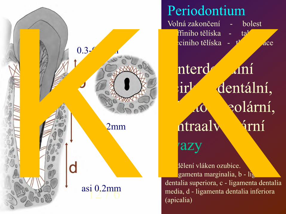
Rozdělení vláken ozubice. a - ligamenta marginalia, b - ligamenta dentalia superiora, c - ligamenta dentalia media, d - ligamenta dentalia inferiora (apicalia)
Interdentální cirkumdentální, dentoalveolární, intraalveolární vazy
0.3-0.5mm
0.1-0.2mm
asi 0.2mm
Periodontium Volná zakončení - bolest Ruffiniho tělíska - tah Pacciniho tělíska - tlak vibrace IKKI

Vazy během vývoje a při prořezávání
Utváření systému závěsných vazů před a během prořezávání zubu. (podle Levyho a Bernicka 1968, upraveno). A - základ zubu ve tvořícím se kostním lůžku, B - počátek prořezávání, C - vytvoření alveolu, stadium, D - plně prořezaný zub
IKKI

Periodontium (Parodontium)
Buňky, vlákna, matrix ligamenta, plasma, cévy a nervy Obsah cév a intercellulární plasma -
“hydroelastický polštář“ IKKI

Jontový výměna mezi pulpou a strukturami vně zubu IKKI

Gingival sulcus (pocket) Sulcus
gingivae
Free gingival groove Paramarginal sulcus
Gingiva = tiskne krček zubu – “cuff (collar) připojení“
volná: Interdentální; cirkumdentální připojená: přilehlá,fixační
IKKI

Alveolarní sliznice = temně červená, parakeratizující
Gingiva volná a připojená = růžová, ďolíčkovaná, keratinizující IKKI

Vazivové smyčky a kruhy přitlačují dáseň k zubu IKKI

Nepravý chobot Pravý chobot
vzniká ztrátou attachementu zubu, je hlubší než 3,5 mm. IKKI

IKKI

Lingua, glossa
svalověepitelový orgán;
uložen v ústní dutině a hltanu apex
corpus (body) dorsum facies inferior
radix (root) margo (margin) sulcus (groove)
terminalis medianus (midline)
foramen caecum tonsilla lingualis
IKKI

Týden 6 - 7
Papillae filiformes fungiformes vallatae foliatae
IKKI
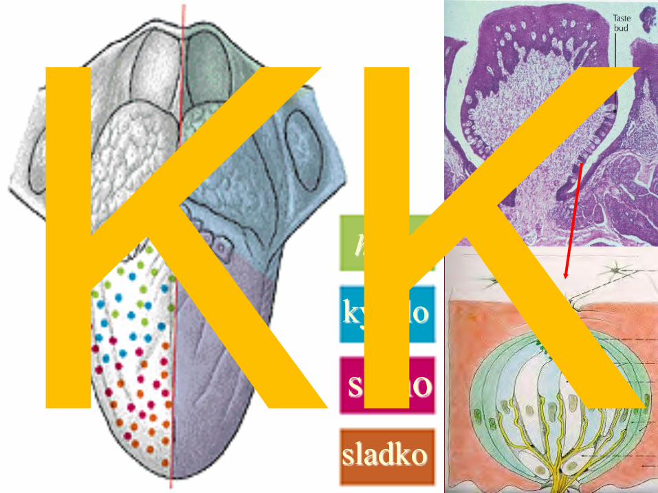
hořko
slano
kyselo
sladko
IKKI
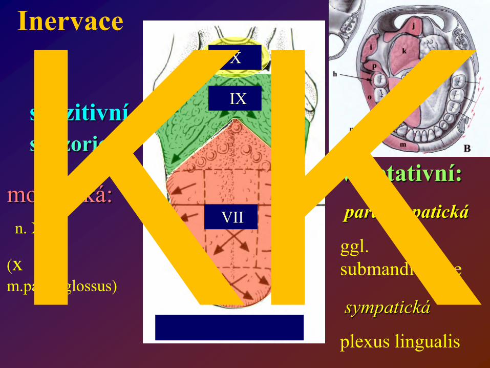
Inervace
motorická: n. XII
(x m.palatoglossus)
senzitivní
V/3
IX
X
senzorická
VII
vegetativní:
parasympatická
ggl. submandibulare
sympatická
plexus lingualis
IKKI

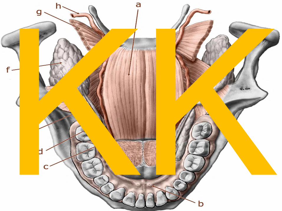
f – plica fimbriata e – frenulum linguae g – vena lingualis profunda b – caruncula sublingualis a – vyústění ductus gl.sublingualis na plica sublingualis IKKI

Ankyloglossia – ovlivňuje utváření dna ústní dutiny
Tongue-tie – Ankyloglossia –
spojení mezi jazkem a
spodinou dutiny ústní,
Jazyková uzdička sahá až ke hrotu jazyka
Kombinováno s různými syndromy (Pierre-Robin, Treacher Collins)
IKKI

Styloglossus Palatoglossus
Hyoglossus
Genioglossus
Zevní svaly jazyka mění
polohu jazyka IKKI

Vnitřní svaly jazyka mění tvar jazyka
mm. Longitudinales superiores et inferiores, transversalis, verticalis IKKI

Mm. genioglossi jsou odděleny od sebe
prostřednictvím septum linguae (linguale)
Mezi m. hyoglossus a m. genioglossus je štěrbina – pro cévy a
nervy
Septum linguae je z řídkého vaziva; může být prosyceno hnisem - absces
IKKI
IKKI

Abscessus v septu
IKKI

Odtok mízy z jazyka
Nodus juguloomohyoideus
Nodus jugulodigastricus
Do hlubokých krčních uzlin IKKI

vestibulum
vestibule
IKKI

c - frenulum linguae sup.
b,a - plicae bucco-alveolares (buccales, gingivobuccales)
IKKI

IKKI

Palatum durum Palatum molle
Hard palate Soft palate
Premaxilla Maxilla Os palatinum
Papilla, rugae (folds) pits (foveolae) Lines (crests)
IKKI

Palatum – povrchová úprava
Lacey
Papilla incisiva
Rugae palatinae
Raphe mediana
Vrchol interalveolárního septa; V úrovni papilla incisiva
Nejventrálnější rugae palatinae jsou mezi alveoly špičáků
IKKI
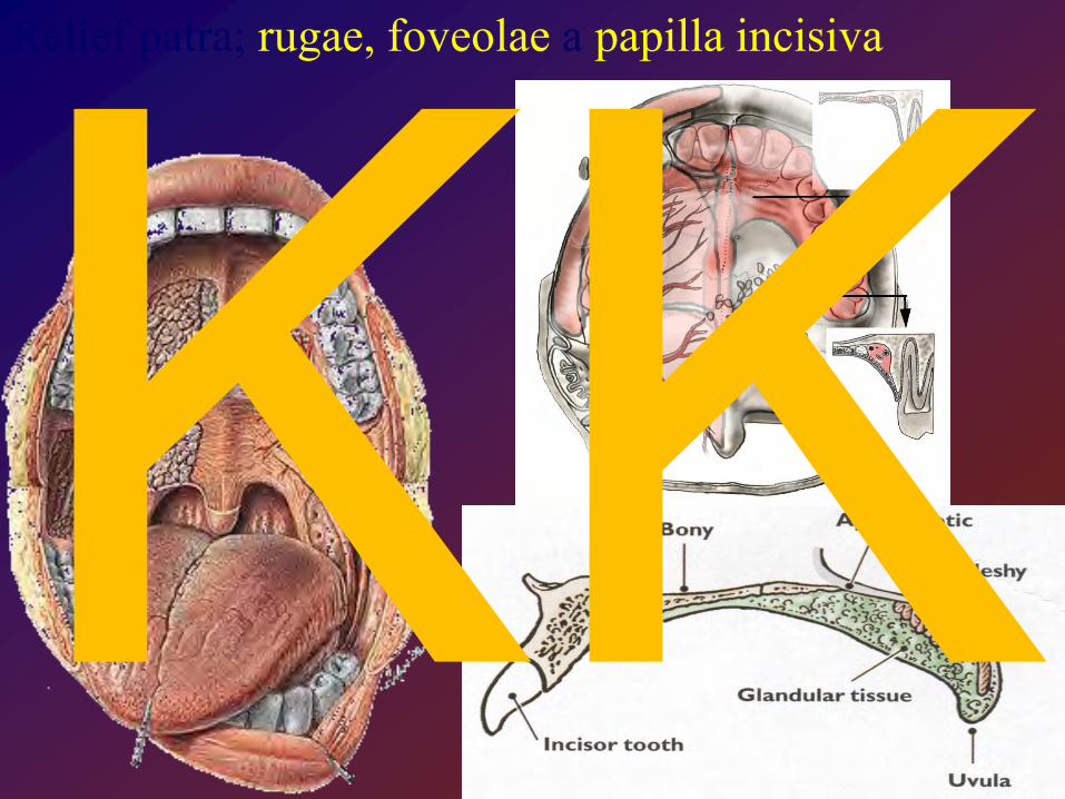
Relief patra; rugae, foveolae a papilla incisiva
IKKI
IKKI

podle Petrovického2002
IKKI

Palatum molle
– pohled zezadu
– pohled zpředu IKKI

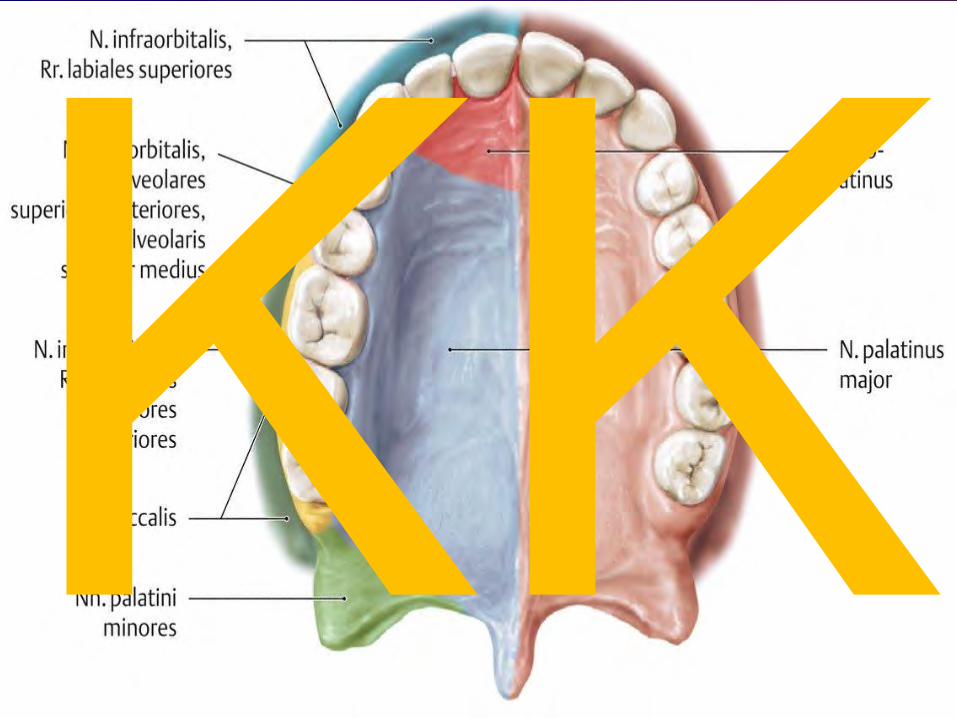
Palate – povrchová úprava
Lacey
Palatum durum
Palatum molle
Raphe mediana
Tuková zóna
Foveolae palatinae
A
H
Hauptmayerova linie – měkké a tvrdé patro
IKKI

Chrápání Snoring
IKKI

Žvýkací svaly
Musculi masticatorii Muscles of mastication
V3 – MANDIBULARIS deriváty 1. žaberního oblouku IKKI
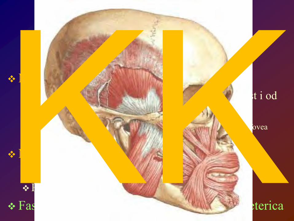
Žvýkací svaly n. mandibularis - 3 větev n. trigeminus
M. masseter Od vnější strany arcus zygomaticus, hluboká část i od
vnitřní Povrchová k angulus mandibulae hluboká do „fovea
zygomaticomandibularis“
M. temporalis Od linea temporalis inferior (+přilehlé kosti) Proc. coronoideus mandibulae
Fascia temporalis et fascia parotideomasseterica
IKKI

M. temporalis et fascia temporalis
Spatium interfasciale IKKI

M. masseter IKKI

Fascia parotideomasseterica
IKKI

Jost , G, Levet, V.: Parotid fascia and Face lifting: A critical Evaluation of the SMAS concept. Plastic and Reconstructive Surg, 74:42-51, 1983
IKKI

Mm. pterygoidei Medialis
Z fossa pterygoidea a od tuber maxillae Tuberositas pterygoidea
Lateralis Od procesus pterygoideus (lamina lateralis) a od facies
infratemporalis alae majoris ossis sphenoidalis Pod caput mandibulae, fovea pterygoidea, a na kloubní
pouzdro IKKI

pterygomandibularis
pterygoideus proprius
pterygospinosus
IKKI

a – lig. pterygospinosum
b – n. alveolaris inferior
c, d – n. lingualis e – lig. pterygomandibulare
(raphe buccopharyngea)
f – sulcus mylohyoideus
g – angulus mandibulae et lig. stylomandibulare
h – lig. sphenomandibulare
IKKI

HLAVNÍ a
Pomocné svaly žvýkací (žvýkací svaly z protetického hlediska)
venter anterior m. digastrici
m. mylohyoideus Inervace:
CN V3
m. geniohyoideus IKKI

JAW MUSCLES There is differentiation of the jaw-closing musculature.
In basal synapsids, the major jaw-closing muscle is the adductor mandibulae (externus). It originates from the back of the skull and inserts on the posterior end of the lower jaw.
In derived synapsids, the adductor mandibulae divides into two major sets of jaw-closing muscles, the temporalis and masseter. The temporalis originates from the skull roof near the sagittal crest and inserts on the coronoid process. The masseter in turn divides into two parts. The deep masseter originates on the zygomatic arch and inserts on the lower jaw; the superficial masseter part arises beneath the eye, passes across the deep masseter, to insert on the angle of the dentary.
Evolution of Synapsid jaw adductor muscles
IKKI

Paleontological evidence for mammalian middle ear evolution. (A) Diagrams of lateral views of jaw skeletal elements showing modifications leading to the mammalian condition (after Allin, ’75). The geological record and occurrence of each animal are indicated on the left. For clarity of comparison, no teeth are shown. Note that a set of postdentary elements (articular, surangular, and angular) and the upper jaw elements (quadrate and quadratojugal), indicated by gray, became separated from the dentary and reduced in size during the transition from pelycosaurs to mammals. The sequence of changes in the fossil record does not represent a true ancestor–descendent relationship, but only structural grades. (B) Changes in jaw articulation during mammalian evolution. In a pelycosaur, Dimetrodon (top), the quadrate and articular formed a functional jaw joint (black arrow). In an ‘‘advanced’’ cynodont, Diarthrognathus (middle), an additional jaw joint was observed between the squamosal and dentary (white arrow). In an extant marsupial, Didelphis (bottom), the functional jaw joint has been taken over only by the squamosal and dentary.
Takechi M, Kuratani S. 2010. History of studies on mammalian middle ear evolution: a comparative morphological and developmental biology perspective. J. Exp. Zool. (Mol. Dev. Evol.) 314B:[page range].
IKKI

Složený kloub Compound joint
Articulatio temporomandibularis
(Temporomandibular
joint TM joint
Temporocranial joint Craniomandibular joint) IKKI

IKKI

IKKI

IKKI

Pozdní triasový savec Spodnokřídový savec ptakopysk
IKKI

rovina okluze occlusal plane protetická rovina Camper plane
x vodorovná rovina horizontal plane
M.Doskočil: Chrupavka ve vývoji mandibuly. (cartilage in the development of the mandible) Cs.Stomatologie, 1:10-18, 1988
Meckelova chrupavka a chrupavčité deriváty v krčku dolní čelisti IKKI

END
Chrupavčitý sloupek – derivát Meckelovy chrupavky ?
IKKI

IKKI

Os tympanicum zvýrazníte dalším kliknutím Tuberc. pharyngicum Foramen lacerum Spina sphenoidalis Foramen spinosum Foramen ovale
Fossa mandibularis Tuberculum articulare
Arcus zygomaticus
Condylus occipitalis
Processus styloideus Foramen jugulare For. stylomastoideum Processus mastoideus Fissura tympanomastoidea Fis.tympanopetrosa Fis.petrosquamosa Fis.tympanosquamosa Porus acusticus ext.
Basis cranii externa – šikmý detailní pohled na os tympanicum
IKKI

Processus postglenoidalis Post-glenoid process
Trigonum suprameatum Suprameatal triangle
IKKI

Articular Capsule is a sac that encloses TMJ. Borders: Superior: Capsule is positioned underneath inferior side of Articular Eminence. Inferior: Capsule wraps around condyle's neck (Collum Mandibulae)
A fibro-cartilageus disc divides synovial cavity of TMJ into: 1. Superior synovial cavity 2. Inferior synovial cavity Both cavities are filled with synovial fluid, secreted by inner side of articular capsule (clear, viscous fluid). Attachments of articular disc:
1. Anterior: a. Anterio-Superior: indirectly to articular eminence through capsule
b. Anterio-inferior: to condyl's neck
2. Posterior: a. Posterio-superior: to post-glenoid process more salient in young spina supra meatum ?
b. Posterio-inferior: to condyl's neck
Gray´s anatomy, The classic collector´s edition IKKI

Salentijn, L. Biology of Mineralized Tissues: Prenatal Skull Development, Columbia University College of Dental Medicine post-graduate dental lecture series, 2007 Moss, ML. The non-existent hinge axis, Am. Inst, Oral Biol. 1972, 59-66 Rodríguez-Vázquez JF, et al., JF; Mérida-Velasco, JR; Mérida-Velasco, JA; Jiménez-Collado, J (1998). "Anatomical considerations on the discomalleolar ligament". J Anat.. 192 (Pt 4): 617–621. PMC 1467815. PMID 9723988. //www.ncbi.nlm.nih.gov/pmc/articles/PMC1467815/. Rodríguez-Vázquez JF, et al. (1993). "Relationships between the temporomandibular joint and the middle ear in human fetuses.". J Dent Res.. 72 (1): 62–66. T Rowicki, J Zakrzewska. (2006). "A study of the discomalleolar ligament in the adult human.". Folia Morphol. (Warsz).. 65 (2): 121–125. S Zhang, N Gersdorff, J Frahm (2011) Real-Time Magnetic Resonance Imaging of Temporomandibular Joint Dynamics. The Open Medical Imaging Journal, 2011, 5, 1-7, [1] Zadik, Yehuda; Aktaş Alper; Drucker Scott; Nitzan W Dorrit (2012). "Aneurysmal bone cyst of mandibular condyle: A case report and review of the literature". J Craniomaxillofac Surg 40.
IKKI

kloubní povrch kondylu tvoří čtyři vrstvy: • Superficial layer: superficial articular layer = connective tissue character • Very cellular layer: • Proliferating layer: • Hypertrophic layer:
discus articularis je v období vzniku bohatě vaskularizován
Ztráta vaskularizace podmiňuje degenerativní změny v disku
IKKI

horní prostor cavitas discosquamosa – 581 mm2
dolní prostor cavitas discocondylaris – 396 mm2 IKKI

Atrophy of the madibular condyle depending age IKKI

A) Fissura tympanosquamosa
B) Stratum superius C) genu vasculosum D) Stratum inferius E) Capsule F) Glandula parotis G) Discus articularis ( Dreger 1994 )
Discus napojen na fascia parotis, a podkožní struktury; pouzdro je slabé
Discus napojen přes pouzdro
Discus je napojen samostatně
Discus a pouzdro jsou pevně spojeny
Dreger H (1994) Untersuchungen zur posterioren Anheftung Des Diskus artikularis im menschlichen Kiefergelenk. Med Diss Kiel
Vasili Naroushvili: Wechselwirkungen zwischen Okklusionsarten und Anheftungsarten des Musculus pterygoideus lateralis bei der Entstehung von Diskus Dislokation des Kiefergelenkes Hamburg 2006 IKKI

Minarelli, AM, DelSanto, M, Liberti, EA: The structure of the human temporomandibular joint disc: A scanning electron microscopy study. J Orof Pain 11:95-98, 1997
Examined discs: 16-39 weeks of intrauterine life Up to 4 months of age 30-39 years 60-69 years IKKI

pars superior, pars inferior, a ´třetí´část m pterygoideus lateralis (vypitvána) 1-Discus articularis, 2-M.pteryg.lat (pars superior), 3-M.pteryg.lat (pars inferior ), 4- třetí část m. pteryg. lat (připojená ke pars inferior)
1
2
3 4
Discus articularis (vypitván) 1-Discus articularis, 2-úpon mm. masseter and temporalis, 3-úpon m. pteryg.lat (pars superior), 4-úpon m. pteryg.lat (pars inferior )
V. Naroushvili 2006
IKKI

IKKI

IKKI

•J. Chen, U. Akyuz, L. Xu, R.M.V. Pidaparti : Stress analysis of the human temporomandibular joint •Medical Engineering & Physics 20/8/: 565-572, October 1998
IKKI

Tenká lamina elastica
silnější lamina fibroelastica
Retroarticulární hydroelastický polštář Zenkerův (obsahuje cévy) IKKI

IKKI

Kondylová dráha Condyle path a Transverse b Longitudinal
Rest position
Central position
Ventral position
habituální (vysoké) při konci žvýkacího cyklu
centrální (zenitové)
diskokondylární komplexy jsou v nejvíce kraniálním
postavení při polknutí sousta
ventrální nízké (klidové)
při nejmenším napětí svalů
IKKI

N. facialis: • lateral surface of the joint capsule IKKI

N. auriculotemporalis nerve is branched into four nerves: • lateral branch •Medial branch • branch from the middle nerve segment • branch from the area where nerve crosses n. temporalis superficialis IKKI

n. massetericus send four branches:
• branch below oval foramen • branch from the first nervous segment closely below skull base • two branches from the first segment below zygomatic bone
m. temporalis profundus:
• supplies rostromedial part of the disc and capsule
Ganglion oticum (otic ganglion):
• supplies dorsal part (pars discosquamalis) of the joint capsule
IKKI

IKKI
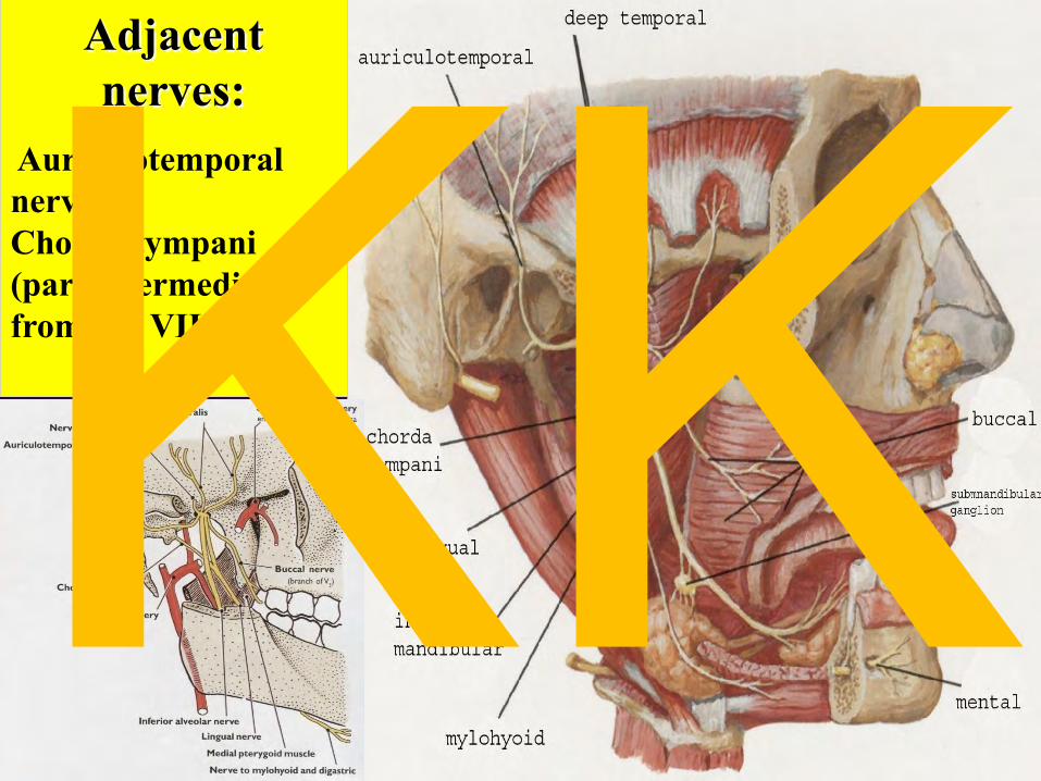
Adjacent nerves:
Auriculotemporal nerve Chorda tympani (pars intermedialis from CN VII.) IKKI

Použitá literatura R. Čihák: Anatomie 1, 2, 3 Grada Publishing 2003
M. Dykes : Anatomy 2th edition, Mosby 2002
S.Snell: Clinical anatomy for Medical Students 6th edition, Lippincott, Williams & Wilkins
I.Klepáček, J.Mazánek et al.: Klinická anatomie ve stomatologii Grada Publishing 2001
G.J.Tortora : Principles of Human Anatomy 4th edition, Williams & Wilkins
K.L.Moore, A.F.Dalley: Clinically Oriented Anatomy 4th edition, Williams & Wilkins
F.H.Netter: anatomický atlas člověka Vlastní archív
IKKI


