Ultrasound medical image recognition with Artificial intelligence for Parkinson´s disease classification Jiří Blahuta * , Tomáš Soukup * , Petr Čermák * , Jiří Rozsypal * and Michal Večerek ** * Silesian University in Opava, Department of Informatics, Opava, CZECH REPUBLIC ** Vysoká škola báňská, Technická univerzita Ostrava, Ostrava, CZECH REPUBLIC [email protected], [email protected], [email protected], [email protected],[email protected]Abstract - This paper shows how to classify the medical ultrasound images by using artificial intelligence with experimental software with MATLAB. The main goal is a classification of ROI substantia nigra in midbrain. This classification of the images is useful to detection Parkinson´s disease. This work is based on image processing and is realized with the help of artificial intelligence which has been experimentally simulated in MATLAB software environment. This method is well applicable with Neural Network Toolbox in MATLAB. I. INTRODUCTION Modern medical imaging methods helps to diagnoses in different branches in medicine. This paper is focused on processing of transcranial (TCS) ultrasound images. It is useful to potential Parkinson´s disease diagnosis. We developed a computer program in MATLAB to area measurement of substantia nigra which is described in next part. This application is based on image processing and neural network approach for artificial intelligence elements. The main goal of designed approach is to measure area of substantia nigra for all levels of echogenicity. The purpose of neural network approach is a positioning of needed ROI in substantia to area computing. Input files are in DICOM format, see chapter II. A. Role of SN and Parkinson´s disease The following part brings some information about role of substantia nigra (SN) in brain stem and Parkinson´s disease diagnosis and features. Parkinson's disease (PD) is caused by the death of dopaminergic neurons. It is a degenerative disease of basal ganglias inside the brain, described by James Parkinson in 19 th century. The main symptoms of PD include muscle rigidity, tremors and changes in speech and gait, bradykinesia, sleep disorders and more. 1 This type of tremor is typical for PD. It is a chronicle disease and initial features sometimes are insignificant for diagnosis. 2 Substantia nigra (SN; in English “black substance”) is a brain structure which is located in the mesencephalon 1 http://www.mdvu.org/library/disease/pd/ 2 http://www2.parkinson.org/Document.Doc?id=618 (midbrain) that plays an important role in reward, addiction, and movement. SN produces important dopamine for correct function of CNS (Central nervous system). The CNS is one of the two major divisions of the nervous system. SN is our ROI for processing and searching pathological defects to Parkinson ´s disease diagnosis. SN is very important part of midbrain and is well recognizable by ultrasound. Ultrasound imaging in neurology is also important for detection of another diagnoses. Detailed information about substantia nigra and echogenicity are available in [1], [2] and [3]. The following figure shows the position of basal ganglias where is located SN. Fig. 1 – Position of basal ganglias where is situated SN II. THE GOAL OF PROCESSING In this section we will describe the goal of this processing and used methods. The following flow-chart shows how to work our designed method. MIPRO 2012/CIS 1139
Transcript
Ultrasound medical image recognition with Artificial intelligence for Parkinson´s disease
classification
Jiří Blahuta*, Tomáš Soukup*, Petr Čermák*, Jiří Rozsypal* and Michal Večerek**
* Silesian University in Opava, Department of Informatics, Opava, CZECH REPUBLIC** Vysoká škola báňská, Technická univerzita Ostrava, Ostrava, CZECH REPUBLIC
Abstract - This paper shows how to classify the medical ultrasound images by using artificial intelligence with experimental software with MATLAB. The main goal is a classification of ROI substantia nigra in midbrain. This classification of the images is useful to detection Parkinson´s disease. This work is based on image processing and is realized with the help of artificial intelligence which has been experimentally simulated in MATLAB software environment. This method is well applicable with Neural Network Toolbox in MATLAB.
I. INTRODUCTION
Modern medical imaging methods helps to diagnoses in different branches in medicine. This paper is focused on processing of transcranial (TCS) ultrasound images. It is useful to potential Parkinson´s disease diagnosis. We developed a computer program in MATLAB to area measurement of substantia nigra which is described in next part. This application is based on image processing and neural network approach for artificial intelligence elements. The main goal of designed approach is to measure area of substantia nigra for all levels of echogenicity. The purpose of neural network approach is a positioning of needed ROI in substantia to area computing. Input files are in DICOM format, see chapter II.
A. Role of SN and Parkinson´s disease
The following part brings some information about role of substantia nigra (SN) in brain stem and Parkinson´s disease diagnosis and features. Parkinson's disease (PD) is caused by the death of dopaminergic neurons. It is a degenerative disease of basal ganglias inside the brain, described by James Parkinson in 19th
century. The main symptoms of PD include muscle rigidity, tremors and changes in speech and gait, bradykinesia, sleep disorders and more. 1 This type of tremor is typical for PD. It is a chronicle disease and initial features sometimes are insignificant for diagnosis.2
Substantia nigra (SN; in English “black substance”) is a brain structure which is located in the mesencephalon
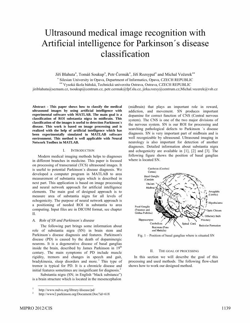
(midbrain) that plays an important role in reward, addiction, and movement. SN produces important dopamine for correct function of CNS (Central nervous system). The CNS is one of the two major divisions of the nervous system. SN is our ROI for processing and searching pathological defects to Parkinson ´s disease diagnosis. SN is very important part of midbrain and is well recognizable by ultrasound. Ultrasound imaging in neurology is also important for detection of another diagnoses. Detailed information about substantia nigra and echogenicity are available in [1], [2] and [3]. The following figure shows the position of basal ganglias where is located SN.
Fig. 1 – Position of basal ganglias where is situated SN
II. THE GOAL OF PROCESSING
In this section we will describe the goal of this processing and used methods. The following flow-chart shows how to work our designed method.
MIPRO 2012/CIS 1139
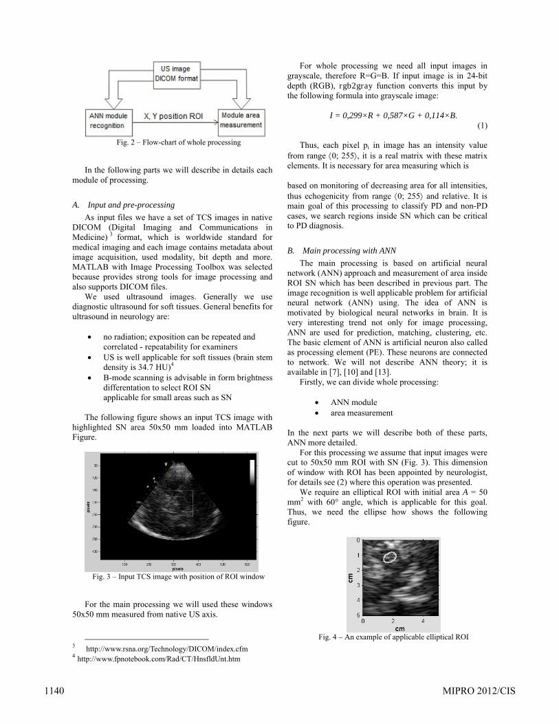
Fig. 2 – Flow-chart of whole processing
In the following parts we will describe in details each module of processing.
A. Input and pre-processing
As input files we have a set of TCS images in native DICOM (Digital Imaging and Communications in Medicine) 3 format, which is worldwide standard for medical imaging and each image contains metadata about image acquisition, used modality, bit depth and more.MATLAB with Image Processing Toolbox was selected because provides strong tools for image processing and also supports DICOM files.
We used ultrasound images. Generally we use diagnostic ultrasound for soft tissues. General benefits for ultrasound in neurology are:
no radiation; exposition can be repeated and correlated - repeatability for examiners
US is well applicable for soft tissues (brain stem density is 34.7 HU)4
B-mode scanning is advisable in form brightness differentation to select ROI SNapplicable for small areas such as SN
The following figure shows an input TCS image with highlighted SN area 50x50 mm loaded into MATLAB Figure.
Fig. 3 – Input TCS image with position of ROI window
For the main processing we will used these windows 50x50 mm measured from native US axis.
For whole processing we need all input images in grayscale, therefore R=G=B. If input image is in 24-bit depth (RGB), rgb2gray function converts this input by the following formula into grayscale image:
I = 0,299×R + 0,587×G + 0,114×B.(1)
Thus, each pixel pi in image has an intensity value from range 0; 255, it is a real matrix with these matrix elements. It is necessary for area measuring which is
based on monitoring of decreasing area for all intensities, thus echogenicity from range 0; 255 and relative. It is main goal of this processing to classify PD and non-PD cases, we search regions inside SN which can be critical to PD diagnosis.
B. Main processing with ANN
The main processing is based on artificial neural network (ANN) approach and measurement of area inside ROI SN which has been described in previous part. The image recognition is well applicable problem for artificial neural network (ANN) using. The idea of ANN is motivated by biological neural networks in brain. It is very interesting trend not only for image processing, ANN are used for prediction, matching, clustering, etc. The basic element of ANN is artificial neuron also called as processing element (PE). These neurons are connected to network. We will not describe ANN theory; it is available in [7], [10] and [13].
Firstly, we can divide whole processing:
ANN module area measurement
In the next parts we will describe both of these parts, ANN more detailed.
For this processing we assume that input images were cut to 50x50 mm ROI with SN (Fig. 3). This dimension of window with ROI has been appointed by neurologist, for details see (2) where this operation was presented.
We require an elliptical ROI with initial area A = 50 mm2 with 60° angle, which is applicable for this goal. Thus, we need the ellipse how shows the following figure.
Fig. 4 – An example of applicable elliptical ROI
1140 MIPRO 2012/CIS
In this part we need to locale this ellipse inside SN in appropriate position, see example on Fig. 4. The aim of our problem is to applicably locate this ellipse inside SN with ANN.
1) ANN module recognition
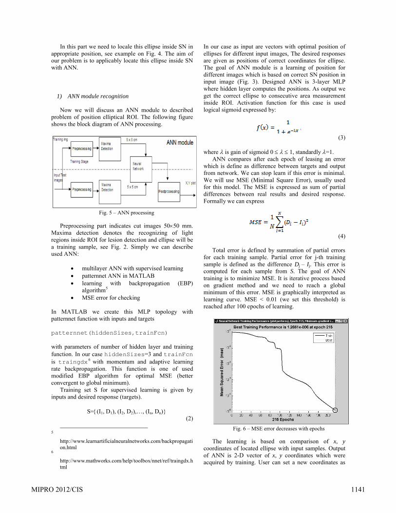
Now we will discuss an ANN module to described problem of position elliptical ROI. The following figure shows the block diagram of ANN processing.
Fig. 5 – ANN processing
Preprocessing part indicates cut images 5050 mm. Maxima detection denotes the recognizing of light regions inside ROI for lesion detection and ellipse will be a training sample, see Fig. 2. Simply we can describe used ANN:
multilayer ANN with supervised learning patternnet ANN in MATLAB learning with backpropagation (EBP)
algorithm5
MSE error for checking
In MATLAB we create this MLP topology with patternnet function with inputs and targets
patternnet(hiddenSizes,trainFcn)
with parameters of number of hidden layer and training function. In our case hiddenSizes=3 and trainFcnis traingdx 6 with momentum and adaptive learning rate backpropagation. This function is one of used modified EBP algorithm for optimal MSE (better convergent to global minimum).
Training set S for supervised learning is given by inputs and desired response (targets).
In our case as input are vectors with optimal position of ellipses for different input images, The desired responses are given as positions of correct coordinates for ellipse. The goal of ANN module is a learning of position for different images which is based on correct SN position in input image (Fig. 3). Designed ANN is 3-layer MLP where hidden layer computes the positions. As output we get the correct ellipse to consecutive area measurement inside ROI. Activation function for this case is used logical sigmoid expressed by:
(3)
where is gain of sigmoid 0 1, standardly =1.ANN compares after each epoch of leasing an error
which is define as difference between targets and output from network. We can stop learn if this error is minimal. We will use MSE (Minimal Square Error), usually used for this model. The MSE is expressed as sum of partial differences between real results and desired response. Formally we can express
(4)
Total error is defined by summation of partial errors for each training sample. Partial error for j-th training sample is defined as the difference Dj – Ij. This error is computed for each sample from S. The goal of ANN training is to minimize MSE. It is iterative process based on gradient method and we need to reach a global minimum of this error. MSE is graphically interpreted as learning curve. MSE < 0.01 (we set this threshold) is reached after 100 epochs of learning.
Fig. 6 – MSE error decreases with epochs
The learning is based on comparison of x, ycoordinates of located ellipse with input samples. Output of ANN is 2-D vector of x, y coordinates which were acquired by training. User can set a new coordinates as
MIPRO 2012/CIS 1141
applicable input followed by re-train of ANN with new coordinates. These coordinates ANN inserts as a new training sample, thus we have more correct samples.Furthermore, we observe descending tendency of MSE (Fig. 5). We set the threshold value for MSE=0.01, thus
if TMSE 0.01 then stop_learning else next_epoch.
Practical testing of this method showed that designed ANN is useful for positioning of ellipse in correct position in ROI window. Accuracy of this method is approximately 70% for 50 tested images. Re-training allows reach higher accuracy for manually set of coordinates and more images as input position. GUI of designed application is presented below. It was implemented with GUIDE in MATLAB.
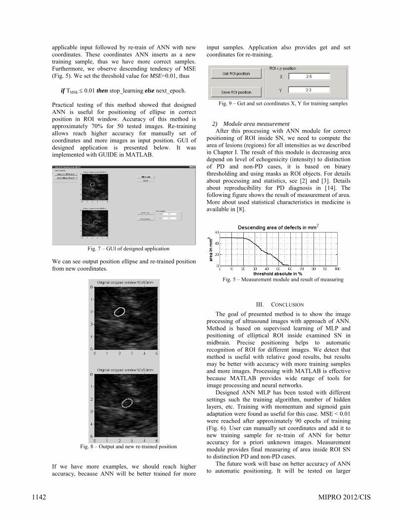
Fig. 7 – GUI of designed application
We can see output position ellipse and re-trained position from new coordinates.
Fig. 8 – Output and new re-trained position
If we have more examples, we should reach higher accuracy, because ANN will be better trained for more
input samples. Application also provides get and set coordinates for re-training.
Fig. 9 – Get and set coordinates X, Y for training samples
2) Module area measurementAfter this processing with ANN module for correct
positioning of ROI inside SN, we need to compute the area of lesions (regions) for all intensities as we described in Chapter I. The result of this module is decreasing area depend on level of echogenicity (intensity) to distinction of PD and non-PD cases, it is based on binary thresholding and using masks as ROI objects. For details about processing and statistics, see [2] and [3]. Details about reproducibility for PD diagnosis in [14]. The following figure shows the result of measurement of area.More about used statistical characteristics in medicine is available in [8].
Fig. 5 – Measurement module and result of measuring
III. CONCLUSION
The goal of presented method is to show the image processing of ultrasound images with approach of ANN. Method is based on supervised learning of MLP and positioning of elliptical ROI inside examined SN in midbrain. Precise positioning helps to automatic recognition of ROI for different images. We detect that method is useful with relative good results, but results may be better with accuracy with more training samples and more images. Processing with MATLAB is effective because MATLAB provides wide range of tools for image processing and neural networks.
Designed ANN MLP has been tested with different settings such the training algorithm, number of hidden layers, etc. Training with momentum and sigmoid gain adaptation were found as useful for this case. MSE < 0.01were reached after approximately 90 epochs of training(Fig. 6). User can manually set coordinates and add it to new training sample for re-train of ANN for better accuracy for a priori unknown images. Measurement module provides final measuring of area inside ROI SN to distinction PD and non-PD cases.
The future work will base on better accuracy of ANN to automatic positioning. It will be tested on larger
1142 MIPRO 2012/CIS
dataset than 100 images and will be examined other ANN topologies with different learning approach.
ACKNOWLEDGMENT
We would like to thank to Fakultní nemocnice v Ostravě (Czech Republic) and Klinik für Neurologie und Poliklinik in Rostock (Germany) to supplying input TCS DICOM images to processing.
REFERENCES
[1] Becker, G.,:“Degeneration of substantia nigra in chronic Parkinson’s disease visualized by transcranial color-coded real-time sonography”, 1995, Journal of Neuroimaging 45.
[2] Bishop, Ch., “Neural Networks for Pattern Recognition”, 1996, Oxford University Press, USA; 1 edition, ISBN-13: 978-0198538646.
[3] Blahuta J., Soukup T., Čermák P., “The image recognition of brain-stem ultrasound images with using a neural network based on PCA”, Medical Measurements and Applications Proceedings (MeMeA), 2011 IEEE International Workshop on BARI, 30-31 May 2011, ISBN: 978-1-4244-9336-4
[4] Blahuta J., Soukup T., Čermák P., “The ROI defect statistical analysis of substantia nigra to reproducibility of designed experimental algorithm for potential PD diagnosis ”,Mathematical Metods and Techniques in Engineering, 3-5.11.2011,Catania,Wseas press,ISBN:978-1-61804-046-6
[5] Diamantaras, K.: “Principal Component Neural Networks: Theory and Applications”, 1996, ISBN:0-471-05436-4.
[6] Gelb, D., O. E. G.-S.: 1999, “Diagnostic criteria for Parkinson´s disease, Archives of Neurology” 56(1), 33–39.
[7] Grossberg, S., Carpenter A.: “Neural Networks for Vision and Image Processing”, 1992, ISBN-13: 978-0262531085.
[8] Montgomery, C. D., Runger, C. G., “Applied Statistics and Probability for Engineers”, 2006, Wiley; 4th Edition, ISBN-13: 978-0471745891.
[10] Principe J. C., Euliano N. R., Lefebvre W. C., “Neural and Adaptive Systems: Fundamentals Through Simulations”, 2000, John Wiley & Sons, Inc.
[11] Schreiber, J., Sojka, E., Ličev, L., Škňouřilová, P., Gaura, J., Školoudík, D.: “A new method for the detection of brain stem in transcranial ultrasound images”, Proceedings of Biosignals 2008, 2008.
[12] Sojka, E.: “A motion estimation method based on possibility theory”, 2006, Proceedings of IEEE ICIP, pp. 1241.
[13] Smagt P. v d., Kröse B., “An introduction to neural networks”, 1996, The University of Amsterdam; 8th Edition.
[14] Školoudík, D.: “Reproducibility of sonographic measurement of the substantia nigra”, Ultrasound in Medicine & Biology (9), 2007, pp. 1347–1352.
[15] Walter U, Wittstock M, Benecke R, Dressler D: “Substantia nigra echogenicity is normal in non-extrapyramidal cerebral disorders but increased in Parkinson's disease.”, J Neural Transm. 2002 , 109:, pp. 191-196.
[16] Webster DD: “Critical analysis of the disability in Parkinson's disease”., Mod Treat, 1968, 5:, pp. 257-282.