297/ ACTA CHIRURGIAE ORTHOPAEDICAE ET TRAUMATOLOGIAE ČECHOSL., 79, 2012, p. 297–306 CURRENT CONCEPTS REVIEW SOUBORNÝ REFERÁT Periprosthetic Fractures around the Knee: Update on Therapeutic Algorithms for Internal Fixation and Revision Arthroplasty Periprotetické zlomeniny v oblasti kolena: nové terapeutické postupy pro vnitřní fixaci a revizní artroplastiku S. MÄRDIAN 1 , F. WICHLAS 1 , K.-D. SCHASER 1 , G. MATZIOLIS 1 , B. FÜCHTMEIER 2 , C. PERKA 1 , P. SCHWABE 1 1 Charité – Universitätsmedizin Berlin, CMSC – Center ofmusculosceletalsurgery, Germany 2 Krankenhaus Barmherzige Brüder Regensburg, Germany SUMMARY Effective treatment of periprosthetic fractures following TKA continues to represent a surgical challenge. The incidence and frequency of these complicated type of fractures is expected to increase as the number of TKA as well as the activi- ty level of these patients steadily rises. A careful and correct analysis of the fracture pattern, its classification, the quality of the existing bone stock and the fixation / loosening of the underlying prosthetic system has to precede decision making for successful conservative or surgical treatment. Therefore, improved diagnostic radiographic imaging of fracture patterns and reliable assessment of prosthesis loosening progressive development of new implant methods and refinement of soft tissue preserving surgical techniques will hold the key for regaining the functional level prior to the fracture. INTRODUCTION During the last decade the number of total knee arth- roplasty (TKA) implantations continues to increase and has exceeded the frequency of surgeries performed for total hip arthroplasty in some countries, e.g. the USA. (58). Due to the demographic changes in the western industrial countries with an aging but active popualtion, the continuously prolonged life expectancy and the inc- reasing survival time of the prostheses, the incidence and rate of periprosthetic fractures is expected to markedly increase. (22, 24, 25, 78). In a systematic study analyzing complications of 415 periprosthetic femur fractures Herrera et al.(35) have observed 9% pseudarthroses/non-unions, 4% mechani- cal complications and 3% infections with an overall revi- sion rate, reaching 13%. To date, the incidence of peri- prosthetic fractures following TKA is considered to center around 1% (0.3% – 2.5%)(2, 81). With a frequ- ency of 0.3 – 2% the periprosthetic femur fractures are most often observed, followed by corresponding peri- prosthetic fractures of the tibia (0,3% – 0,5%) (9) and patella 0,6% (4, 9). The rate of postoperative periprost- hetic fractures is known to be comparably higher follo- wing revision arthroplasty as opposed to primary implantations (81). Fracture patterns of intra- and postoperative peri- prosthetic fractures do not resemble the typical rules as known from dia-/metadiaphyseal long bone fractures in general traumatology. For this reasons various authors have developed valid classifications aiming to syste- matically subgroup different fracture types of the femur, tibia and patella in relation to a fixed or loosened pro- sthesis. Most of these postoperative periprosthetic frac-
Transcript
297/ ACTA CHIRURGIAE ORTHOPAEDICAEET TRAUMATOLOGIAE ČECHOSL., 79, 2012, p. 297–306 CURRENT CONCEPTS REVIEW
SOUBORNÝ REFERÁT
Periprosthetic Fractures around the Knee: Updateon Therapeutic Algorithms for Internal Fixation and Revision Arthroplasty
Periprotetické zlomeniny v oblasti kolena: nové terapeutické postupy pro vnitřnífixaci a revizní artroplastiku
S. MÄRDIAN1, F. WICHLAS1, K.-D. SCHASER1, G. MATZIOLIS1, B. FÜCHTMEIER2, C. PERKA1,P. SCHWABE1
Effective treatment of periprosthetic fractures following TKA continues to represent a surgical challenge. The incidenceand frequency of these complicated type of fractures is expected to increase as the number of TKA as well as the activi-ty level of these patients steadily rises. A careful and correct analysis of the fracture pattern, its classification, the qualityof the existing bone stock and the fixation / loosening of the underlying prosthetic system has to precede decision makingfor successful conservative or surgical treatment. Therefore, improved diagnostic radiographic imaging of fracture patternsand reliable assessment of prosthesis loosening progressive development of new implant methods and refinement of softtissue preserving surgical techniques will hold the key for regaining the functional level prior to the fracture.
INTRODUCTION
During the last decade the number of total knee arth-roplasty (TKA) implantations continues to increase andhas exceeded the frequency of surgeries performed fortotal hip arthroplasty in some countries, e.g. the USA.(58). Due to the demographic changes in the westernindustrial countries with an aging but active popualtion,the continuously prolonged life expectancy and the inc-reasing survival time of the prostheses, the incidence andrate of periprosthetic fractures is expected to markedlyincrease. (22, 24, 25, 78).
In a systematic study analyzing complications of 415periprosthetic femur fractures Herrera et al.(35) haveobserved 9% pseudarthroses/non-unions, 4% mechani-cal complications and 3% infections with an overall revi-sion rate, reaching 13%. To date, the incidence of peri-prosthetic fractures following TKA is considered to
center around 1% (0.3% – 2.5%)(2, 81). With a frequ-ency of 0.3 – 2% the periprosthetic femur fractures aremost often observed, followed by corresponding peri-prosthetic fractures of the tibia (0,3% – 0,5%) (9) andpatella 0,6% (4, 9). The rate of postoperative periprost-hetic fractures is known to be comparably higher follo-wing revision arthroplasty as opposed to primaryimplantations (81).
Fracture patterns of intra- and postoperative peri-prosthetic fractures do not resemble the typical rules asknown from dia-/metadiaphyseal long bone fractures ingeneral traumatology. For this reasons various authorshave developed valid classifications aiming to syste-matically subgroup different fracture types of the femur,tibia and patella in relation to a fixed or loosened pro-sthesis. Most of these postoperative periprosthetic frac-
The treatment goals of the periprosthetic femoral frac-tures are as follows (21, 33, 81): – stable joint without significant malalignment– uneventful and completed fracture healing (within
6 months)– „range of motion“ and restored knee function to the
level prior to the trauma.A functionally favourable result is also assumed if
a minimum range of motion of 90°, a shortening of lessthan 1 cm, a varus-/valgus-misalignment of less than 5°,a minimal change in torsion and ante-/retroflexion ofless than 10° is reached/accomplished (21, 81). Mal -alignments exceeding the mentioned limitations com-pellingly lead to excentric overload of the prosthesis andperiprosthetic bone stock, which in turn, typically andrather sooner than later result in early implant loosening(81).
TherapyConservative approach Conservative therapy, which invariably results in
reduced knee function, is rarely indicated and exceptio-nally restricted to undisplaced, stable fractures (10, 19,20, 56, 59, 77, 79). Long lasting traction treatment isnowadays completely obsolet (21, 58). Exceptional indi-cations are incidentally reported for patients with veryreduced general health status and unjustifiable risk ofanaesthesia and surgery.
Surgical approachConcepts of surgical therapy are guided by the under-
lying fracture type/classification, the type of prosthesisand essentially depend on assessing the loosening beha-viour of the prosthesis. Exact and valid analysis of thequestion whether the prosthesis is loose or not is man-datory and of utmost importance. Preoperative assess-ment/exclusion of a loose prosthesis component may bedifficult and requires detailed clinical analysis and suf-ficient radiographic/imaging studies. Pain reported to bepresent already prior to the fracture is sometimes theonly sign indicating a subclinical prosthesis loosening,not clearly visible on radiographs. To which extent thispain may have contributed to uncontrolled loss of musc-le performance with consecutively increased tendencyfor stumbling can only be argued and remains specula-tory.
tures result from low energy injuries or stress/fatigueproblems. Only few are observed in response to a highenergy trauma. As these specific fractures are usuallyseen in older patients with multiple comorbidities andsurgical repair per se is always a revision surgery withimplant fixation around prosthetic implants in mostlyosteoporotic bone or even revision arthroplasty effecti-ve surgical therapy is tremendously complicated. The-refore, adequate treatment of periprosthetic fracturesfollowing TKA remains a surgical challenge requiringbiomechanical and operative knowledge, experience aswell as skills from difficult osteosynthetic techniquesto the whole program needed for revision arthroplasty.Consequently, this paper is aimed to systemicallyreview contemporary literature for periprosthetic frac-tures following TKA in order to unravel the differentsurgical therapy options and provide corresponding treatment algorithms as guided by the underlying clas-sifications.
Risk factorsSeveral authors were able to demonstrate the relati-
ve importance and impact of underlying, patient spe -cific risk factors known to promoteperiprosthetic frac-tures. Among others, osteoporosis (10, 56, 75),rheumatoid arthritis (19, 55, 56, 77), enduring steroid//corticoid therapy (35, 36, 55, 77), pre-existing neuro-logical disorders such as e. g. cerebral ataxia, Parkin-son s disease, myasthenia gravis and many others (19),previous revision arthroplasty (10, 56), arthrofibrosis(due to reduced knee flexion and to increased lever armforces during walking) (2) as well as progressive localperiprosthetic osteolysis due to septic or aseptic loose-ning (65, 72).
PERIPROSTHETIC FRACTURES OF THE FEMUR
Periprosthetic femoral fractures are the most frequentsubgroup (0.3% - 2.5%) of periprosthetic fractures fol-lowing TKA. Apart from the above mentioned generalrisk factors notching of the anterior femoral cortex isknown to increase the risk of periprostheticfemoralfractures (10, 50, 56, 75). Biomechanical studies clear-ly have shown that femoral notching reduces femoralstrength and resistance to fracture (50) while large cli-nical studies failed to confirm these results, possibly dueto ongoing bony remodelling (70, 81).
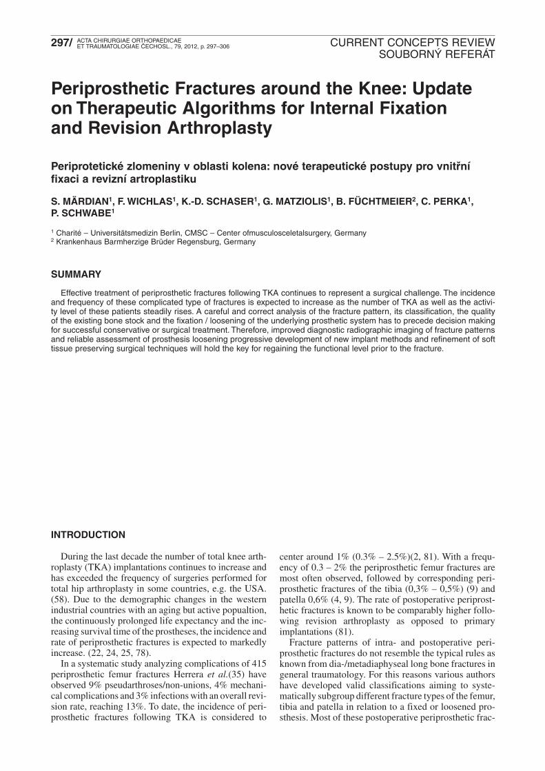
Several classification for periprosthetic femoral frac-tures following TKA have been described, which diffe-rentially consider and refer to either the fracture lineextension or the fixation type/loosening behaviour of theprosthesis (21, 43, 73, 81). For the clinical practice anddaily use the classification systems acc. to Rorabeck et al.(73) and/or Su et al. (81) have found wide acceptance (seetable 1). As to the time point of sustained periprostheticfracture intraoperative fractures can be differentiatedfrom postoperative fractures. Both classification systemsare designed and directed towards standardized decisionmaking, reliable guidance of periprosthetic fracture careand establishment of corresponding treatment algorithms.
Rorabeck-Classification Su-ClassificationType I undisplaced fracture with fracture proximal to the
fixed prosthesis femoral prosthesis component (no extension distal to the anterior shield)
Type II displaced fracture with fracture starts at the levelfixed prosthesis of the femoral prosthesis
shield and extends proximalType III loose prosthesis, all fracture elements/ lines
un-/ displaced fracture involve the segment below/ distal the femoral prosthesis shield
Tab. 1. Classification of periprosthetic femur fractures acc. toRorabeck and Su
Osteosynthetic reconstruction is feasible and indica-ted only in cases without implant loosening. Difficultsituation may arise when e.g., the main fracture zoneclosely extends to or partly involves the meta-/diaphy-seal segment responsible for prosthesis fixation. Peri-prosthetic fractures with loose components of prosthe-ses require removal of the component according toprescribed rules of orthopaedic revision arthroplasty(54). In most cases, however, the prosthesis is not loo-se and osteosynthetic reconstruction can be performedusing an – closed reduction and internal fixation (using extra-
/intramedullar implants),– open reduction and internal fixation using extrame-
dullar implants. The indication for primary bon grafting is still under
controversial discussion but in most primary cases notnecessary (33, 47, 82, 86). It is mandatory, that perfor-mance of different methods for osteosynthetic recon-structions and types of extra-/intramedullar implant fixa-tion need to preserve the soft tissue envelope around theknee. Any „iatrogeneic soft tissue damage“, i.e., unne-cessarily large approaches, stripping of fragments, arepredictors for impeded knee function, susceptibility toinfection, hardware failure and non-unions. Concerningsoft tissue-preserving, biologicalosteosynthesises withminimal/less invasive approaches and primary restora-tion of length, axis and rotation rather than anatomicreduction of meta-/diaphyseal, non-articular fracturesconcepts of classic fracture care do not differ from tho-se for periprosthetic fractures.
Nevertheless, every surgeon needs to be aware ofpotential, inevitable intraoperative changes in terms ofsurgical techniques and therefore, should be able to com-plete any infeasible osteosynthetic reconstruction witha correct revision arthroplasty. Pursuing that approach,however, means also that these surgeries should be per-formed in Centers which hold availability for all requi-red instruments and implants.
Whenever possible soft tissues around the knee shouldbe preserved and reduction should be performed indi-rectly. If needed and direct exposure is indispensablynecessary an open fracture reduction has to be perfor-med.
– Closed/less open/open reduction and internal fixa-tion
Today, new generations of anatomically preshaped,angular stable implants (angular stable locking plates)which act as an internal fixator, can be combined withpercutaneous screw fixation and applied in a mini -mal/less invasive way, are available (58, 81). Many stu-dies have convincingly revealed the benefits of thesetypes of implants, in particular for osteoporotic bone,when compared to classic conventional plates (1, 45, 46,53, 63, 66). Among the factors which contribute to thesuperiority of angular stable implants the preservationof the local periosteal blood flow (internal fixator, nocompression to the cortex) together with the opportuni-ty to place unicortical locking screws with biomechani-cal advantages in terms of stress distribution (particula-
rily in interprosthetic fractures) are thought to be mostimportant (54).
Apart from extramedullar fixation there are also intra-medullar techniques like retrograde nailing availablethat have shown to be useful for treatment of periprost-hetic femur fractures following TKA (29, 40, 55, 60, 71,78, 87). From a biomechanical point of view it could bedemonstrated that intramedullar fixation techniques aresuperior to conventional non-angular stable extrame-dullar plating techniques (2, 71). Moreover in a cadavermodel that simulates a periprosthetic femur fractureBong et al. were able to show that due to the central loa-ding conditions of the nail the retrograde nailing tech-nique seemed to reach better biomechanical results thanangular stable implants like the L.I.S.S.-System (LessInvasive Stabilising System, Synthes) (7). Retrogradenailing uses the same surgical approach, is minimal inva-sive and does not expose the fracture zone (no touch).Large reviews, analysing 415 cases of periprostheticfemur fractures following TKA report a decreased riskfor evolving non-union of 87%, combined with declinedrate of revision surgeries by 70% as opposed to „clas-sic“ conventional plate osteosynthesis (35). However,retrograde nailing technique has also some inherentdrawbacks as very distal fractures cannot sufficientlystabilized due to the lacking distal interlocking options.Therefore, it is recommended that at least two bicorti-cally fixed interlocking bolts should be placed in thedistal fragment. Nevertheless, malalignment of the distalfragment resulting from the impaired ability of the nailto keep the reduction in the metaphysis is a frequentlyobserved phenomenon profoundly complicating theretrograde nailing technique. The more distal the frac-ture extends the more obvious this problem becomes.Several techniques have been designed to circumventthat problem, as spiral blades for interlocking keep an -gular stability, poller screws should prevention loss ofreduction and angular stable locking bolts may offermultidirectional angular stable fixation(83). Furthermo-re, retrograde nailing is limited to prostheses with „openbox“ designs allowing for intercondylar insertion of theretrograde nail. „Closed box designs“, „open boxdesigns“ with small intercondylar distance or thick nailsof certain companies interfering/impinging with thehardware of the femoral prosthesis component are cri-teria that make retrograde nailing impossible (57).
– External fixationThe external fixation of periprosthetic fracture follo-
wing TKA as a definitive treatment option is definitelyobsolete (27). The external fixation with pin tracks per-forating the medullary canal and thereby contacting theprosthesis tremendously increases the risk of prostheticinfections (27). Temporary external fixation of peri-prosthetic fractures is therefore limited to very rare andexceptional cases like severe open fractures, massive softtissue damage and polytraumatized patients (6, 38, 76).
Revision arthroplastyPrincipal and obligatory indication for revision arth-
roplasty is a TKA component that shows signs of asep-
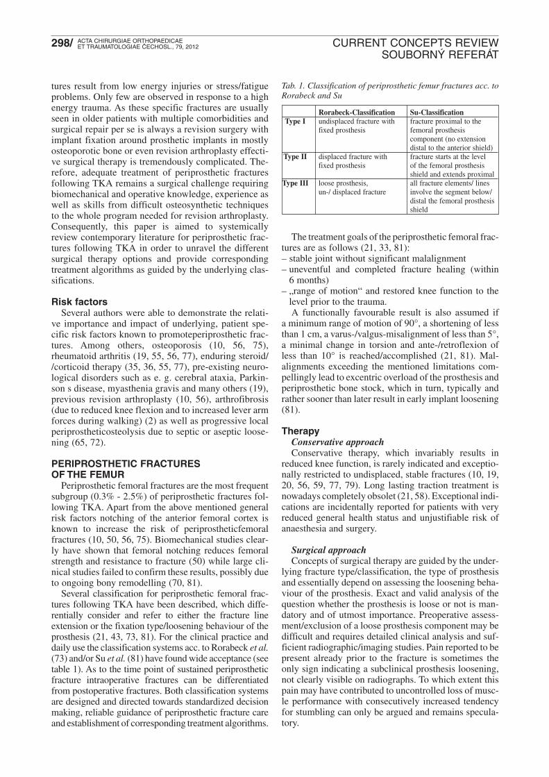
the tibial component, the forced application of impactiontechniques for the tibial head cancellous bone areas aswell as large osteolysis (9, 58). The classification of Felixet al. (Tab. 2), published in 1997 has attracted major inte -rest and found wide acceptance as it considers and refersto the time point of fracture (intra- versus postoperative),topography of the fracture to the tibial component as wellas the anchorage of the prosthesis (fixed versus loose)(25). Most likely due to the low incidence and the fre -quency of periprosthetic tibia fractures there is no validand generally approved treatment regimen available (25,43, 80). Again, loose and malpositioned prostheses re -quire exchange of the prosthesis and revision arthroplas-ty. Any extensive dislocation of fragments, or unstabledisplaced fractures are indications for operative interven-tion (24). Unstable intraoperative fractures nearly alwaysneed immediate fixation (58).
Therapy Conservative approachIntraoperatively caused undisplaced fractures which
are stable and first seen/diagnosed at the postoperativeradiograph do not necessarily need operative interven-tion. In those cases, an adaptation of the postoperativeweight bearing and radiographic controls are indicated(9). There is sufficient evidence to suggest that undis-placed fractures type II may also be treated successful-ly in a conservative manner (25).
tic/septic loosening prior to the trauma or that becameloose in response to the fracture. However, in selectedcases of very distal fracture zones (Typ III according toSu, Typ I or II according to Rorabeck) a revision withexchange of the prosthesis may be indicated as there isinsufficient space and bone stock for implant/screw fixa-tion in the distal fragment of any type of osteosyntheticreconstruction. Modular prosthesis designs with variab-le shaft and plateau designs, however, allow sometimeexchange of only one component with stable fracturebridging and fixation and direct postoperative fullweight bearing (14, 17, 81). According to our experien-ce it seems mandatory and helpful to have alternativeoptions available including also different types of semi- and constrained models in order to be preparedand equipped for any conceivable change, incl. total revi-sion and exchange of the entire system (14, 17, 54, 81).
Differential indication and correspondingtreatment algorithms based on the underlyingfracture classification
Exact assessment of possible/potential loosening ofthe prosthesis is a precondition for successful surgicaltreatment. In addition, information about previous sur-geries, other implants in situ (total hip arthroplasty, pro-ximal femur nails, screws and plates) decisively deter-mine the choice of implant and fracture fixation. Basedon the accepted classification system by Su et al.(81)and the design of the prosthesis together with the fixa-tion/loosening of the prosthesis we developed an algo-rithm for decision making and treatment planning ofperiprosthetic femur fractures following TKA.
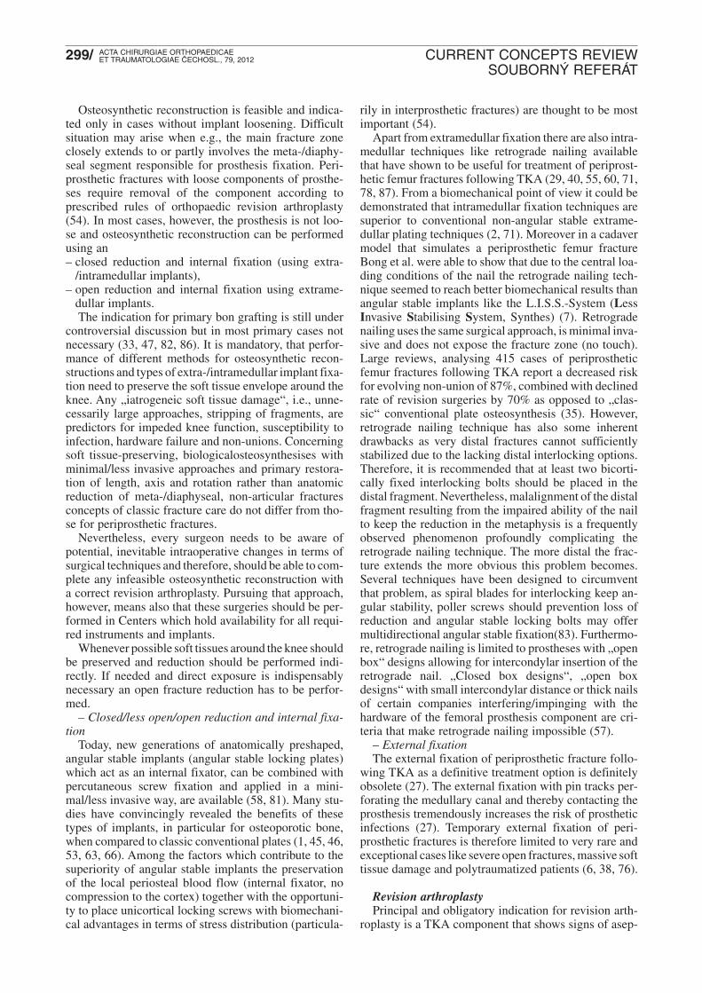
All fracture types from type I – II acc. to Su as wellas proximal type III-fractures are optimum candidatesfor osteosynthetic reconstructions, preferably usingangular stable plates that extramedullary overlap theintramedullar length of the femoral stem. However,encouraging results have also been reported for type I-und II-fractures by various authors using the retrogradeintramedullary nail (2, 29, 35, 40,55, 60, 71, 78, 87). However, un -known prosthesis systems whichmay only questionably have an openbox design or those with closed boxdesign (e.g. PS-variants) angularstable plate fixation techniquesrepresent the recommended treat-ment of choice (57, 58) (Fig. 1).
PERIPROSTHETIC TIBIALFRACTURES
Periprosthetic fractures of the tibiafollowing TKA are significantly ra -rely and infrequently observed whencompared to corresponding femurfractures, rating between 0.3 – 0.5%of all implanted TKA (9). Additio-nally to general above mentioned riskfactors specific factors also includevarus positioning and malrotation of
Felix-classificationType I fracture of the tibial head with involvement
of the prosthesis-implant interfaceType II fracture of the meta-/diaphyseal transitionType III fracture distal to the tibial componentType IV fractures of the tibial tuberosity
Subtype A stable prosthesisSubtype B loose prosthesisSubtype C intraoperative fracture
Tab. 2. Classification of periprosthetic tibial fractures acc. toFelix
Fig. 1. Treatment algorithm for periprosthetic femoral fractures (modified after [58]).
Surgical approachWhether the tibial component is tightly fixed or loo-
se is decisively guiding the resulting treatment concept.Similarly to the femoral situation any loose or malposi-tioned tibial component requires revision arthroplastywith exchange of the component. This may be perfor-med in combination with a osteosynthetic reconstruc -tion or using a long stem revision implant (43, 58). Inrare cases with periprosthetic fractures around alreadyimplanted revision arthroplasty systems additional aug-mentation techniques with either auto- or homologousbone may be necessary. In particular patients who al -ready suffer from a longlasting loosening of the tibialcomponent may have large bone defects (43, 64). In ana-logy to the femoral fractures there are similar techniqu-es and implant systems available for the management ofperiprosthetic tibial fractures.
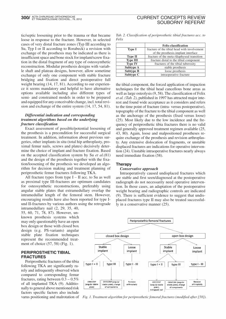
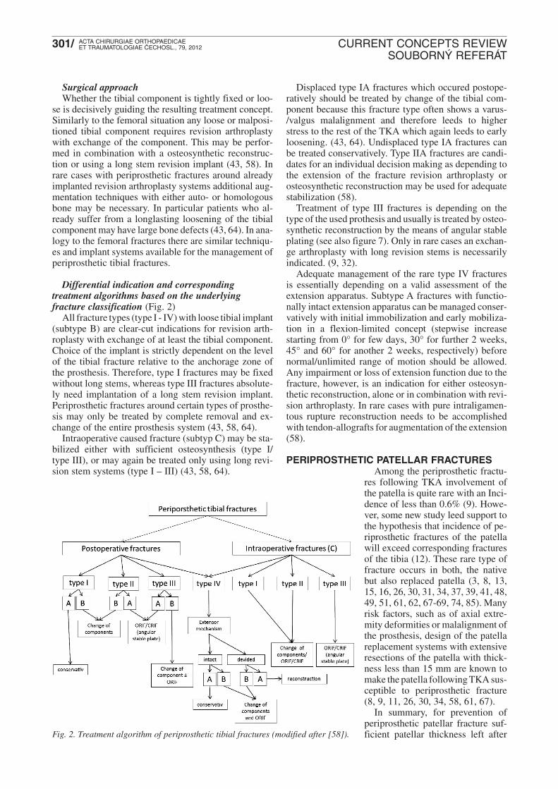
Differential indication and correspondingtreatment algorithms based on the underlyingfracture classification (Fig. 2)
All fracture types (type I - IV) with loose tibial implant(subtype B) are clear-cut indications for revision arth-roplasty with exchange of at least the tibial component.Choice of the implant is strictly dependent on the levelof the tibial fracture relative to the anchorage zone ofthe prosthesis. Therefore, type I fractures may be fixedwithout long stems, whereas type III fractures absolute-ly need implantation of a long stem revision implant.Periprosthetic fractures around certain types of prosthe-sis may only be treated by complete removal and ex -change of the entire prosthesis system (43, 58, 64).
Intraoperative caused fracture (subtyp C) may be sta-bilized either with sufficient osteosynthesis (type I/type III), or may again be treated only using long revi-sion stem systems (type I – III) (43, 58, 64).
Displaced type IA fractures which occured postope-ratively should be treated by change of the tibial com-ponent because this fracture type often shows a varus-/valgus malalignment and therefore leeds to higherstress to the rest of the TKA which again leeds to earlyloosening. (43, 64). Undisplaced type IA fractures canbe treated conservatively. Type IIA fractures are candi-dates for an individual decision making as depending tothe extension of the fracture revision arthroplasty orosteosynthetic reconstruction may be used for adequatestabilization (58).
Treatment of type III fractures is depending on thetype of the used prothesis and usually is treated by osteo -synthetic reconstruction by the means of angular stableplating (see also figure 7). Only in rare cases an exchan-ge arthroplasty with long revision stems is necessarilyindicated. (9, 32).
Adequate management of the rare type IV fracturesis essentially depending on a valid assessment of theextension apparatus. Subtype A fractures with functio-nally intact extension apparatus can be managed conser -vatively with initial immobilization and early mobiliza-tion in a flexion-limited concept (stepwise increasestarting from 0° for few days, 30° for further 2 weeks,45° and 60° for another 2 weeks, respectively) beforenormal/unlimited range of motion should be allowed.Any impairment or loss of extension function due to thefracture, however, is an indication for either osteosyn -thetic reconstruction, alone or in combination with revi-sion arthroplasty. In rare cases with pure intraligamen-tous rupture reconstruction needs to be accomplishedwith tendon-allografts for augmentation of the exten sion(58).
PERIPROSTHETIC PATELLAR FRACTURESAmong the periprosthetic fractu-
res following TKA involvement ofthe patella is quite rare with an Inci-dence of less than 0.6% (9). Howe-ver, some new study leed support tothe hypothesis that incidence of pe -riprosthetic fractures of the patellawill exceed corresponding fracturesof the tibia (12). These rare type offracture occurs in both, the nativebut also replaced patella (3, 8, 13,15, 16, 26, 30, 31, 34, 37, 39, 41, 48,49, 51, 61, 62, 67-69, 74, 85). Manyrisk factors, such as of axial extre-mity deformities or malalignment ofthe prosthesis, design of the patellareplacement systems with extensiveresections of the patella with thick-ness less than 15 mm are known tomake the patella following TKA sus-ceptible to periprosthetic fracture (8, 9, 11, 26, 30, 34, 58, 61, 67).
In summary, for prevention ofperiprosthetic patellar fracture suf -ficient patellar thickness left afterFig. 2. Treatment algorithm of periprosthetic tibial fractures (modified after [58]).
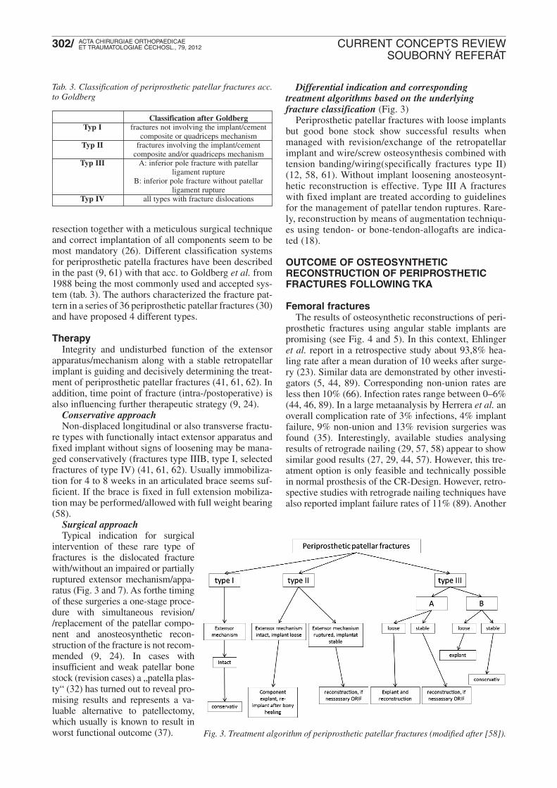
Differential indication and correspondingtreatment algorithms based on the underlyingfracture classification (Fig. 3)
Periprosthetic patellar fractures with loose implantsbut good bone stock show successful results whenmanaged with revision/exchange of the retropatellarimplant and wire/screw osteosynthesis combined withtension banding/wiring(specifically fractures type II)(12, 58, 61). Without implant loosening anosteosynt-hetic reconstruction is effective. Type III A fractureswith fixed implant are treated according to guidelinesfor the ma nagement of patellar tendon ruptures. Rare-ly, reconstruction by means of augmentation techniqu-es using tendon- or bone-tendon-allogafts are indica-ted (18).
OUTCOME OF OSTEOSYNTHETIC RECONSTRUCTION OF PERIPROSTHETICFRACTURES FOLLOWING TKA
Femoral fracturesThe results of osteosynthetic reconstructions of peri-
prosthetic fractures using angular stable implants arepromising (see Fig. 4 and 5). In this context, Ehlingeret al. report in a retrospective study about 93,8% hea-ling rate after a mean duration of 10 weeks after surge-ry (23). Similar data are demonstrated by other investi-gators (5, 44, 89). Corresponding non-union rates areless then 10% (66). Infection rates range between 0–6%(44, 46, 89). In a large metaanalysis by Herrera et al. anoverall complication rate of 3% infections, 4% implantfailure, 9% non-union and 13% revision surgeries wasfound (35). Interestingly, available studies analysingresults of retrograde nailing (29, 57, 58) appear to showsimilar good results (27, 29, 44, 57). However, this tre-atment option is only feasible and technically possiblein normal prosthesis of the CR-Design. However, retro -spective studies with retrograde nailing techniques havealso reported implant failure rates of 11% (89). Another
resection together with a meticulous surgical techniqueand correct implantation of all components seem to bemost mandatory (26). Different classification systemsfor periprosthetic patella fractures have been describedin the past (9, 61) with that acc. to Goldberg et al. from1988 being the most commonly used and accepted sys-tem (tab. 3). The authors characterized the fracture pat-tern in a series of 36 periprosthetic patellar fractures (30)and have proposed 4 different types.
TherapyIntegrity and undisturbed function of the extensor
apparatus/mechanism along with a stable retropatellarimplant is guiding and decisively determining the treat-ment of periprosthetic patellar fractures (41, 61, 62). Inaddition, time point of fracture (intra-/postoperative) isalso influencing further therapeutic strategy (9, 24).
Conservative approachNon-displaced longitudinal or also transverse fractu-
re types with functionally intact extensor apparatus andfixed implant without signs of loosening may be mana-ged conservatively (fractures type IIIB, type I, selectedfractures of type IV) (41, 61, 62). Usually immobiliza-tion for 4 to 8 weeks in an articulated brace seems suf-ficient. If the brace is fixed in full extension mobiliza -tion may be performed/allowed with full weight bearing(58).
Surgical approachTypical indication for surgical
intervention of these rare type offractures is the dislocated fracturewith/without an impaired or partiallyruptured extensor mechanism/appa-ratus (Fig. 3 and 7). As forthe timingof these surgeries a one-stage proce-dure with simultaneous revision//replacement of the patellar compo-nent and anosteosynthetic recon-struction of the fracture is not recom-mended (9, 24). In cases withinsufficient and weak patellar bonestock (revision cases) a „patella plas-ty“ (32) has turned out to reveal pro-mising results and represents a va -luable alternative to patellectomy,which usually is known to result inworst functional outcome (37).
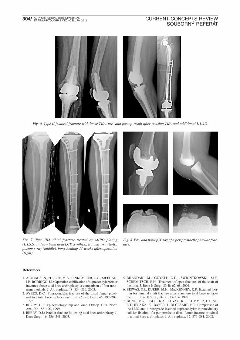
problem, frequently coming up during insertion of thenail and reduction of the fracture, is axial malalignmentof the distal femur, due to loss of reduction in the fun-nel-shaped metaphyseal distal femur bone segment (88,89). As mentioned before, in loose TKAs the revisionarthroplasty is the gold standard. In some cases an addi-tional plate osteosynthesis may be required in order toachive stable conditions for early mobilisation (seeFig. 6).
Tibial fractures To date, for periprosthetic tibia fractures following
TKA there are no valid data available. Most likely thisis the result of the infrequency incidence of these frac-tures. Valid data with recommendations for treatmentstrategies are also lacking (25, 84).
Patellar fracturesOnly a limited number of studies have analyzed the
treatment results of periprosthetic patella fractures . Orti-guera and Berry present patient data with a healing rate
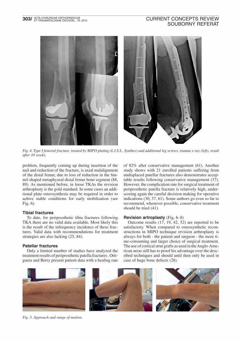
Fig. 4. Type I femoral fracture, treated by MIPO plating (L.I.S.S., Synthes) and additional leg screws, trauma x-ray (left), resultafter 10 weeks.
Fig. 5. Approach and range of motion.
of 82% after conservative management (61). Anotherstudy shows with 21 enrolled patients suffering fromundisplaced patellar fractures also demonstrates accep-table results following conservative management (37).However, the complication rate for surgical treatment ofperiprosthetic patella fracture is relatively high, under -scoring again the careful decision making for operativeindications (30, 37, 61). Some authors go even so far torecommend, whenever possible, conservative treatmentshould be tried (41).
Revision artroplasty (Fig. 6–8)Outcome results (17, 19, 42, 52) are reported to be
satisfactory. When compared to osteosynthetic recon-structions in MIPO technique revision arthroplasty isalways for both - the patient and surgeon - the more ti -me-consuming and larger choice of surgical treatment.The use of cortical strut grafts as used in the Anglo-Ame-rican areas still has to proof his advantage over the desc-ribed techniques and should until then only be used incase of huge bone defects (28).
Fig. 7. Type IIIA tibial fracture treated by MIPO plating(L.I.S.S. and low bend tibia LCP, Synthes), trauma x-ray (left),postop x-ray (middle), bony healing 11 weeks after operation(right).
5. BHANDARI M., GUYATT, G.H., SWIONTKOWSKI, M.F.,SCHEMITSCH, E.H.: Treatment of open fractures of the shaft ofthe tibia. J. Bone Jt Surg., 83-B: 62–68, 2001.
6. BISWAS, S.P., KURER, M.H., MacKENNEY, R.P.: External fixa-tion for femoral shaft fracture after Stanmore total knee replace-ment. J. Bone Jt Surg., 74-B: 313–314, 1992.
7. BONG, M.R., EGOL, K.A., KOVAL, K.J., KUMMER, F.J., SU,E.T., IESAKA, K., BAYER, J., DI CESARE, P.E.: Comparison ofthe LISS and a retrograde-inserted supracondylar intramedullarynail for fixation of a periprosthetic distal femur fracture proximalto a total knee arthroplasty. J. Arthroplasty, 17: 876–881, 2002.
References
1. ALTHAUSEN, P.L., LEE, M.A., FINKEMEIER, C.G., MEEHAN,J.P., RODRIGO, J.J.: Operative stabilization of supracondylar femurfractures above total knee arthroplasty: a comparison of four treat-ment methods. J. Arthroplasty, 18: 834–839, 2003.
2. AYERS, D.C.: Supracondylar fracture of the distal femur proxi-mal to a total knee replacement. Instr. Course Lect., 46: 197–203,1997.
3. BERRY, D.J.: Epidemiology: hip and knee. Orthop. Clin. NorthAm., 30: 183–190, 1999.
4. BERRY, D.J.: Patellar fracture following total knee arthroplasty. J.Knee Surg., 16: 236–241, 2003.
Fig. 6. Type II femoral fracture with loose TKA, pre- and postop result after revision TKA and additional L.I.S.S.
17. CORDEIRO, E.N., COSTA, R.C., CARAZZATO. J.G., SILVAJDOS, S.: Periprosthetic fractures in patients with total knee arth-roplasties. Clin. Orthop. Relat. Res., 252: 182–189, 1990.
18. CROSSETT, L.S., SINHA, R.K., SECHRIEST, V.F., RUBASH,H.E.: Reconstruction of a ruptured patellar tendon with achillestendon allograft following total knee arthroplasty. J. Bone Jt Surg.,84-A: 1354–1361, 2002.
20. DELPORT, P.H., VAN AUDEKERCKE, R., MARTENS, M.,MULIER, J.C.: Conservative treatment of ipsilateral supracondy-lar femoral fracture after total knee arthroplasty. J. Trauma, 24:846–849, 1984.
21. DIGIOIA, A.M., 3rd, RUBASH, H.E.: Periprosthetic fractures ofthe femur after total knee arthroplasty. A literature review and tre-atment algorithm. Clin. Orthop. Relat. Res., 271: 135–142, 1991.
22. DORR, L.D.: Fractures following total knee arthroplasty. Ortho-pedics, 20: 848–850, 1997.
23. EHLINGER, M., ADAM, P., ABANE, L., RAHME, M., MOOR,B.K., ARLETTAZ, Y., BONNOMET, F.: Treatment of peri -prosthetic femoral fractures of the knee. Knee Surg. Sports Trau-matol. Arthrosc., 2011.
25. FELIX, N.A., STUART, M.J., HANSSEN, A.D.: Periprostheticfractures of the tibia associated with total knee arthroplasty. Clin.Orthop. Relat. Res., 345: 113–124, 1997.
26. FIGGIE, H.E., 3rd, GOLDBERG, V.M., FIGGIE, M.P., INGLIS,A.E., KELLY, M., SOBEL, M.: The effect of alignment of theimplant on fractures of the patella after condylar total knee arth-roplasty. J. Bone Jt Surg., 71-A: 1031–1039, 1989.
27. FIGGIE, M.P., GOLDBERG, V.M., FIGGIE, H.E., 3rd,SOBEL, M.: The results of treatment of supracondylar fractureabove total knee arthroplasty. J. Arthroplasty, 5: 267–276, 1990.
28. FONT-VIZCARRA, L., FERNANDEZ-VALENCIA, J.A., GAL-LART, X., SEGUR. J.M., PRAT. S., RIBA, J.: Cortical strut allo-graft as an adjunct to plate fixation for periprosthetic fractures ofthe femur. Hip Int., 20: 43–49, 2010.
29. GLIATIS, J., MEGAS, P., PANAGIOTOPOULOS, E., LAMBI-RIS, E.: Midterm results of treatment with a retrograde nail forsupracondylar periprosthetic fractures of the femur following totalknee arthroplasty. J. Orthop. Trauma, 19: 164–170, 2005.
30. GOLDBERG, V.M., FIGGIE, H.E., 3rd, INGLIS, A.E., FIG-GIE, M.P., SOBEL, M., KELLY, M., KRAAY, M.: Patellar frac-ture type and prognosis in condylar total knee arthroplasty. Clin.Orthop. Relat. Res., 236: 115–122, 1988.
31. GRACE, J.N., SIM, F.H.: Fracture of the patella after total kneearthroplasty. Clin. Orthop. Relat. Res., 230: 168–175, 1988.
33. HEALY, W.L., SILISKI, J.M., INCAVO, S.J.: Operative treatmentof distal femoral fractures proximal to total knee replacements. J.Bone Jt Surg., 75-A: 27–34, 1993.
34. HEALY, W.L., WASILEWSKI, S.A., TAKEI, R., OBERLAN-DER, M.: Patellofemoral complications following total knee arth-roplasty. Correlation with implant design and patient risk factors.J. Arthroplasty, 10: 197–201, 1995.
35. HERRERA, D.A., KREGOR, P.J., COLE, P.A., LEVY, B.A.,JONSSON, A., ZLOWODZKI, M.: Treatment of acute distalfemur fractures above a total knee arthroplasty: systematic reviewof 415 cases (1981–2006). Acta Orthop., 79: 22–27, 2008.
36. HIRSH, D.M., BHALLA, S., ROFFMAN, M.: Supracondylarfracture of the femur following total knee replacement. Report offour cases. J. Bone Jt Surg., 63-A: 162–163, 1981.
37. HOZACK, W.J., GOLL, S.R., LOTKE, P.A., ROTHMAN, R.H.,BOOTH, R.E., Jr.: The treatment of patellar fractures after totalknee arthroplasty. Clin. Orthop. Relat. Res., 236: 123–127, 1988.
38. HURSON, C., SYNNOTT, K., McCORMACK, D.: Above-kneeIlizarov external fixation for early periprosthetic supracondylarfemoral fracture -a case report. Knee, 12: 145–147, 2005.
39. INSALL, J.N., LACHIEWICZ, P.F., BURSTEIN, A.H.: The poste-rior stabilized condylar prosthesis: a modification of the total con-dylar design. Two to four-year clinical experience. J. Bone Jt Surg.,64-A: 1317–1323, 1982.
40. JABCZENSKI, F.F., CRAWFORD, M.: Retrograde intramedulla-ry nailing of supracondylar femur fractures above total knee arth-roplasty. A preliminary report of four cases. J. Arthroplasty, 10:95–101, 1995.
43. KIM, K.I., EGOL, K.A., HOZACK, W.J., PARVIZI, J.: Peri-prosthetic fractures after total knee arthroplasties. Clin. Orthop.Relat. Res., 446: 167–175, 2006.
44. KOLB, W., GUHLMANN, H., WINDISCH, C., MARX, F., KOL-LER, H., KOLB, K.: Fixation of periprosthetic femur fracturesabove total knee arthroplasty with the less invasive stabilizationsystem: a midterm follow-up study. J. Trauma, 69: 670–676, 2010.
45. KOVAL, K.J., HOEHL, J.J., KUMMER, F.J., SIMON, J.A.: Distalfemoral fixation: a biomechanical comparison of the standard con-dylar buttress plate, a locked buttress plate, and the 95-degree bla-de plate. J. Orthop. Trauma, 11: 521–524, 1997.
46. KREGOR, P.J., HUGHES, J.L., COLE, P.A.: Fixation of distalfemoral fractures above total knee arthroplasty utilizing the LessInvasive Stabilization System (L.I.S.S.). Injury 32: SC64–75, 2001.
47. KUMAR, A., CHAMBERS, I., MAISTRELLI, G., WONG, P.:Management of periprosthetic fracture above total knee arthro-plasty using intramedullary fibular allograft and plate fixation. J.Arthroplasty, 23: 554–558, 2008.
48. LARSON, C.M., McDOWELL, C.M., LACHIEWICZ, P.F.: One-peg versus three-peg patella component fixation in total knee arth-roplasty. Clin. Orthop. Relat. Res., 392: 94–100, 2001.
49. LE, A.X., CAMERON, H.U., OTSUKA, N.Y., HARRINGTON,I.J., BHARGAVA, M.: Fracture of the patella following total kneearthroplasty. Orthopedics 22: 395–398; discussion 398–399, 1999.
50. LESH, M.L., SCHNEIDER, D.J., DEOL, G., DAVIS, B.,JACOBS, C.R., PELLEGRINI, V.D., Jr.: The consequences ofanterior femoral notching in total knee arthroplasty. A biomecha-nical study. J. Bone Jt Surg., 82-A: 1096–1101, 2000.
51. LYNCH, A.F., RORABECK, C.H., BOURNE, R.B.: Extensormechanism complications following total knee arthroplasty. J.Arthroplasty, 2: 135–140, 1987.
73. RORABECK, C.H., TAYLOR, J.W.: Classification of periprost-hetic fractures complicating total knee arthroplasty. Orthop. Clin.North Am., 30: 209–214, 1999.
74. SCOTT, R.D., TUROFF, N., EWALD, F.C.: Stress fracture of thepatella following duopatellar total knee arthroplasty with patellarresurfacing. Clin. Orthop. Relat. Res., 170: 147–151, 1982.
75. SHAWEN, S.B., BELMONT, P.J., JR., KLEMME, W.R., TOPO-LESKI, L.D., XENOS, J.S., ORCHOWSKI, J.R.: Osteoporosisand anterior femoral notching in periprosthetic supracondylarfemoral fractures: a biomechanical analysis. J. Bone Jt Surg., 85-A: 115–121, 2003.
76. SIMON, R.G., BRINKER, M.R.: Use of Ilizarov external fixati-on for a periprosthetic supracondylar femur fracture. J. Arthro-plasty, 14: 118–121, 1999.
78. SMITH, W.J., MARTIN, S.L., MABREY, J.D.: Use of a supra-condylar nail for treatment of a supracondylar fracture of the femurfollowing total knee arthroplasty. J. Arthroplasty, 11: 210–213,1996.
79. SOCHART, D.H., HARDINGE, K.: Nonsurgical management ofsupracondylar fracture above total knee arthroplasty. Still the nine-ties option. J. Arthroplasty, 12: 830–834, 1997.
80. STUART, M.J., HANSSEN, A.D.: Total knee arthroplasty: peri-prosthetic tibial fractures. Orthop. Clin. North Am.: 30: 279–286,1999.
81. SU, E.T., DEWAL, H., DI CESARE, P.E.: Periprosthetic femoralfractures above total knee replacements. J. Am. Acad. Orthop.Surg., 12: 12–20, 2004.
82. TANI, Y., INOUE. K., KANEKO, H., NISHIOKA, J., HUKUDA,S.: Intramedullary fibular graft for supracondylar fracture of thefemur following total knee arthroplasty. Arch. Orthop. TraumaSurg., 117: 103–104, 1998.
83. THELEN, S., BETSCH, M., GRASSMANN, J.P., SPOOR, V.,EICHLER, C., KOEBKE, J., WINDOLF, J., HAKIMI, M.,WILD, M.: Angle stable locking nails versus conventionally loc-ked intramedullary nails in proximal tibial shaft fractures: a bio-mechanical study. Arch. Orthop. Trauma Surg., [Epub 2011 Aug30.], 132: 57–63, 2012.
84. THOMPSON, N.W., MCALINDEN, M.G., BRESLIN, E., CRO-NE, M.D., KERNOHAN, W.G., BEVERLAND, D.E.: Periprost-hetic tibial fractures after cementless low contact stress total kneearthroplasty. J. Arthroplasty, 16: 984–990, 2001.
86. WANG, J.W., WANG, C.J.: Supracondylar fractures of the femurabove total knee arthroplasties with cortical allograft struts. J. Arth-roplasty, 17: 365–372, 2002.
88. WICK, M., MULLER, E.J., MUHR, G.: [Supracondylar femoralfractures in knee endoprostheses. Stabilizing with retrograde inter-locking nail]. Unfallchirurg, 104: 410–413, 2001.
89. WICK, M., MULLER, E.J., KUTSCHA-LISSBERG, F., HOPF,F., MUHR, G.: [Periprosthetic supracondylar femoral fractures:LISS or retrograde intramedullary nailing? Problems with the useof minimally invasive technique]. Unfallchirurg, 107: 181–188,2004.
Corresponding author:Sven Märdian, M. D.Charité – Universitätsmedizin BerlinCMSC – Center ofmusculosceletalsurgeryAugustenburger Platz 1D – 13353 Berlin, GermanyE-mail: [email protected]
52. MANIAR, R.N., UMLAS, M.E., RODRIGUEZ, J.A., RANAWAT,C.S.: Supracondylar femoral fracture above a PFC posterior cru-ciate-substituting total knee arthroplasty treated with supracondy-lar nailing. A unique technical problem. J. Arthroplasty, 11:637–639, 1996.
53. MARTI, A., FANKHAUSER, C., FRENK, A., CORDEY, J., GAS-SER, B.: Biomechanical evaluation of the less invasive stabiliza-tion system for the internal fixation of distal femur fractures. J.Orthop. Trauma, 15: 482–487, 2001.
54. McGRAW, P., KUMAR, A.: Periprosthetic fractures of the femurafter total knee arthroplasty. J. Orthop. Traumatol., 11: 135–141,2010.
55. McLAREN, A.C., DUPONT, J.A., SCHROEBER, D.C.: Openreduction internal fixation of supracondylar fractures above totalknee arthroplasties using the intramedullary supracondylar rod.Clin. Orthop. Relat. Res., 302: 194–198, 1994.
56. MERKEL, K.D., JOHNSON, E.W., Jr.: Supracondylar fracture ofthe femur after total knee arthroplasty. J. Bone Jt Surg., 68-A:29–43, 1986.
60. MURRELL, G.A., NUNLEY, J.A.: Interlocked supracondylarintramedullary nails for supracondylar fractures after total kneearthroplasty. A new treatment method. J. Arthroplasty, 10: 37–42,1995.
61. ORTIGUERA, C.J., BERRY, D.J.: Patellar fracture after total kneearthroplasty. J. Bone Jt Surg., 84-A: 532–540, 2002.
62. PARVIZI, J., KIM, K.I., OLIASHIRAZI, A., ONG, A., SHAR-KEY, P.F.: Periprosthetic patellar fractures. Clin. Orthop. Relat.Res., 446: 161–166, 2006.
63. RAAB, G.E., DAVIS, C.M., 3rd: Early healing with locked con-dylar plating of periprosthetic fractures around the knee. J. Arth-roplasty, 20: 984–989, 2005.
64. RAND, J.A., COVENTRY, M.B.: Stress fractures after total kneearthroplasty. J. Bone Jt Surg., 62-A: 226–233, 1980.
65. RAND, J.A.: Supracondylar fracture of the femur associated withpolyethylene wear after total knee arthroplasty. A case report. J.Bone Jt Surg., 76-A: 1389–1393, 1994.
66. RICCI, W.M., LOFTUS, T., COX, C., BORRELLI, J.: Locked pla-tes combined with minimally invasive insertion technique for thetreatment of periprosthetic supracondylar femur fractures abovea total knee arthroplasty. J. Orthop. Trauma, 20: 190–196, 2006.
67. RITTER, M.A., CAMPBELL, E.D.: Postoperative patellar com-plications with or without lateral release during total knee arthro-plasty. Clin. Orthop. Relat. Res., 219: 163–168, 1987.
68. RITTER, M.A., HERBST, S.A., KEATING, E.M., FARIS, P.M.,MEDING, J.B.: Patellofemoral complications following total kneearthroplasty. Effect of a lateral release and sacrifice of the superi-or lateral geniculate artery. J. Arthroplasty, 11: 368–372, 1996.
70. RITTER, M.A., THONG, A.E., KEATING, E.M., FARIS, P.M.,MEDING, J.B., BEREND, M.E., PIERSON, J.L., DAVIS, K.E.:The effect of femoral notching during total knee arthroplasty onthe prevalence of postoperative femoral fractures and on clinicaloutcome. J. Bone Jt Surg., 87-A: 2411–2414, 2005.
71. ROLSTON, L.R., CHRIST, D.J., HALPERN, A., O’CONNOR,P.L., RYAN, T.G., UGGEN, W.M.: Treatment of supracondylarfractures of the femur proximal to a total knee arthroplasty. A reportof four cases. J. Bone Jt Surg., 77-A: 924–931, 1995.
72. RORABECK, C.H., TAYLOR, J.W.: Periprosthetic fractures of thefemur complicating total knee arthroplasty. Orthop. Clin. NorthAm., 30: 265–277, 1999.